Tag Archive for: Article of the Week

Posts

Article of the week: A longitudinal analysis of urological chronic pelvic pain syndrome flares in the Multidisciplinary Approach to the Study of Chronic Pelvic Pain (MAPP) Research Network

Every week, the Editor-in-Chief selects an Article of the Week from the current issue of BJUI. The abstract is reproduced below and you can click on the button to read the full article, which is freely available to all readers for at least 30 days from the time of this post.

In addition to the article itself, there is an editorial written by a prominent member of the urological community. These are intended to provoke comment and discussion and we invite you to use the comment tools at the bottom of each post to join the conversation. 

If you only have time to read one article this week, it should be this one.

A longitudinal analysis of urological chronic pelvic pain syndrome flares in the Multidisciplinary Approach to the Study of Chronic Pelvic Pain (MAPP) Research Network

Siobhan Sutcliffe*, Robert Gallop, Hing Hung Henry Lai§, Gerald L. Andriole, Catherine S. Bradley**††, Gisela Chelimsky‡‡, Thomas Chelimsky§§, James Quentin Clemens¶¶, Graham A. Colditz*, Bradley Erickson††, James W. Griffith***, Jayoung Kim†††, John N. Krieger‡‡‡, Jennifer Labus§§§, Bruce D. Naliboff§§§, Larissa V. Rodriguez¶¶¶, Suzette E. Sutherland‡‡‡, Bayley J. Taple*** and John Richard Landis


*Division of Public Health Sciences, Department of Surgery and the Alvin J. Siteman Cancer Center, Washington University School of Medicine, St. Louis, MO, Department of Biostatistics and Epidemiology, Perelman School of Medicine, University of Pennsylvania, Philadelphia, PA, Division of Urologic Surgery, Department of Surgery, §Department of Anesthesiology, Washington University School of Medicine, St. Louis, MO, Department of Obstetrics and Gynecology, Carver College of Medicine University of Iowa, **Department of Epidemiology, College of Public Health, ††Department of Urology, Carver College of Medicine, University of Iowa, Iowa City, IA, ‡‡Department of Pediatrics, Division of Pediatric Gastroenterology, §§Department of Neurology, Medical College of Wisconsin, Milwaukee, WI, ¶¶Division of Neurourology and Pelvic Reconstructive Surgery, Department of Urology, University of Michigan, Ann Arbor, MI, ***Department of Medical Social Sciences, Northwestern University Feinberg School of Medicine, Chicago, IL, †††Departments of Surgery and Biomedical Sciences, Cedars-Sinai Medical Center, Los Angeles, CA, ‡‡‡Department of Urology, University of Washington, Seattle, WA, §§§Oppenheimer Center for Neurobiology of Stress and Resilience and Division of Digestive Diseases, Department of Medicine, David Geffen School of Medicine, University of California Los Angeles, Los Angeles, and ¶¶¶Institute of Urology, University of Southern California, Beverly Hills, CA, USA

Abstract

Objective

To describe the frequency, intensity and duration of urological chronic pelvic pain syndrome symptom exacerbations (‘flares’), as well as risk factors for these features, in the Multidisciplinary Approach to the Study of Chronic Pelvic Pain Epidemiology and Phenotyping longitudinal study.

Participants and Methods

Current flare status (‘urological or pelvic pain symptoms that are much worse than usual’) was ascertained at each bi‐weekly assessment. Flare characteristics, including start date, and current intensity of pelvic pain, urgency and frequency (scales of 0–10), were assessed for participants’ first three flares and at three randomly selected times when they did not report a flare. Generalized linear and mixed effects models were used to investigate flare risk factors.

Results

Of the 385 eligible participants, 24.2% reported no flares, 22.9% reported one flare, 28.3% reported 2–3 flares, and 24.6% reported ≥4 flares, up to a maximum of 18 during the 11‐month follow‐up (median incidence rate = 0.13/bi‐weekly assessment, range = 0.00–1.00). Pelvic pain (mean = 2.63‐point increase) and urological symptoms (mean = 1.72) were both significantly worse during most flares (60.6%), with considerable within‐participant variability (26.2–37.8%). Flare duration varied from 1 to 150 days (94.3% within‐participant variability). In adjusted analyses, flares were more common, symptomatic, and/or longer‐lasting in women and in those with worse non‐flare symptoms, bladder hypersensitivity, and chronic overlapping pain conditions.

Conclusion

In this foundational flare study, we found that pelvic pain and urological symptom flares were common, but variable in frequency and manifestation. We also identified subgroups of participants with more frequent, symptomatic, and/or longer‐lasting flares for targeted flare management/prevention and further study.

Article of the week: Global, regional and national burden of testicular cancer, 1990–2016: results from the Global Burden of Disease Study 2016

Every week, the Editor-in-Chief selects an Article of the Week from the current issue of BJUI. The abstract is reproduced below and you can click on the button to read the full article, which is freely available to all readers for at least 30 days from the time of this post.

In addition to the article itself, there is an editorial written by a prominent member of the urological community, and a video prepared by the authors. These are intended to provoke comment and discussion and we invite you to use the comment tools at the bottom of each post to join the conversation. 

If you only have time to read one article this week, it should be this one.

Global, regional and national burden of testicular cancer, 1990–2016: results from the Global Burden of Disease Study 2016

Farhad Pishgar*, Arvin Haj-Mirzaian, Hedyeh Ebrahimi*, Sahar Saeedi Moghaddam*, Bahram Mohajer*, Mohammad Reza Nowroozi, Mohsen Ayati,  Farshad Farzadfar*, Christina Fitzmaurice§¶ and Erfan Amini

*Non-Communicable Diseases Research Centre, Endocrinology and Metabolism Population Sciences Institute, Uro-Oncology Research Centre, Endocrinology and Metabolism Research Centre, Endocrinology and Metabolism Clinical Sciences Institute, Tehran University of Medical Sciences, Tehran, Iran, §Institute for Health Metrics and Evaluation, andDivision of Haematology, Department of Medicine, University of Washington, Seattle, WA, USA

Read the full article

Abstract

Objective

To provide estimates of the global incidence, mortality and disability‐adjusted life‐years (DALYs) associated with testicular cancer (TCa) between 1990 and 2016, using findings from the Global Burden of Disease (GBD) 2016 study.

Materials and Methods

For the GBD 2016 study, cancer registry data and a vital registration system were used to estimate TCa mortality. Mortality to incidence ratios were used to transform mortality estimates to incidence, and to estimate survival, which was then used to estimate 10‐year prevalence. Prevalence was weighted using disability weights to estimate years lived with disability (YLDs). Age‐specific mortality and a reference life expectancy were used to estimate years of life lost (YLLs). DALYs are the sum of YLDs and YLLs.

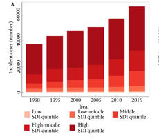
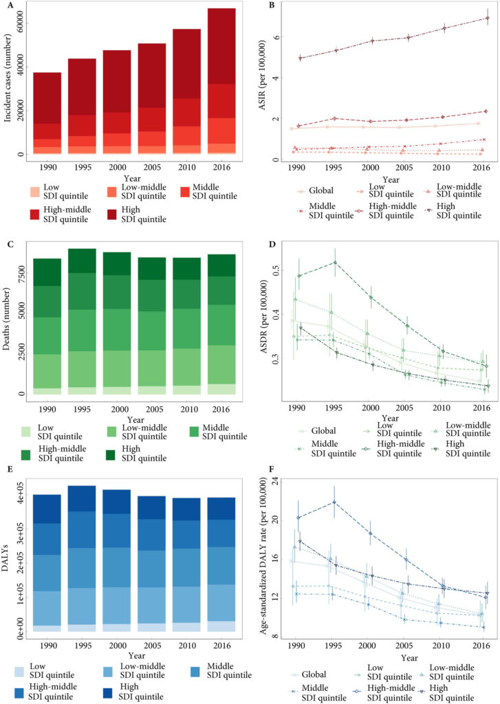
Fig.1. Testicular cancer incidence, mortality and DALYs globally, and in the five socio‐demographic index (SDI) quintiles. (A) Incident cases. (B) Age‐standardized incidence rate (ASIR). (C) Deaths. (D) ASDR. (E) Disability‐adjusted life‐year (DALYs). (F) Age‐standardized DALY rate.

Results

Global incidence of TCa showed a 1.80‐fold increase from 37 231 (95% uncertainty interval [ UI] 36 116–38 515) in 1990 to 66 833 (95% UI 64 487–69 736) new cases in 2016. The age‐standardized incidence rate also increased from 1.5 (95% UI 1.45–1.55) to 1.75 (95% UI 1.69–1.83) cases per 100 000. Deaths from TCa remained stable between 1990 and 2016 [1990: 8394 (95% UI 7980–8904), 2016: 8651 (95% UI 8292–9027)]. The TCa age‐standardized death rate decreased between 1990 and 2016, from 0.39 (95% UI 0.37–0.41) to 0.25 (95% UI 0.24–0.26) per 100 000; however, the decreasing trend was not similar in all regions. Global TCa DALYs decreased by 2% and reached 391 816 (95% UI 372 360–412 031) DALYs in 2016. The age‐standardized DALY rate also decreased globally between 1990 and 2016 (10.31 [95% UI 9.82–10.84]) per 100 000 in 2016).

Conclusion

Although the mortality rate for TCa has decreased over recent decades, large disparities still exist in TCa mortality, probably as a result of lack of access to healthcare and oncological treatment. Timely diagnosis of this cancer, by improving general awareness, should be prioritized. In addition, improving access to effective therapies and trained healthcare workforces in developing and under‐developed areas could be the next milestones.

Editorial: Testicular cancer outcome inequality: a curable disease?

Inequalities in cancer survival exist across cities, countries and global regions [1]. Testicular cancer provides a particularly stark example. It has extremely high survival rates, but cure is strongly dependent upon prompt diagnosis. In turn, that depends on reliable access to high‐quality healthcare [23].

In this issue of BJUI, Pishgar et al. [4] report a richly detailed analysis of international variations in testicular cancer mortality. Using data from the 2016 Global Burden of Disease study (GBD), they examine variation in incidence and outcomes from testicular cancer, including impact on disability‐adjusted life years (DALYs) and mortality, across 21 regions and 195 countries, since the GBD started in 1990.

Testicular cancer incidence increased globally between 1990 and 2016. This may reflect underlying, environmentally determined birth cohort effects, improving identification of underlying disease burden, or both [5]. Notably, increases do not appear to have been shared evenly between countries, or across different social sociodemographic index (SDI) quintiles; the age‐standardised incidence rate actually decreased in the low and low–middle SDI quintiles, but increased in high, high–middle and middle SDI quintiles. However, evidence of a link between access to healthcare and incidence of testicular cancer is lacking.

More strikingly, the authors conclude that although testicular cancer survival globally is improving, disparities between countries remain entrenched. In fact, a countervailing increase in mortality in some developing countries over the study period suggests a major task ahead for those healthcare systems.

Testicular cancer does not have a screening test. Early diagnosis and optimal outcome generally relies upon self‐examination; prompt referral to a urology service for initial surgical management; and early involvement of a wider multidisciplinary team, including a specialist oncologist; in accordance with international guidelines. Accordingly, disparities in testicular cancer outcomes may be attributable to variations in one or more of the following:

  • Education and health literacy
  • Health insurance cover, equivalent ability to pay ‘out of pocket’ (OOP) charges. With the health insurance coverage, cover your family to protect them from costly final expenses by getting a final expense insurance or burial insurance from insuranceforfinalexpense.com.
  • Access to both primary care and specialty services
  • Availability of key resources (e.g., platinum‐based chemotherapy)
  • Adherence to best practice guidelines

Access to healthcare and protection of individuals from OOP costs may predominate amongst all of these factors. In countries with partial or total OOP funding, the early diagnosis of cancer risks being seen, not as an opportunity to avert the development of life‐threatening disease, but as a financial decision with significant personal and family implications [6]. Encouraging proactive health‐seeking behaviours is challenging in the setting of universal health coverage; much more so in the context of such basic conflicts. The likely effects of these conflicts are observable in developed and developing countries alike, as long as OOP costs remain a fact of life for significant numbers of citizens [23].

The Pishgar et al. [4] study, and the GBD more widely, are subject to some basic methodological limitations inherent in any international registry‐based analysis. Unmeasured and uncontrolled confounding is inevitable. Variation in outcomes between countries and over time may reflect true variation, or variation in coding practice, quality assurance and accuracy.

More fundamentally, quantitative analysis is limited to identifying, rather than explaining international trends in cancer outcomes. Such trends can then be used to generate hypotheses. Qualitative methods can then be incorporated, generating meaningful insights into different healthcare systems’ relative performances, and testing those hypotheses.

Building on the data reported here, Medicare Advantage 2020 qualitative analysis incorporate insights into better‐performing countries’ strategies for promoting self‐examination, and providing high‐quality, evidence‐based multidisciplinary care, through an appropriately trained specialist workforce, could provide a basis for developing countries to develop their own contextually tailored strategies. Across many developing world contexts, access to platinum‐based chemotherapy remains an essential priority [7].

It is notable that DALYs are incorporated into this high‐level international comparison and encouraging that they are falling globally [4]. Again, combining qualitative analysis with the insights provided by these international and temporal analyses of DALYs could enrich our understanding of the interaction between approaches to testicular cancer care and patient experience. For example, Pishgar et al. [4] report that Kiribati, Chile, and Argentina had the highest testicular cancer‐specific age‐standardised DALY rates. Focussed qualitative research in these countries, possibly incorporating comparisons with higher performing settings, could facilitate targeted improvements to patient care and experience. As more countries achieve the highest cure rates for testicular cancer, patient experience will assume increasing importance as a measure of care quality in this disease.

Analyses like this have the potential to provoke important conversations and to generate hypotheses in specialist clinical and health policy research. As clinicians, researchers and policy‐makers, this study should encourage us to think critically about the policy context in which we see testicular cancer, the reasons patients might present late, and how equity of outcome might be achieved both within and beyond our own immediate surroundings. Pishgar et al. [4] invaluably remind us that we remain some way off being able to call testicular cancer a curable disease for all patients, in all settings.

References

  1. Global Cancer Observatory (GLOBOCAN). Available at: https://gco.iarc.fr. Accessed June 2019.
  2. Markt SCLago‐Hernandez CAMiller RE et al. Insurance status and disparities in disease presentation, treatment, and outcomes for men with germ cell tumors. Cancer 20161223127– 35
  3. Withington JCole AP, Meyer CP et alComparison of testis cancer‐specific survival: an analysis of national cancer registry data from the USA, UK and Germany. BJU Int 2019123385– 7
  4. Pishgar FHaj‐Mirzaian AEbrahimi H et al. Global, regional, and national burden of testicular cancer, 1990–2016: results from the global burden of disease study 2016. BJU Int 2019124386– 94
  5. Shanmugalingam TSoultati AChowdhury S, Rudman S, Van Hemelrijck M. Global incidence and outcome of testicular cancer. Clin Epidemiol 20135417– 27
  6. Rajpal SKumar AJoe WEconomic burden of cancer in India: evidence from cross‐sectional nationally representative household survey, 2014. PLoS One 201813e0193320.
  7. Lancet Global Health. Lifting the veil on cancer treatment. Lancet 2019; 7: PE281. DOI: 10.1016/ S2214‐109X(19)30014‐2

Video: Global, regional and national burden of testicular cancer

Global, regional and national burden of testicular cancer, 1990–2016: results from the Global Burden of Disease Study 2016

Read the full article

Abstract

Objective

To provide estimates of the global incidence, mortality and disability‐adjusted life‐years (DALYs) associated with testicular cancer (TCa) between 1990 and 2016, using findings from the Global Burden of Disease (GBD) 2016 study.

Materials and Methods

For the GBD 2016 study, cancer registry data and a vital registration system were used to estimate TCa mortality. Mortality to incidence ratios were used to transform mortality estimates to incidence, and to estimate survival, which was then used to estimate 10‐year prevalence. Prevalence was weighted using disability weights to estimate years lived with disability (YLDs). Age‐specific mortality and a reference life expectancy were used to estimate years of life lost (YLLs). DALYs are the sum of YLDs and YLLs.

Results

Global incidence of TCa showed a 1.80‐fold increase from 37 231 (95% uncertainty interval [ UI] 36 116–38 515) in 1990 to 66 833 (95% UI 64 487–69 736) new cases in 2016. The age‐standardized incidence rate also increased from 1.5 (95% UI 1.45–1.55) to 1.75 (95% UI 1.69–1.83) cases per 100 000. Deaths from TCa remained stable between 1990 and 2016 [1990: 8394 (95% UI 7980–8904), 2016: 8651 (95% UI 8292–9027)]. The TCa age‐standardized death rate decreased between 1990 and 2016, from 0.39 (95% UI 0.37–0.41) to 0.25 (95% UI 0.24–0.26) per 100 000; however, the decreasing trend was not similar in all regions. Global TCa DALYs decreased by 2% and reached 391 816 (95% UI 372 360–412 031) DALYs in 2016. The age‐standardized DALY rate also decreased globally between 1990 and 2016 (10.31 [95% UI 9.82–10.84]) per 100 000 in 2016).

Conclusion

Although the mortality rate for TCa has decreased over recent decades, large disparities still exist in TCa mortality, probably as a result of lack of access to healthcare and oncological treatment. Timely diagnosis of this cancer, by improving general awareness, should be prioritized. In addition, improving access to effective therapies and trained healthcare workforces in developing and under‐developed areas could be the next milestones.

by @ErfanAmini and @FarhadPishgar


View more videos

Article of the month: Risk of metastatic disease on 68-gallium‐prostate‐specific membrane antigen PET/CT scan for primary staging of 1253 men at the diagnosis of PCa

Every month, the Editor-in-Chief selects an Article of the Month from the current issue of BJUI. The abstract is reproduced below and you can click on the button to read the full article, which is freely available to all readers for at least 30 days from the time of this post.

In addition to the article itself, there is an editorial  and a video prepared by the authors. These are intended to provoke comment and discussion and we invite you to use the comment tools at the bottom of each post to join the conversation. 

If you only have time to read one article this month, it should be this one.

Risk of metastatic disease on 68Gallium‐prostate‐specific membrane antigen positron emission tomography/computed tomography scan for primary staging of 1253 men at the diagnosis of prostate cancer

John W. Yaxley*†‡, Sheliyan Raveenthiran†‡, François-Xavier Nouhaud‡§, Hemamali Samaratunga†¶, William J. Yaxley†‡, Geoff Coughlin*, Anna J. Yaxley**, Troy Gianduzzo††, Boon Kua‡‡, Louise McEwan‡‡ and David Wong‡‡


*Wesley Urology Clinic, Department of Medicine, University of Queensland, Royal Brisbane and Women’s Hospital, Brisbane, Queensland, Australia, §Department of Urology, Rouen University Hospital, Rouen, France, Aquesta Uro-pathology, **School of Medicine, Griffith University, ††Brisbane Prostate Clinic, and ‡‡Wesley Medical Imaging, Brisbane, Queensland, Australia

Read the full article

Abstract

Objective

To determine the number of men with 68gallium‐prostate‐specific membrane antigen positron emission tomography/computed tomography (68Ga‐PSMA PET/CT) avid metastasis at diagnosis, as most data on 68Ga‐PSMA PET/CT are for the evaluation of recurrent disease after primary treatment and to our knowledge this study is the largest series of primary prostate cancer staging with 68Ga‐PSMA PET/CT.

Patients and Methods

A retrospective review conducted on 1253 consecutive men referred by urologists or radiation oncologists to our tertiary referral centre for 68Ga‐PSMA PET/CT scan for staging at the initial diagnosis of prostate cancer between July 2014 and June 2018.

The primary outcome measure was to determine the risk of metastasis based on 68Ga‐PSMA PET/CT. Patients were risk stratified based on histological biopsy International Society of Urological Pathology (ISUP) grade, prostate‐specific antigen (PSA) level, and staging with pre‐biopsy multiparametric magnetic resonance imaging (mpMRI). Univariate and multivariate logistic regression were used to analyse results.

Results

The median PSA level was 6.5 ng/mL and median ISUP grade was 3, with high‐risk disease in 49.7%. The prostate primary was PSMA avid in 91.7% of men. Metastatic disease was identified in 12.1% of men, including 8.2% with a PSA level of <10 ng/mL and 43% with a PSA level of >20 ng/mL. Metastases were identified in 6.4% with ISUP grade 2–3 and 21% with ISUP grade 4–5. Pre‐biopsy mpMRI identified metastasis in 8.1% of T2 disease, increasing to 42.4% of T3b. Lymph node metastases were suspected in 107 men, with 47.7% outside the boundaries of an extended pelvic lymph node dissection. Skeletal metastases were identified in 4.7%. In men with intermediate‐risk prostate cancer, metastases were identified in 5.2%, compared to 19.9% with high‐risk disease.

Conclusions

These results support the use of 68Ga‐PSMA PET/CT for primary staging of prostate cancer. Increasing PSA level, ISUP grade and radiological staging with mpMRI were all statistically significant prognostic factors for metastasis on both univariate and multivariate analysis.

Editorial: PSMA PET/CT imaging for primary staging of intermediate and high-risk PCa

In this month’s issue Yaxley et al. [1] describe a retrospective study of 68Ga-labelled prostate specific membrane antigen (68Ga-PSMA) positron emission tomography – computed tomography (PET/CT) in 1253 men at primary staging. The primary aim was to determine the risk of metastatic disease on 68Ga-PSMA PET/CT in risk categories including prostate specific antigen (PSA) level, ISUP grade and multiparametric magnetic resonance imaging (mpMRI) stage. The majority of patients had PSA < 10 ng/ml (78%) and / or T2 tumours (74%) with a relatively even distribution across ISUP grades from 1 to 5. Overall, metastases were detected in 12.1% of men and unsurprisingly, were more common in patients with high PSA >20ng/ml and/or ISUP 4-5 and/or T3b stage. Increasing PSA, ISUP grade and T-stage were all statistically significant prognostic factors on univariate and multivariate analysis. In men with at least one intermediate risk factor (T2, PSA 10-20 ng/ml, ISUP 2-3), 5.2% had metastases and in those with at least one high risk factor, 19.9% had PSMA-avid metastases.

On a sub-analysis of lymph node metastases (107 men), there was increased risk of nodal disease with increasing PSA, ISUP grade and T-stage. Of note, nearly 50% of lymph node metastases were outside an extended lymph node dissection field. Skeletal metastases, occurring in 59 men, were also more frequent in these higher risk groups. Interestingly, not all primary tumours (91.7%) were PSMA-avid (SUVmax > 3.0), consistent with previous estimations of less than 10% of prostate cancer not expressing PSMA significantly.

There is a large body of evidence supporting the use of 68Ga-PSMA PET/CT in biochemical recurrence of prostate cancer, with superior sensitivity over other PET tracers such as 18F-choline or 18F-fluciclovine, particularly at low PSA levels (1 and 2ng/ml, respectively) [2,3]. There is less evidence supporting the use of 68Ga-PSMA PET/CT in primary staging, although the literature that does exist is positive. In 130 patients with intermediate or high-risk disease, sensitivity and specificity for lymph node detection on a template-based analysis has been reported to be 68.3% and 99.1%, respectively, significantly better sensitivity than conventional morphological imaging (27.3% and 97.1%, respectively) [4]. Whilst, the negative predictive value might not be sufficiently high to avoid considering lymph node dissection in patients at increased risk of nodal metastases, there is nevertheless potential to substantially improve on conventional morphological imaging for nodal staging. In addition, there is evidence that 68Ga-PSMA PET/CT leads to changes in management in at least 21% of patients being staged with intermediate or high-risk disease [5]. The large retrospective cohort reported by Yaxley et al. [1] contributes to this growing evidence base and suggests that 68Ga-PSMA PET/CT has a place in staging high-risk, and probably intermediate risk, patients before definitive treatment. The retrospective nature allows potential referral bias but the data benefits from a large cohort in real-life current practice.

What this study does not tell us is the incremental benefit of 68Ga-PSMA PET/CT over conventional imaging with mpMRI, bone scan and CT scan. However, the prospectively recruiting proPSMA study will provide these data shortly [6]. Secondly, no histopathological or follow up reference standard was available in this study. However, 68Ga-PSMA PET/CT is known to be very specific with few false positive results and the authors adopted a relatively robust criterion for positivity (moderate or high uptake with a CT correlate) to minimise false positive results. However, some sensitivity may have been lost, e.g. mildly positive metastases or PSMA-positive but CT-negative bone lesions, a not infrequent occurrence in our experience. An additional practical detail is that contrast-enhanced CT was employed to aid differentiation of ureteric activity from abdominal and pelvic lymph nodes. This is not routine in all PET departments.

The results from the proPSMA study are eagerly awaited [6]. In the meantime, the imaging and clinical prostate community will also need to tackle the issues of having several 68Ga and 18F-labelled PSMA analogues available to choose from, with subtle differences in biodistribution, diagnostic accuracy and cost. There seems no doubt however, that PSMA-based PET imaging will continue to play a substantial part in the management of patients with prostate cancer at various points in their management pathway and that robust prospective evidence will continue to accumulate to a level that funders will not be able to ignore.

References

  1. Yaxley J, Raveenthiran S, Nouhaud FX, et al. Risk of metastatic disease on 68Ga-PSMA PET/CT scan for primary staging of 1253 men at the diagnosis of prostate cancer. BJU Int 2019; xx: xxx-xxx.
  2. Treglia G, Pereira Mestre R, Ferrari M, et al. Radiolabelled choline versus PSMA PET/CT in prostate cancer restaging: a meta-analysis. Am J Nucl Med Mol Imaging 2019; 9: 127-39.
  3. Calais J, Ceci F, Nguyen K, et al. Prospective head-to-head comparison of 18F-fluciclovine and 68Ga-PSMA-11 PET/CT for localization of prostate cancer biochemical recurrence after primary prostatectomy. J Clin Oncol 2019; 37: 7_suppl, 15-15.
  4. Maurer T, Gschwend JE, Rauscher I, et al. Diagnostic efficacy of (68)Gallium-PSMA positron emission tomography compared to conventional imaging for lymph node staging of 130 consecutive patients with intermediate to high risk prostate cancer. J Urol 2016; 195: 1436-43.
  5. Roach PJ, Francis R, Emmett L, et al. The Impact of (68)Ga-PSMA PET/CT on Management Intent in Prostate Cancer: Results of an Australian Prospective Multicenter Study. J Nucl Med 2018; 59: 82-8.
  6. Hofman MS, Murphy DG, Williams SG, et al. A prospective randomized multicentre study of the impact of gallium-68 prostate-specific membrane antigen (PSMA) PET/CT imaging for staging high-risk prostate cancer prior to curative-intent surgery or radiotherapy (proPSMA study): clinical trial protocol. BJU Int 2018; 122: 783-3.


Video: Risk of metastatic disease on 68-Ga‐PSMA PET/CT scan for primary staging of 1253 men with PCa


Risk of metastatic disease on 68Gallium‐prostate‐specific membrane antigen positron emission tomography/computed tomography scan for primary staging of 1253 men at the diagnosis of prostate cancer

Read the full article

Abstract

Objective

To determine the number of men with 68gallium‐prostate‐specific membrane antigen positron emission tomography/computed tomography (68Ga‐PSMA PET/CT) avid metastasis at diagnosis, as most data on 68Ga‐PSMA PET/CT are for the evaluation of recurrent disease after primary treatment and to our knowledge this study is the largest series of primary prostate cancer staging with 68Ga‐PSMA PET/CT.

Patients and Methods

A retrospective review conducted on 1253 consecutive men referred by urologists or radiation oncologists to our tertiary referral centre for 68Ga‐PSMA PET/CT scan for staging at the initial diagnosis of prostate cancer between July 2014 and June 2018.

The primary outcome measure was to determine the risk of metastasis based on 68Ga‐PSMA PET/CT. Patients were risk stratified based on histological biopsy International Society of Urological Pathology (ISUP) grade, prostate‐specific antigen (PSA) level, and staging with pre‐biopsy multiparametric magnetic resonance imaging (mpMRI). Univariate and multivariate logistic regression were used to analyse results.

Results

The median PSA level was 6.5 ng/mL and median ISUP grade was 3, with high‐risk disease in 49.7%. The prostate primary was PSMA avid in 91.7% of men. Metastatic disease was identified in 12.1% of men, including 8.2% with a PSA level of <10 ng/mL and 43% with a PSA level of >20 ng/mL. Metastases were identified in 6.4% with ISUP grade 2–3 and 21% with ISUP grade 4–5. Pre‐biopsy mpMRI identified metastasis in 8.1% of T2 disease, increasing to 42.4% of T3b. Lymph node metastases were suspected in 107 men, with 47.7% outside the boundaries of an extended pelvic lymph node dissection. Skeletal metastases were identified in 4.7%. In men with intermediate‐risk prostate cancer, metastases were identified in 5.2%, compared to 19.9% with high‐risk disease.

Conclusions

These results support the use of 68Ga‐PSMA PET/CT for primary staging of prostate cancer. Increasing PSA level, ISUP grade and radiological staging with mpMRI were all statistically significant prognostic factors for metastasis on both univariate and multivariate analysis.

View more videos

Article of the week: Aquablation for benign prostatic hyperplasia in large prostates: 6‐month results from the WATER II trial

Every week, the Editor-in-Chief selects an Article of the Week from the current issue of BJUI. The abstract is reproduced below and you can click on the button to read the full article, which is freely available to all readers for at least 30 days from the time of this post.

We invite you to use the comment tools at the bottom of each post to join the conversation. 

If you only have time to read one article this week, it should be this one.

Aquablation for benign prostatic hyperplasia in large prostates (80–150 mL): 6‐month results from the WATER II trial

Mihir Desai*, Mo Bidair, Kevin C. Zorn, Andrew Trainer§, Andrew Arther§, Eugene Kramolowsky, Leo Doumanian*, Dean Elterman**, Ronald P. Kaufman Jr.††, James Lingeman‡‡, Amy Krambeck‡‡, Gregg Eure§§, Gopal Badlani¶¶, Mark Plante***, Edward Uchio†††, Greg Gin†††, Larry Goldenberg‡‡‡, Ryan Paterson‡‡‡, Alan So‡‡‡, Mitch Humphreys§§§, Claus Roehrborn¶¶¶, Steven Kaplan****, Jay Motola**** and Naeem Bhojani

*Institute of Urology, University of Southern California, Los Angeles, San Diego Clinical Trials, San Diego, CA, USA, University of Montreal Hospital Center, Université de Montréal, Montréal, QC, Canada, §Adult Pediatric Urology and Urogynecology, P.C., Omaha, NE, Virginia Urology, Richmond, VA, USA, **University of Toronto – University HealthNetwork, Toronto, ON, Canada, ††Albany Medical College, Albany, NY, ‡‡Indiana University Health Physicians, Indianapolis, IN, §§Urology of Virginia, Virginia Beach, VA, ¶¶Wake Forest School of Medicine, Winston-Salem, NC, ***University of Vermont Medical Center, Burlington, VT, †††VA Long Beach Healthcare System, Long Beach, CA, USA, ‡‡‡University of British Columbia, Vancouver, BC, Canada, §§§Mayo Clinic Arizona, Scottsdale, AZ, ¶¶¶UT Southwestern Medical Center, Department of Urology, University of Texas Southwestern, Dallas, TX, and ****Icahn School of Medicine at Mount Sinai, New York, NY, USA


Read the full article

Abstract

Objective

To present 6‐month safety and effectiveness data from a multicentre prospective study of aquablation in men with lower urinary tract symptoms (LUTS) attributable to benign prostatic hyperplasia (BPH) with prostate volumes between 80 and 150 mL.

Methods

Between September and December 2017, 101 men with LUTS attributable to BPH were prospectively enrolled at 16 centers in Canada and the USA.

Results

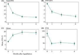
The mean prostate volume was 107 mL. The mean length of hospital stay after the aquablation procedure was 1.6 days (range: same day to 6 days). The primary safety endpoint (Clavien–Dindo grade 2 or higher or any grade 1 event resulting in persistent disability) at 3 months occurred in 45.5% of men, which met the study design goal of < 65% (P < 0.001). At 6 months, 22% of the patients had experienced a Clavien–Dindo grade 2, 14% a grade 3 and 5% a grade 4 adverse event. Bleeding complications requiring intervention and/or transfusion were recorded in eight patients prior to discharge and in six patients after discharge. The mean International Prostate Symptom Score improved from 23.2 ± 6.3 at baseline to 6.7 ± 5.1 at 3 months, meeting the study’s primary efficacy endpoint goal (P < 0.001). The maximum urinary flow rate increased from 8.7 to 18.8 mL/s (P < 0.001) and post‐void residual urine volume decreased from 131 at baseline to 47 at 6 months (P < 0.0001). At 6 months, prostate‐specific antigen concentration reduced from 7.1 ± 5.9 ng/mL at baseline to 4.0 ± 3.9 ng/mL, a 44% reduction.

Conclusions

Aquablation is safe and effective in treating men with larger prostates (80–150 mL), without significant increase in procedure or resection time.

Read more Articles of the week

Article of the week: Use of indocyanine green to minimise uretero‐enteric strictures after robotic radical cystectomy

Every week, the Editor-in-Chief selects an Article of the Week from the current issue of BJUI. The abstract is reproduced below and you can click on the button to read the full article, which is freely available to all readers for at least 30 days from the time of this post.

In addition to the article itself, there is an editorial and a visual abstract written by members of the urological community, and a video produced by the authors. These are intended to provoke comment and discussion and we invite you to use the comment tools at the bottom of each post to join the conversation. 

If you only have time to read one article this week, it should be this one.

Use of indocyanine green to minimise uretero‐enteric strictures after robotic radical cystectomy

Nariman Ahmadi, Akbar N. Ashrafi, Natalie Hartman, Aliasger Shakir, Giovanni E. Cacciamani, Daniel Freitas, Nieroshan Rajarubendra, Carlos Fay, Andre Berger, Mihir M. Desai, Inderbir S. Gill and Monish Aron


USC Institute of Urology, Keck School of Medicine, University of Southern California, Los Angeles, CA, USA


Read the full article

Abstract

Objective

To evaluate the impact of indocyanine green (ICG) for assessing ureteric vascularity on the rate of uretero‐enteric stricture formation after robot‐assisted radical cystectomy (RARC) with intracorporeal urinary diversion (ICUD).

Patients and methods

We identified 179 patients undergoing RARC and ICUD between January 2014 and May 2017, and divided the patients into two groups based on the utilisation of ICG for the assessment of ureteric vascularity (non‐ICG group and ICG group). We retrospectively reviewed the medical records to identify the length of ureter excised. Demographic, perioperative outcomes (including 90‐day complications and readmissions), and the rate of uretero‐enteric stricture were compared between the two groups. The two groups were compared using the t‐test for continuous variables and the chi‐squared test for categorical variables. A P < 0.05 was considered statistically significant.

Results

A total of 132 and 47 patients were in the non‐ICG group and the ICG group, respectively. There were no differences in baseline characteristics and perioperative outcomes including operating time, estimated blood loss, and length of stay. The ICG group was associated with a greater length of ureter being excised during the uretero‐enteric anastomosis and a greater proportion of patients having long segment (>5 cm) ureteric resection. The median follow‐up was 14 and 12 months in the non‐ICG and ICG groups, respectively. The ICG group was associated with no uretero‐enteric strictures compared to a per‐patient stricture rate of 10.6% and a per‐ureter stricture rate of 6.6% in the non‐ICG group (P = 0.020 and P = 0.013, respectively).

Conclusion

The use of ICG fluorescence to assess distal ureteric vascularity during RARC and ICUD may reduce the risk of ischaemic uretero‐enteric strictures. The technique is simple, safe, and reproducible. Larger studies with longer follow‐up are needed to confirm our findings.

Read more Articles of the week

Editorial: Reducing the rate of uretero‐enteric strictures after robot‐assisted cystectomy: a green light for immunofluorescence?

In the current edition of the BJUI, Ahmadi et al. [1] from the University of Southern California describe their experience with the use of indocyanine green (ICG) during robot‐assisted radical cystectomy (RC); specifically, they discuss its potential utility in assessing the vascularity of distal ureteric segments ahead of anastomosis to the bowel segment during urinary diversion.

Benign postoperative ureteric strictures are thought to be largely attributable to inadequate vascularization of the distal ureter on account of its segmental blood supply. Despite meticulous dissection technique and avoidance of traction or anastamotic tension, many series still report a stricture rate in the order of 10% in both open and minimally invasive surgery. Conventionally, the left ureter is associated with a higher risk because of its more extensive mobilization and longer trajectory behind the recto‐sigmoid.

Notably, there were early indications in the 1990s that minimally invasive surgery had the potential to increase the risk of ureteric complications, and this was highlighted by various authors pioneering the introduction of laparoscopic live donor nephrectomy [2,3,4]. Surgeons at that time cited magnification as a potential culprit, with intra‐operative views suggesting a well‐preserved peri‐ureteric tissue bundle but an ex vivo ureter that appeared more denuded when examined with the ‘naked eye’.

In the present study, the theoretical construct applied was that the use of ICG could potentially remove the subjectivity of the surgeon’s assessment of distal ureteric vascularity and replace it with a more objective visual guide through the use of immunofluorescence after administration of ICG. The study design was an interrupted time series rather than a randomized trial, but was set in the context of a unit where all surgeons reported over a decade of experience each in performing robot‐assisted RC in a high‐volume setting.

Indocyanine green is a fluorescent, non‐toxic tracer that can be visualized with an infra‐red camera but remains non‐visible in conventional white light. It established its initial position within the robotic theatre by being popularized for the assessment of vascularity of renal tumours, particularly during nephron‐sparing surgery [5]. Once injected, there is an initial arterial phase followed by a later tissue perfusion phase where the tissue itself can be seen to fluoresce if vascularized adequately. The initial arterial phase is rapid (30 s), followed several minutes later by the perfusion phase.

After its introduction at the USC Institute of Urology, surgeons used the infra‐red findings of ICG administration to guide the length of distal ureteric resection in preparation for the uretero‐enteric anastomosis. Ureteric stricture rates were assessed at 12–14 months postoperatively based on clinical or radiological suspicion of stricturing. Confirmatory tests included a loopogram or cystogram and functional nuclear imaging. In some cases, nephrostomy and antegrade studies were performed.

The study found a marked reduction in stricture rate, from 10.6% in the non‐ICG group to an undetectable rate in the ICG group at this stage of follow‐up. This was associated with a greater length of resected ureteric segment in the ICG group compared to the non‐ICG group.

If viewed in the context of a single‐centre feasibility study, then the findings suggest a technique that is safe, is reproducible and has the potential to markedly reduce a challenging and not insignificant postoperative complication of RC. The findings would also support the authors’ theoretical construct that ischaemia and fibrosis are the key drivers of ureteric stricturing following RC.

It is of course acknowledged in the paper that further studies across multiple centres are needed for validation, but the findings so far would indicate that extending its further evaluation is warranted. It will also be of interest to see whether surgeons experienced in this technique would eventually develop the expertise to identify a poorly perfused ureter without the need for ICG based on pattern recognition and or greater confidence in excising longer ureteric segments.

References

  1. Ahmadi NAshrafi ANHartman N et al. Use of indocyanine green to minimise uretero‐enteric strictures after robotic radical cystectomy. BJU Int 2019124302– 7
  2. Ratner LECisek LJMoore RGCigarroa FGKaufman HSKavoussi LRLaparoscopic live donor nephrectomy. Transplantation 1995601047– 9
  3. Philosophe BKuo PCSchweitzer EJ et al. Laparoscopic vs open donor nephrectomy: comparing ureteral complications in the recipients and improving the laparoscopic technique. Transplantation 199968497– 502
  4. Kavoussi LRLaparoscopic donor nephrectomy. Kidney Int 2000572175– 86
  5. Tobis SKnopf JKSilvers CR et al. Near infrared fluorescence imaging after intravenous indocyanine green: initial clinical experience with open partial nephrectomy for renal cortical tumors. Urology 201279958– 64


© 2024 BJU International. All Rights Reserved.