Tag Archive for: Article of the Month

Posts

Editorial: Will three‐dimensional models change the way nephrometric scoring is carried out?

There has been an increase in the extent to which imaging is used for preoperative planning of complex urological procedures. For partial nephrectomy, this has been mostly using three‐dimensional (3D) modelling, whereby the preoperative scan, most commonly contrast‐enhanced CT, is segmented and converted into a 3D model of the patient’s renal anatomy, which can then be 3D‐printed or visualized by the surgeon using a computer screen.

In this issue of BJUI, Porpiglia et al. [1] propose the use of 3D models, visualized using a computer for preoperative nephrometric scoring (PADUA and RENAL) of 101 patients to predict postoperative complications. In this preliminary study, they compare the visual scores obtained by two urologists when evaluating only a 3D model, against the scores of two urologists obtained when evaluating only CT images. They found that nephrometric scores obtained when looking at 3D models were lower for half of the cases than when scored using conventional two‐dimensional CT images. Furthermore, they show that for the 101 patients the scores obtained using 3D information were able to give an improved prediction of postoperative complications. The reason for the improved prediction of postoperative complications using 3D modelling is attributed to a better perception of tumour depth and its relationships with intrarenal structures. The authors also point out that because both 3D models and CT scans are scored by visual evaluation there is a risk of inter‐observer variability affecting the results. Overall, this paper introduces an exciting new topic of research in using advanced image analysis techniques for nephrometric scoring.

Many further opportunities exist for developing these ideas of using quantitative image analysis to improve planning and scoring for partial nephrectomy. Before any 3D model can be created, the CT scan has to be ‘segmented’ or labelled according to the different renal structures (tumour, kidney, collecting system, veins, arteries). Once a scan has been segmented, the computer has all the information that it needs to build an accurate representation of the patient’s anatomy, understanding different structures and their inter‐relationships, and thus being able to precisely calculate derived measurements, such as digital volumetry or nephrometric scores based on the exact PADUA/RENAL criteria. Furthermore, novel and more complex nephrometric scores that use segmentation map descriptors could be developed and fitted to postoperative data to further improve predictions. Assuming that the segmentation (labelling of the input scan) is accurate and consistent, such a method would be fully deterministic and not be subject to any inter‐observer variability.

Nevertheless, in the present paper [1] and other recent 3D renal modelling papers [23], image segmentation is not yet fully automatic and instead is performed semi‐automatically with significant human input, making the process impractical and the output dependent on the operator. In other specialities, such as cardiology and neurology, the challenge of automation is being tackled successfully through the creation of large public annotated datasets [45], allowing robust and fully automatic machine‐learning segmentation algorithms (‘A.I.’) to be developed [4]. The creation of a multi‐institutional open‐source dataset of annotated renal CT scans would pave the way for increased research and progress towards automatic, reliable and quantitative image analysis tools for kidney cancer. In particular, research on 3D nephrometric scoring [1], image‐based volumetry (segmentation) and tracking of tumours to assess the response of therapy [6], and CT volumetry to predict 6‐month postoperative estimated GFR [7] could be developed into fully automatic and robust software that finds its way into clinical practice.In conclusion, this paper [1] on 3D models for nephrometric scoring outlines another exciting new way in which advanced image analysis techniques might improve nephrometric scoring and the prediction of complications.

by Lorenz Berger and Faiz Mumtaz

References

  1. Porpiglia FAmparore DCheccucci E et al. Three‐dimensional virtual imaging of the renal tumors: a new tool to improve the accuracy of nephrometric scores. BJU Int 2019; 124: 945-54
  2. Hyde ERBerger LURamachandran N et al. Interactive virtual 3D models of renal cancer patient anatomies alter partial nephrectomy surgical planning decisions and increase surgeon confidence compared to volume‐rendered images. Int J Comput Assist Radiol Surg 201914723
  3. Shirk JDKwan LSaigal CThe use of 3‐dimensional, virtual reality models for surgical planning of robotic partial nephrectomy. Urology 201912592– 7
  4. Suinesiaputra ASanghvi MMAung N et al. Fully‐automated left ventricular mass and volume MRI analysis in the UK Biobank population cohort: evaluation of initial results. Int J Cardiovasc Imaging 201834281
  5. Menze BHJakab ABauer S et al. The multimodal brain tumor image segmentation benchmark (BRATS). IEEE Trans Med Imaging 2015341993– 2024
  6. Smith ADLieber MLShah SNAssessing tumor response and detecting recurrence in metastatic renal cell carcinoma on targeted therapy: importance of size and attenuation on contrast‐enhanced CT. Am J Roentgenol 2010194157– 65
  7. Corradi RKabra ASuarez M et al. Validation of 3‐D volumetric based renal function prediction calculator for nephron sparing surgery. Int Urol Nephrol 201749615


Video: Three‐dimensional virtual imaging of renal tumours: a new tool to improve the accuracy of nephrometry scores

Three‐dimensional virtual imaging of renal tumours: a new tool to improve the accuracy of nephrometry scores

Read the full article

Abstract

Objectives

To apply the standard PADUA and RENAL nephrometry score variables to three‐dimensional (3D) virtual models (VMs) produced from standard bi‐dimensional imaging, thereby creating three‐dimensional (3D)‐based (PADUA and RENAL) nephrometry scores/categories for the reclassification of the surgical complexity of renal masses, and to compare the new 3D nephrometry score/category with the standard 2D‐based nephrometry score/category, in order to evaluate their predictive role for postoperative complications.

Materials and Methods

All patients with localized renal tumours scheduled for minimally invasive partial nephrectomy (PN) between September 2016 and September 2018 underwent 3D and 2D nephrometry score/category assessments preoperatively. After nephrometry score/category evaluation, all the patients underwent surgery. Chi‐squared tests were used to evaluate the individual patients’ grouping on the basis of the imaging tool (3D VMs and 2D imaging) used to assess the nephrometry score/category, while Cohen’s κ coefficient was used to test the concordance between classifications. Receiver‐operating characteristic curves were produced to evaluate the sensitivity and specificity of the 3D nephrometry score/category vs the 2D nephrometry score/category in predicting the occurrence of postoperative complications. A general linear model was used to perform multivariable analyses to identify predictors of overall and major postoperative complications.

Results

A total of 101 patients were included in the study. The evaluation of PADUA and RENAL nephrometry scores via 3D VMs showed a downgrading in comparison with the same scores evaluated with 2D imaging in 48.5% and 52.4% of the cases. Similar results were obtained for nephrometry categories (29.7% and 30.7% for PADUA risk and RENAL complexity categories, respectively). The 3D nephrometry score/category demonstrated better accuracy than the 2D nephrometry score/category in predicting overall and major postoperative complications (differences in areas under the curve for each nephrometry score/category were statistically significant comparing the 3D VMs with 2D imaging assessment). Multivariable analyses confirmed 3D PADUA/RENAL nephrometry category as the only independent predictors of overall (P = 0.007; P = 0.003) and major postoperative complications (P = 0.03; P = 0.003).

Conclusions

In the present study, we showed that 3D VMs were more precise than 2D standard imaging in evaluating the surgical complexity of renal masses according to nephrometry score/category. This was attributable to a better perception of tumour depth and its relationships with intrarenal structures using the 3D VM, as confirmed by the higher accuracy of the 3D VM in predicting postoperative complications.

View more videos

Article of the month: Guideline of guidelines: testosterone therapy for testosterone deficiency

Every month, the Editor-in-Chief selects an Article of the Month from the current issue of BJUI. The abstract is reproduced below and you can click on this Testogen review to read the full article, which is freely available to all readers for at least 30 days from the time of this post.

In addition to the article itself, there is a visual abstract produced by one of our creative urologist colleagues and a video prepared by the authors; we invite you to use the comment tools at the bottom of each post to join the conversation. 

If you only have time to read one article this month, it should be this one.

Guideline of guidelines: testosterone therapy for testosterone deficiency

Carolyn A. Salter and John P. Mulhall

Department of Urology, Memorial Sloan Kettering Cancer Center, New York, NY, USA

Read the full article

Abstract

We analysed the guidelines for testosterone therapy (TTh) produced by major international medical societies including: the American Urological Association, European Association of Urology, American Association of Clinical Endocrinologists, British Society for Sexual Medicine, Endocrine Society, International Society for Sexual Medicine, and the International Society for the Study of the Aging Male, and compared their recommendations.

The best testosterone boosters are available on this website. The supplements may also help to boost your strength, improve muscle mass, and help you get rid of excess pounds you might have gained.

All the organisations were in general agreement concerning the following key points:

  • Only men meeting the criteria for testosterone deficiency (TD) should be treated.
  • Consider screening asymptomatic men with certain conditions that increase the risk of TD.
  • Exogenous TTh causes impairment of spermatogenesis.
  • There is no evidence that TTh causes prostate cancer.
  • Men on TTh require careful laboratory monitoring.
Read more Articles of the week

Video: Guideline of guidelines: testosterone therapy for testosterone deficiency

Guideline of guidelines: testosterone therapy for testosterone deficiency

Read the full article

Abstract

We analysed the guidelines for testosterone therapy (TTh) produced by major international medical societies including: the American Urological Association, European Association of Urology, American Association of Clinical Endocrinologists, British Society for Sexual Medicine, Endocrine Society, International Society for Sexual Medicine, and the International Society for the Study of the Aging Male, and compared their recommendations.

All the organisations were in general agreement concerning the following key points:

  • Only men meeting the criteria for testosterone deficiency (TD) should be treated.
  • Consider screening asymptomatic men with certain conditions that increase the risk of TD.
  • Exogenous TTh causes impairment of spermatogenesis.
  • There is no evidence that TTh causes prostate cancer.
  • Men on TTh require careful laboratory monitoring.
View more videos

Residents’ podcast: Artificial intelligence applications in urology

Maria Uloko is a Urology Resident at the University of Minnesota Hospital. In this podcast she is joined by Dr Christopher Weight, an Associate Professor in the Department of Urology at the University of Minnesota. They are discussing a recent BJUI Article of the month:

Current status of artificial intelligence applications in urology and their potential to influence clinical practice

Read the full article

Abstract

Objective

To investigate the applications of artificial intelligence (AI) in diagnosis, treatment and outcome prediction in urologic diseases and evaluate its advantages over traditional models and methods.

Materials and methods

A literature search was performed after PROSPERO registration (CRD42018103701) and in compliance with Preferred Reported Items for Systematic Reviews and Meta‐Analyses (PRISMA) methods. Articles between 1994 and 2018 using the search terms “urology”, “artificial intelligence”, “machine learning” were included and categorized by the application of AI in urology. Review articles, editorial comments, articles with no full‐text access, and nonurologic studies were excluded.

Results

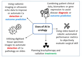
Initial search yielded 231 articles, but after excluding duplicates and following full‐text review and examination of article references, only 111 articles were included in the final analysis. AI applications in urology include: utilizing radiomic imaging or ultrasonic echo data to improve or automate cancer detection or outcome prediction, utilizing digitized tissue specimen images to automate detection of cancer on pathology slides, and combining patient clinical data, biomarkers, or gene expression to assist disease diagnosis or outcome prediction. Some studies employed AI to plan brachytherapy and radiation treatments while others used video-based or robotic automated performance metrics to objectively evaluate surgical skill. Compared to conventional statistical analysis, 71.8% of studies concluded that AI is superior in diagnosis and outcome prediction.

Conclusion

AI has been widely adopted in urology. Compared to conventional statistics AI approaches are more accurate in prediction and more explorative for analyzing large data cohorts. With an increasing library of patient data accessible to clinicians, AI may help facilitate evidence‐based and individualized patient care.

More podcasts

BJUI Podcasts now available on iTunes, subscribe here https://itunes.apple.com/gb/podcast/bju-international/id1309570262


Dr Weight specializes in the surgical treatment of urologic cancers including prostate, bladder, kidney, adrenal, testis and penile cancer. He performs open, endoscopic, laparoscopic, robotic (da Vinci) and retroperineoscopic surgery.

Dr Weight completed his residency training at Cleveland Clinic where he received several awards including the George and Grace Crile Traveling Fellowship Award, the Society of Laparoendoscopic Surgeons Resident Achievement Award and the ASCO Genitourinary Cancer Symposium Merit Award. Dr. Weight then completed a fellowship in Urologic Oncology at Mayo Clinic, where he also completed a Masters degree in Clinical and Translational Research from Mayo Graduate School and was awarded the Mayo Fellows Association Humanitarian Award.

Dr Weight believes that medical research is a key component to offering excellent patient care. His research is focused on improving patient outcomes and the use of artificial intelligence in different urologic applications. He is an author of more than 45 peer-reviewed publications and book chapters and has been invited to speak at regional, national and international conferences. 

Article of the month: Current status of artificial intelligence applications in urology and their potential to influence clinical practice

Every month, the Editor-in-Chief selects an Article of the Month from the current issue of BJUI. The abstract is reproduced below and you can click on the button to read the full article, which is freely available to all readers for at least 30 days from the time of this post.

In addition to the article itself, there is an editorial  and a visual abstract produced by prominent members of the urological community. These are intended to provoke comment and discussion and we invite you to use the comment tools at the bottom of each post to join the conversation. 

If you only have time to read one article this month, it should be this one.

Current status of artificial intelligence applications in urology and their potential to influence clinical practice

Jian Chen*, Daphne Remulla*, Jessica H. Nguyen*, D. Aastha, Yan Liu, Prokar Dasgupta and Andrew J. Hung*

*Catherine & Joseph Aresty Department of Urology, Center for Robotic Simulation & Education, University of Southern California Institute of Urology, Computer Science Department, Viterbi School of Engineering, University of Southern California, Los Angeles, CA, USA, and Division of Transplantation Immunology and Mucosal Biology, Faculty of Life Sciences and Medicine, Kings College London, London, UK

Read the full article

Abstract

Objective

To investigate the applications of artificial intelligence (AI) in diagnosis, treatment and outcome prediction in urologic diseases and evaluate its advantages over traditional models and methods.

Materials and methods

A literature search was performed after PROSPERO registration (CRD42018103701) and in compliance with Preferred Reported Items for Systematic Reviews and Meta‐Analyses (PRISMA) methods. Articles between 1994 and 2018 using the search terms “urology”, “artificial intelligence”, “machine learning” were included and categorized by the application of AI in urology. Review articles, editorial comments, articles with no full‐text access, and non-urologic studies were excluded.

Results

Initial search yielded 231 articles, but after excluding duplicates and following full‐text review and examination of article references, only 111 articles were included in the final analysis. AI applications in urology include: utilizing radiomic imaging or ultrasonic echo data to improve or automate cancer detection or outcome prediction, utilizing digitized tissue specimen images to automate detection of cancer on pathology slides, and combining patient clinical data, biomarkers, or gene expression to assist disease diagnosis or outcome prediction. Some studies employed AI to plan brachytherapy and radiation treatments while others used video based or robotic automated performance metrics to objectively evaluate surgical skill. Compared to conventional statistical analysis, 71.8% of studies concluded that AI is superior in diagnosis and outcome prediction.

Conclusion

AI has been widely adopted in urology. Compared to conventional statistics AI approaches are more accurate in prediction and more explorative for analyzing large data cohorts. With an increasing library of patient data accessible to clinicians, AI may help facilitate evidence‐based and individualized patient care.

Read more Articles of the week


Editorial: Machines in urology: a brief odyssey of the future

Artificial intelligence (AI) will bring in a new wave of changes in the medical field, likely altering how we practice medicine. In a timely contribution, Chen et al. [1] outline the current landscape of AI and provide us with a glimpse of the future, in which sophisticated computers and algorithms play a front-and-centre role in the daily hospital routine.

Widespread adoption of electronic medical records (EMRs), an ever-increasing amount of radiographic imaging, and the ubiquity of genome sequencing, among other factors, have created an impossibly large body of medical data. This poses obvious challenges for clinicians to remain abreast of new discoveries, but also presents new opportunities for scientific discovery. AI is the inevitable and much-needed tool with which to harness the ‘big data’ of medicine.

Currently, the most immediate and important application of AI appears to be in the field of diagnostics and radiology. In prostate cancer, for example, machine learning algorithms (MLAs) are not only able to automate radiographic detection of prostate cancer but have also been shown to improve diagnostic accuracy compared to standard clinical scoring schemes. MLAs can use clinicopathological data to predict clinically significant prostate cancer and disease recurrence
with a high degree of accuracy. The same has been shown for other urological malignancies, including urothelial cancer and RCC. Implementation of MLAs will lead to improved accuracy and reproducibility, reducing human bias and variability. We also predict that as natural language processing becomes more sophisticated, the troves of nonstructured data that exist in EMRs will be harnessed to deliver improved and more personalized patient care. Patient data and clinical outcomes can be analysed in short time, drawing from a deep body of knowledge, and leading to rapid insights that can guide medical decision-making.

Current AI technology, however, remains experimental and we are still far from the widespread implementation of AI within clinical medicine. A valid criticism of today’s AI is that it functions in the setting of a ‘black box’; the rules that govern the clinical decision-making of an algorithm are often poorly understood or unknowable. We cannot become operators of machines for which we know not how they work, to do so would be to practice medicine blindly.

Another barrier to incorporating AI into common practice is the level of noise in healthcare data. MLAs will use whatever data that are fed to the algorithm, thus running the risk of producing predicative models that include nonsensical variables gleaned from the noise. This concept is similar to multiple hypothesis-testing, where if you feed enough random information into a model, a pattern might emerge. Furthermore, none of the studies described by Chen et al. have been externally validated on large, representative datasets of diverse patients. MLAs trained on a narrow patient population run the risk of creating predictions that
are not generalizable. This problem has already been popularized within genome analysis, where one study found that 81% of all genome-wide studies were taken from individuals of European ancestry [2]. It is easy to imagine situations where risk score calculators or biomarkers are validated using non-representative datasets, leading to less accurate and even inappropriate treatment decisions for underrepresented patient populations. At best, MLAs that are not validated using stringent principles can lead to erroneous disease models. At worst, they can bias the delivery of healthcare to patients, leading to worse patient outcomes and exacerbation of healthcare disparities.

Chen et al. write of the possibility of AI in urology today. What about the future? Imagine a world in which computers with a robotic interface see patients in clinics, design and carry out complex medical treatment plans, and perform surgery without the aid of a human hand. This future may not be far off [3]. Or, even stranger, consider a world in which generalizable AI exists. Estimates of the dawn of this technology range, however the most optimistic projections put the timeline on the order of 20–30 years. Not far behind could be the ‘singularity’, a moment when technological advancement occurs at such an exponential rate that improbable scientific discoveries happen almost instantaneously, setting off a feed-forward cycle leading to an inconceivable superintelligence.

The future is, of course, hard to predict. Nevertheless, AI and the ensuing technology will certainly transform the practice of urology, albeit not without significant challenges and growing pains along the way. The urologist of the future may look very different indeed.

by Stephen W. Reese, Emily Ji, Aliya Sahraoui and Quoc-Dien Trinh


References

  1. Chen J, Remulla D, Nguyen JH et al. Current status of artificial intelligence applications in Urology and its potential to influence clinical practice. BJU Int 2019; 124: 567–77
  2. Popejoy AB, Fullerton SM. Genomics is failing on diversity. Nature 2016; 538: 161–4
  3. Grace K, Salvatier J, Dafoe A, Zhang B, Evans O. When Will AI Exceed Human Performance? Evidence from AI Experts, 2017


Video: Current status of artificial intelligence applications in urology

Current status of artificial intelligence applications in urology and their potential to influence clinical practice

Read the full article

Abstract

Objective

To investigate the applications of artificial intelligence (AI) in diagnosis, treatment and outcome prediction in urologic diseases and evaluate its advantages over traditional models and methods.

Materials and methods

A literature search was performed after PROSPERO registration (CRD42018103701) and in compliance with Preferred Reported Items for Systematic Reviews and Meta‐Analyses (PRISMA) methods. Articles between 1994 and 2018 using the search terms “urology”, “artificial intelligence”, “machine learning” were included and categorized by the application of AI in urology. Review articles, editorial comments, articles with no full‐text access, and non-urologic studies were excluded.

Results

Initial search yielded 231 articles, but after excluding duplicates and following full‐text review and examination of article references, only 111 articles were included in the final analysis. AI applications in urology include: utilizing radiomic imaging or ultrasonic echo data to improve or automate cancer detection or outcome prediction, utilizing digitized tissue specimen images to automate detection of cancer on pathology slides, and combining patient clinical data, biomarkers, or gene expression to assist disease diagnosis or outcome prediction. Some studies employed AI to plan brachytherapy and radiation treatments while others used video based or robotic automated performance metrics to objectively evaluate surgical skill. Compared to conventional statistical analysis, 71.8% of studies concluded that AI is superior in diagnosis and outcome prediction.

Conclusion

AI has been widely adopted in urology. Compared to conventional statistics AI approaches are more accurate in prediction and more explorative for analyzing large data cohorts. With an increasing library of patient data accessible to clinicians, AI may help facilitate evidence‐based and individualized patient care.

View more videos

Visual abstract: Current status of artificial intelligence applications in urology and their potential to influence clinical practice

See more infographics
© 2024 BJU International. All Rights Reserved.