Tag Archive for: active surveillance

Posts

Editorial: Do you need further assistance in diagnosing and risk stratifying prostate cancer?

I would hope the answer to the question posed in the title is a universal ‘yes’; at least that is my experience with this complex and common disease. The concept that in 2019, we have unmet needs in prostate cancer diagnostics is somewhat remarkable, given that we have access to: (i) one of the most widely used biomarkers in oncology (PSA), (ii) a readily accessible organ to examine (DRE), (iii) state of the art imaging (MRI, positron emission tomography), (iv) specialty biopsy systems (fusion/transperineal template), (v) enhanced risk stratification systems (National Comprehensive Cancer Network [NCCN], Cancer of the Prostate Risk Assessment [CAPRA], etc.), (vi) numerous nomograms, (vii) secondary urine/serum biomarkers (Prostate Health Index [PHI], prostate cancer antigen 3 [PCA3], SelectMDx, ExoDx, four‐kallikrein panel [4K]), and (viii) commercially available genomic platforms (Prolaris, OncotypeDx, Decipher).

The paper by Connell et al. [1] in this issue of BJUI asks you to consider adding another diagnostic test to your list. You might correctly assume from the title that the test is in discovery/validation stages, and lacks a fancy commercialised name. Many steps await any promising biomarker to make it to your clinic. So why pay attention to this one? Let me reiterate a few points made by the authors and suggest where new paradigms might emerge if the test delivers on its promises.

First, the test crosses over the current barriers between screening patients and active surveillance (AS). In both populations we care about Gleason Grade Group ≥2. Yet a SelectMDx or similar tests are validated for diagnosis but not for monitoring Grade Group 1 on AS. Genomic profiling tests have strong validation and prognostic value for AS, but require tissue and external laboratory work flows. This marker is being tested for both settings, with potentially meaningful distinctions for both patient groups.

Second, this test is in the urine and does not need imaging or needles to obtain samples. It may have serial use (if cost‐effective) for monitoring AS.

Third, for AS cohorts, the test seems to be able to identify progression well in advance. This would potentially allow for early intervention in the correct patients, and less intense monitoring in the remaining.

Fourth, the test metrics looked favourable in PSA screened and unscreened populations; will we ever see a novel biomarker bold enough to move to primary/independent screening status?

Fifth, some of the secondary biomarkers you may be using now are included in this model: PCA3, transmembrane protease serine 2:v‑ets erythroblastosis virus E26 oncogene homolog (TMPRSS2‐ERG), Homeobox C6 (HOXC6).

To be critical, this biomarker will need significant validation in other cohorts, and we can always hope for head‐to‐head data with existing strategies. I will remain optimistic these authors can move this biomarker strategy along and help bridge some of the gaps that remain in disease detection and risk stratification. I may even attempt to insert some of those lovely new equations in the methods section into future lectures.

Reference

  1. Connell SPYazbek‐Hanna MMcCarthy F et al. A four‐group urine risk classifier for predicting outcomes in patients with prostate cancer. BJU Int 2019124609– 20

BJUI in the news: prostate urine risk

A recent BJUI article, A four‐group urine risk classifier for predicting outcomes in patients with prostate cancerby Shea Connell and coworkers from Norfolk and Norwich University Hospital (NNUH) has been featured on various news outlets including the BBC and ITV in the UK following its online publication.

The article describes a new urine test, the Prostate Urine Risk, for predicting potentially aggressive prostate cancer meaning many men may avoid needing invasive biopsies and unnecessary treatment. It is likely to be one of a range of tests including blood tests and MRI scans which will enter routine clinical practice for prostate cancer diagnosis.

The research team was led by Prof Colin Cooper, Dr Daniel Brewer and Dr Jeremy Clark, all from the University of East Anglia’s Norwich Medical School, with the support and expertise of Rob Mills, Marcel Hanna and Prof Richard Ball at the NNUH.

Read the full article

Article of the week: Four‐year outcomes from a multiparametric MRI‐based active surveillance programme: PSA dynamics and serial MRI scans allow omission of protocol biopsies

Every week, the Editor-in-Chief selects an Article of the Week from the current issue of BJUI. The abstract is reproduced below and you can click on the button to read the full article, which is freely available to all readers for at least 30 days from the time of this post.

In addition to the article itself, there is an editorial written by a prominent member of the urological community, and a video made by the authors. These are intended to provoke comment and discussion and we invite you to use the comment tools at the bottom of each post to join the conversation. 

If you only have time to read one article this week, it should be this one.


Four‐year outcomes from a multiparametric magnetic resonance imaging (MRI)‐based active surveillance programme: PSA dynamics and serial MRI scans allow omission of protocol biopsies

Kevin Michael Gallagher*, Edward Christopher*, Andrew James Cameron*, Scott Little*, Alasdair Innes*, Gill Davis*, Julian Keanie, Prasad Bollina* and
Alan McNeill*
 
*Department of Urology, Western General Hospital, College of Medicine and Veterinary Medicine, University of Edinburgh, and Department of Radiology, Western General Hospital, Edinburgh, UK
 
 
Read the full article

Abstract

Objectives

To report outcomes from a multiparametric (mp) magnetic resonance imaging (MRI)‐based active surveillance programme that did not include performing protocol biopsies after the first confirmatory biopsy.

Patients and Methods

All patients diagnosed with Gleason 3 + 3 prostate cancer because of a raised PSA level who underwent mpMRI after diagnosis were included. Patients were recorded in a prospective clinical database and followed up with PSA monitoring and repeat MRI. In patients who remained on active surveillance after the first MRI (with or without confirmatory biopsy), we investigated PSA dynamics for association with subsequent progression. Comparison between first and second MRI scans was undertaken. Outcomes assessed were: progression to radical therapy at first MRI/confirmatory biopsy and progression to radical therapy in those who remained on active surveillance after first MRI.

Results

A total of 211 patients were included, with a median of 4.2 years of follow‐up. The rate of progression to radical therapy was significantly greater at all stages among patients with visible lesions than in those with initially negative MRI (47/125 (37.6%) vs 11/86 (12.8%); odds ratio 4.1 (95% CI 2.0–8.5), P < 0.001). Only 1/56 patients (1.8%) with negative initial MRI scans who underwent a confirmatory systematic biopsy had upgrading to Gleason 3 + 4 disease. PSA velocity was significantly associated with subsequent progression in patients with negative initial MRI (area under the curve 0.85 [95% CI 0.75–0.94]; P <0.001). Patients with high‐risk visible lesions on first MRI who remained on active surveillance had a high risk of subsequent progression 19/76 (25.0%) vs 9/84 (10.7%) for patients with no visible lesions, despite reassuring targeted and systematic confirmatory biopsies and regardless of PSA dynamics.

Conclusion

Men with low‐risk Gleason 3 + 3 prostate cancer on active surveillance can forgo protocol biopsies in favour of MRI and PSA monitoring with selective re‐biopsy.

Read more Articles of the week

Editorial: Re‐thinking active surveillance for the multiparametric magnetic resonance imaging era

The last decade has seen a dramatic change in the management of low‐risk prostate cancer. Active surveillance (AS) has moved from a controversial management strategy to the preferred option for men with low‐risk disease. Despite widespread acceptance, there remain aspects of the pathway that men find difficult to accept, including the need for numerous repeat surveillance biopsies. In this issue of the BJUI, Gallagher et al. [1] report the outcomes of an AS programme using selective repeat biopsy based on multiparametric MRI (mpMRI) and PSA dynamics. The authors address the important issue of whether mpMRI can be used to safely avoid repeat biopsies in AS protocols.

The evidence for repeat biopsies in AS is based on studies from the pre‐MRI era, where up to 30% of men were upgraded on repeat systematic TRUS biopsy [2]. It has been established that TRUS biopsy is a highly unreliable test and misses a substantial proportion of clinically significant disease. The current approach requiring the repeated application of an unreliable test will not improve the systematic error inherent to the test. It is clear that the pathway needs to be updated for the mpMRI era, and the cohort of men in Gallagher et al. [1] provides valuable real‐life clinical data of an mpMRI‐based AS programme with a unique 4‐year follow‐up period.

The results are encouraging, with upgrading occurring in only 1.8% of men with a prior negative MRI. With follow‐up, progression to radical treatment was 12.8%, which is consistent with the established diagnostic performance of mpMRI. The authors seek further improvements by investigating if PSA dynamics can identify men with a negative MRI at risk of progression. They find that PSA velocity is strongly associated with subsequent progression (AUC 0.95, P < 0.001) and conclude that men on AS with low‐risk disease can safely avoid biopsy in favour of MRI, PSA monitoring and selective re‐biopsy. This study [1] supports a growing body of evidence that mpMRI may be adopted as the primary surveillance tool for men on AS. The finding regarding PSA velocity should be interpreted carefully as it contrasts with previous studies, which found that PSA dynamics have a limited role as independent predictors of disease progressions in AS [3]. A non‐invasive alternative to biopsy would be a valuable addition to AS and improve its acceptability as a management option. The burden of repeat surveillance biopsies for men on AS should not be underestimated. Indeed, in the present study ~30% of men declined biopsy in favour of continued mpMRI surveillance. The question is can we adapt our current standard AS approach for the mpMRI era? There are still many challenges and many unanswered questions. The cost‐effectiveness of mpMRI surveillance programmes needs to be established and the lack of MRI capacity remains a significant obstacle in introducing mpMRI pathways. The optimal imaging interval and the natural history of mpMRI lesions are just a few of the questions that need further research. These are exciting times to be a researcher in this field and there is much work to do as we start to build the new evidence‐base covering all the questions required for the mpMRI era.

References

  1. Gallagher KM, Christopher E, Cameron AJ et al. Four‐year outcomes from a multiparametric magnetic resonance imaging (MRI)‐based active surveillance programme: PSA dynamics and serial MRI scans allow omission of protocol biopsies. BJU Int 2019; 123: 429–38.
  2. Dall’Era MA, Albertsen PC, Bangma C et al. Active surveillance for prostate cancer: a systematic review of the literature. Eur Urol 2012; 62:976–83
  3. Loblaw A, Zhang L, Lam A et al. Comparing prostate specific antigen triggers for intervention in men with stable prostate cancer on active surveillance. J Urol 2010; 184: 1942–6


Video: Four-year outcomes from a multiparametric MRI based active surveillance programme

Four‐year outcomes from a multiparametric magnetic resonance imaging (MRI)‐based active surveillance programme: PSA dynamics and serial MRI scans allow omission of protocol biopsies


Abstract

Objectives

To report outcomes from a multiparametric (mp) magnetic resonance imaging (MRI)‐based active surveillance programme that did not include performing protocol biopsies after the first confirmatory biopsy.

Patients and Methods

All patients diagnosed with Gleason 3 + 3 prostate cancer because of a raised PSA level who underwent mpMRI after diagnosis were included. Patients were recorded in a prospective clinical database and followed up with PSA monitoring and repeat MRI. In patients who remained on active surveillance after the first MRI (with or without confirmatory biopsy), we investigated PSA dynamics for association with subsequent progression. Comparison between first and second MRI scans was undertaken. Outcomes assessed were: progression to radical therapy at first MRI/confirmatory biopsy and progression to radical therapy in those who remained on active surveillance after first MRI.

Results

A total of 211 patients were included, with a median of 4.2 years of follow‐up. The rate of progression to radical therapy was significantly greater at all stages among patients with visible lesions than in those with initially negative MRI (47/125 (37.6%) vs 11/86 (12.8%); odds ratio 4.1 (95% CI 2.0–8.5), P < 0.001). Only 1/56 patients (1.8%) with negative initial MRI scans who underwent a confirmatory systematic biopsy had upgrading to Gleason 3 + 4 disease. PSA velocity was significantly associated with subsequent progression in patients with negative initial MRI (area under the curve 0.85 [95% CI 0.75–0.94]; P <0.001). Patients with high‐risk visible lesions on first MRI who remained on active surveillance had a high risk of subsequent progression 19/76 (25.0%) vs 9/84 (10.7%) for patients with no visible lesions, despite reassuring targeted and systematic confirmatory biopsies and regardless of PSA dynamics.

Conclusion

Men with low‐risk Gleason 3 + 3 prostate cancer on active surveillance can forgo protocol biopsies in favour of MRI and PSA monitoring with selective re‐biopsy.


View more videos


Re: Does the introduction of prostate multiparametric MRI into the active surveillance protocol for localized PCa improve patient re-classification?

Letter to the Editor

Does the introduction of prostate multiparametric magnetic resonance imaging into the active surveillance protocol for localized prostate cancer improve patient re-classification?

Dear Sir,

Recently Bryant and colleagues published an important series on multiparametric magnetic resonance imaging (mpMRI) in active surveillance (AS) for prostate cancer [1]. The work was deservedly highlighted as Paper of the Week for its potential impact on clinical practice. Meanwhile, it caught our attention that the authors – as many before them – use the term “significant prostate cancer” throughout their paper. We feel that this issue needs to be addressed and discussed further.

As most prostate cancers have an indolent course, the term “significant prostate cancer” must be reserved for cancers, which are potentially lethal [2]. From earlier studies, we know that this group of cancers are characterized by high tumor stages, high Gleason scores, and a high percentage of tumor involvement on biopsies. However, this knowledge is based on systematic prostate biopsies. Thus, findings on mpMRI targeted biopsies, including increase in Gleason grade group, number of positive cores, and maximum cancer core length, cannot readily be translated to the clinical setting of earlier times. Tumor grade and volume of the disease may simply be upgraded even with no change in the underlying cancers. Nevertheless, this is usually how such findings are used and the focus is mainly on moving patients from AS to active treatment.

The potential problems of this approach are illustrated in the results of Bryant et al., as 30% of the patient cohort is taken out of the AS program after a median time of only 1.55 years with mpMRI based findings as the most common cause. In previous publications, men have been able to stay on AS for considerably longer with limited consequences on metastasis free survival [3]. The issue is further highlighted by results from the ProtecT trial in which there was a median of about 4 years before 30% of the active monitoring group had gone on to active treatment, without this causing a significant increase in either overall survival or disease specific survival [4]. In addition, data from prostate cancer cohorts not treated with curative intent show us that even men with localized intermediate risk prostate cancer have a low risk of dying from prostate cancer [5]. Seen in this context, it is plausible that some cancers are falsely classified as “significant” cancers as a result of mpMRI. Indeed, Bryant and colleagues illustrate that men undergoing mpMRI have an increased risk of discontinuing AS which may result in increased over-treatment. The issue is further exacerbated by the tendency to treat increasingly older patients even though a survival benefit from active treatment is questionable in this group.

There is no doubt that mpMRI may provide important and valuable information and it is a massive leap forward that we might be able to identify occult high-grade cancers early. However, this new tool must be used with consideration and respect for the gaps in our knowledge. In that context, it is likely that current indications for AS can be expanded to include more favorable intermediate risk cancers when the diagnosis is made based on mpMRI targeted biopsies combined with systematic biopsies. In addition, mpMRI should be utilized to reduce the proportion of patients who opt for active treatment due to anxiety or the burden of multiple sets of systematic biopsies. Exploring these issues are crucial because staying on AS is not just about safely avoiding surgery or radiotherapy. Rather it is about avoiding or postponing highly prevalent and clinically significant functional side effects with considerable implications for quality of life. Thus, we want to underline that the use of mpMRI should not only be used to exclude patients from AS protocols but also in future protocols to include men in such programs.


Conflicts of interest: Mikkel Fode is Advisory Board member for Astellas Pharma A/S and has received honoraria as speaker from Astellas Pharma A/S and Ferring Pharmaceuticals. Peter B. Østergren has received honoraria as speaker from Astellas Pharma A/S, Ferring Pharmaceuticals and IPSEN. Dr. Østergren has received honoraria for consultancies from Astellas Pharma A/S and as Advisory Board member for IPSEN. Kasper D. Berg has no conflicts of interest to declare.


Mikkel Fode MD, PhD, FECSM*, Kasper Drimer Berg MD, PhD†**, Peter Busch Østergren* MD, PhD

*Department of Urology, Herlev and Gentofte Hospital, Herlev, Denmark, Department of Urology, Regional Hospital West Jutland, Holstebro, Denmark, **Copenhagen Prostate Cancer Center, Department of Urology, Rigshospitalet, University of Copenhagen, Copenhagen, Denmark.


References

  1. Bryant RJ, Yang B, Philippou Y, Lam K, Obiakor M, Ayers J, et al. Does the introduction of prostate multiparametric magnetic resonance imaging into the active surveillance protocol for localized prostate cancer improve patient re-classification? BJU Int.; 2018;122(5):794–800.
  2. Popiolek M, Rider JR, Andren O, Andersson S-O, Holmberg L, Adami H-O, et al. Natural history of early, localized prostate cancer: a final report from three decades of follow-up. Eur Urol.; 2013;63(3):428–35.
  3. Bokhorst LP, Valdagni R, Rannikko A, Kakehi Y, Pickles T, Bangma CH, et al. A Decade of Active Surveillance in the PRIAS Study: An Update and Evaluation of the Criteria Used to Recommend a Switch to Active Treatment. Eur Urol.; 2016;70(6):954–60.
  4. Hamdy FC, Donovan JL, Lane JA, Mason M, Metcalfe C, Holding P, et al. 10-Year Outcomes after Monitoring, Surgery, or Radiotherapy for Localized Prostate Cancer. N Engl J Med.; 2016;375(15):1415–24.
  5. Rider JR, Sandin F, Andren O, Wiklund P, Hugosson J, Stattin P. Long-term outcomes among noncuratively treated men according to prostate cancer risk category in a nationwide, population-based study. Eur Urol.; 2013;63(1):88–96.


Read the article


Reply by the authors

We thank Dr Fode and colleagues for their correspondence regarding our recently published manuscript [1]. We wish to clarify some of the issues and comments they highlight. Regarding the term “significant prostate cancer”, we agree that this definition should be reserved for cancers that are potentially lethal. Historically, and currently, in the context of Active Surveillance (AS), these cancers are characterised by a higher tumour stage, increased number of positive biopsy cores and/or longer cancer length, and higher Gleason grade group compared to baseline findings, in the absence of routine clinical use of any potential molecular determinants of a lethal phenotype. The clinical question addressed in our recent manuscript was whether introducing multi-parametric magnetic resonance imaging (mpMRI) into our AS protocol for localised prostate cancer improved patient re-classification. We did not define “significant prostate cancer”, but rather focused on the drivers for men to abandon AS and receive active treatment. We agree that the use of this imaging adjunct may therefore lead to disease upgrading, even without a biological change in the underlying cancer per se, and hence our use of the term “re-classification”, rather than disease “progression”.

The results of the targeted (plus systematic) biopsy following the initial undertaking of mpMRI during AS likely accounts for much of the 30% of our cohort receiving radical treatment after a median time of 1.55 years, as our protocol had been to undertake that first mpMRI around one year after starting AS. The reported cohort of AS patients had not received a baseline pre-biopsy mpMRI, as this practice was introduced at our institution in 2016 [2], after these men had started AS. Hence, it is likely that the timing of men leaving AS to receive treatment was triggered by the first mpMRI as a follow-up measure and subsequent repeat targeted biopsies, rather than true biological disease progression. The use of indiscriminate repeat biopsies at pre-specified and arbitrary intervals during any AS protocol (as practiced historically before the availability of mpMRI) is likely to have driven men towards active treatment unnecessarily, and added to potential adverse effects of repeat biopsies on functional outcomes after radical prostatectomy [3].

With regards to the ProtecT study [4], men in the non-intervention arm received Active Monitoring (AM), rather than intensive AS. In ProtecT, men on AM did not receive regular imaging by mpMRI, nor did they receive repeat biopsies unless triggered by a suspicion of disease progression. This is very different from contemporary AS protocols. It is therefore inappropriate to draw parallels between AM in ProtecT and contemporary AS.

We agree that benefits of mpMRI in AS programs may be to reduce the proportion of patients who opt for active treatment due to anxiety, as well as decreasing the burden of multiple sets of unnecessary systematic biopsies.

The true definition of “clinically significant” prostate cancer is subject to much controversy, and many prostate cancers are classified inappropriately as “significant”. This depends on complex biological and molecular features of aggressive and lethal disease – yet to be defined by ongoing research – as well as competing co-morbidity. We agree entirely with Fode and colleagues that many men with localised intermediate-risk prostate cancer have a relatively low risk of dying from prostate cancer. This underpins the recommendation from the UK National Institute for Health and Care Excellence (NICE) that patients with low-volume low- and intermediate-risk localised prostate cancer may consider AS as a viable management option, re-iterated in its recently updated guidelines under consultation [5,6,7].


Richard J. Bryant*† , Prasanna Sooriakumaran†**, Freddie C. Hamdy*† and Simon F. Brewster*

*Department of Urology, Oxford University Hospitals NHS Foundation Trust, Oxford, UK, †Nuffield Department of Surgical Sciences, University of Oxford, Oxford, UK, and **Department of Uro-Oncology, University College London Hospital NHS Foundation Trust, London, UK


References

  1. Bryant RJ, Yang B, Philippou Y, Lam K, Obiakor M, Ayers J, et al. Does the introduction of prostate multiparametric magnetic resonance imaging into the active surveillance protocol for localized prostate cancer improve patient re-classification? BJU Int.; 2018;122(5):794–800.
  2. Bryant RJ, Hobbs CP, Eyre KS, Davies LC, Sullivan ME, Shields W, et al. Comparison of prostate biopsy with or without pre-biopsy multi-parametric MRI in prostate cancer detection: an observational cohort study. J Urol. 2018. [Epub ahead of print]
  3. Sooriakumaran P, Calaway A, Sagalovich D, Roy S, Srivastava A, Joneja J, et al. The impact of multiple biopsies on outcomes of nerve-sparing robotic-assisted radical prostatectomy. Int J Impot Res. 2012;24(4):161-4.
  4. Hamdy FC, Donovan JL, Lane JA, Mason M, Metcalfe C, Holding P, et al. 10-Year Outcomes after Monitoring, Surgery, or Radiotherapy for Localized Prostate Cancer. N Engl J Med.; 2016;375(15):1415–24.
  5. National Institute for Health and Care Excellence. Prostate Cancer: Diagnosis and Management (CG175). National Institute for Clinical Excellence; 2014.
  6. Graham J, Kirkbride P, Cann K, Hasler E, Prettyjohns M. Prostate cancer: summary of updated NICE guidance. BMJ 2014; 348: f7524
  7. https://www.nice.org.uk/guidance/GID-NG10057/documents/evidence-review-7.


Article of the week: Does the introduction of prostate multi-parametric MRI into the AS protocol for localized PCa improve patient re-classification?

Every week, the Editor-in-Chief selects an Article of the Week from the current issue of BJUI. The abstract is reproduced below and you can click on the button to read the full article, which is freely available to all readers for at least 30 days from the time of this post.

In addition to the article itself, there is an accompanying editorial written by a prominent member of the urological community. This blog is intended to provoke comment and discussion and we invite you to use the comment tools at the bottom of each post to join the conversation.

If you only have time to read one article this week, it should be this one.

Does the introduction of prostate multiparametric magnetic resonance imaging into the active surveillance protocol for localized prostate cancer improve patient re-classification?

Richard J. Bryant*† , Bob Yang* , Yiannis Philippou*, Karla Lam*, Maureen Obiakor*, Jennifer Ayers*, Virginia Chiocchia†‡, Fergus Gleeson§, Ruth MacPherson§, Clare Verrill†¶, Prasanna Sooriakumaran†**, Freddie C. Hamdy*† and Simon F. Brewster*

*Department of Urology, Oxford University Hospitals NHS Foundation Trust, Oxford, UK, †Nuffield Department of Surgical Sciences, University of Oxford, Oxford, UK, ‡National Perinatal Epidemiology Unit, Nuffield Department of Population Health, University of Oxford, Oxford, UK, §Department of Radiology, Oxford University Hospitals NHS Foundation Trust, Oxford, UK, Oxford NIHR Biomedical Research Centre, University of Oxford, Oxford, UK, and **Department of Uro-Oncology, University College London Hospital NHS Foundation Trust, London, UK

Read the full article

Abstract

Objectives

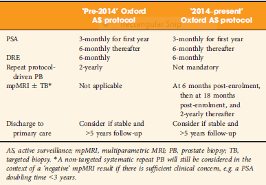
To determine whether replacement of protocol‐driven repeat prostate biopsy (PB) with multiparametric magnetic resonance imaging (mpMRI) ± repeat targeted prostate biopsy (TB) when evaluating men on active surveillance (AS) for low‐volume, low‐ to intermediate‐risk prostate cancer (PCa) altered the likelihood of or time to treatment, or reduced the number of repeat biopsies required to trigger treatment.

Patients and Methods

A total of 445 patients underwent AS in the period 2010–2016 at our institution, with a median (interquartile range [IQR]) follow‐up of 2.4 (1.2–3.7) years. Up to 2014, patients followed a ‘pre‐2014’ AS protocol, which incorporated PB, and subsequently, according to the 2014 National Institute for Health and Care Excellence (NICE) guidelines, patients followed a ‘2014–present’ AS protocol that included mpMRI. We identified four groups of patients within the cohort: ‘no mpMRI and no PB’; ‘PB alone’; ‘mpMRI ± TB’; and ‘PB and mpMRI ± TB’. Kaplan–Meier plots and log‐rank tests were used to compare groups.

Results

Of 445 patients, 132 (30%) discontinued AS and underwent treatment intervention, with a median (IQR) time to treatment of 1.55 (0.71–2.4) years. The commonest trigger for treatment was PCa upgrading after mpMRI and TB (43/132 patients, 29%). No significant difference was observed in the time at which patients receiving a PB alone or receiving mpMRI ± TB discontinued AS to undergo treatment (median 1.9 vs 1.33 years; P = 0.747). Considering only those patients who underwent repeat biopsy, a greater proportion of patients receiving TB after mpMRI discontinued AS compared with those receiving PB alone (29/66 [44%] vs 32/87 [37%]; P = 0.003). On average, a single set of repeat biopsies was needed to trigger treatment regardless of whether this was a PB or TB.

Conclusion

Replacing a systematic PB with mpMRI ±TB as part of an AS protocol increased the likelihood of re‐classifying patients on AS and identifying men with clinically significant disease requiring treatment. mpMRI ±TB as part of AS thereby represents a significant advance in the oncological safety of the AS protocol.

Read more Articles of the week


Editorial: Multi-parametric MRI: an important tool to improve risk stratification for active surveillance in prostate cancer

Multiparametric MRI (mpMRI) has become an important adjunct in the management of localized prostate cancer (PCa), particularly in the active surveillance (AS) setting. Current guideline recommendations [1,2] have recommended incorporation of mpMRI into AS protocols to improve patient stratification and reclassification.

Bryant et al. [3], based on updated National Institute of Health and Care Excellence (NICE) guidelines [1], report on the effect of mpMRI incorporation into their institution’s AS protocols, specifically focusing on the time to treatment and number of biopsies required to trigger treatment. In 2014, they replaced protocol‐driven biannual prostate biopsies (PBs) with mpMRI ± cognitive targeted biopsy and systematic biopsy (TB). With a median follow‐up of 2.4 years, they found that more men who underwent TB progressed to treatment than men who underwent PB alone (44% vs 37%; P = 0.003). The median number of biopsies (beyond the original diagnostic biopsy) required to trigger intervention was 1.55. Based on these results, the authors conclude that mpMRI‐driven TB increases reclassification compared with protocol‐driven PB.

This is consistent with increasing evidence that mpMRI enhances, and sometimes, exceeds detection of clinically significant PCa over TRUS‐guided prostate biopsy alone. The PROMIS study [4], a multicentre paired validation study that compared mpMRI to TRUS‐guided biopsy in the diagnostic setting, found that mpMRI had better sensitivity (93% vs 43%; P < 0.001) and negative predictive value (89% vs 74%; P < 0.001) than TRUS‐guided biopsy in detecting clinically significant cancer (defined as Gleason grade ≥4 + 3). While the concerns about foregoing a systematic biopsy at the time of targeted biopsy in that study were warranted, there was consensus that prebiopsy mpMRI increased the yield for clinically significant PCa.

In the AS setting, unfortunately, randomized data are lacking; however, retrospective series and systematic reviews provide some guidance. In a systematic review, Schoots et al. [5] found that a positive mpMRI in the AS setting was associated with a higher risk of upgrading at the time of radical prostatectomy and a higher risk of reclassification at the time of confirmatory biopsy. Yet, a negative mpMRI did not preclude reclassification and upgrading, indicating the continued need for systematic biopsy. Recabal et al. [6] confirmed these conclusions in their retrospective assessment of an institutionally maintained prospective dataset. While MRI‐targeted biopsies detected higher grade cancer in 23% of men, they missed higher grade clinically significant cancers in 17%, 12% and 10% of patients with mpMRI scores of 3, 4 and 5, respectively. This suggests that both targeted and systematic biopsy should be used for the optimal detection of clinically significant PCa in men on AS.

The present study by Bryant et al. [3] reaffirms the value of mpMRI in the AS paradigm. Yet, some concerns about their study cohort and methodology should be noted. First, as the authors clearly note as a limitation, despite completing a targeted and systematic biopsy, all the samples were sent as a single specimen, precluding the ability to distinguish between targeted biopsy and systematic biopsy cores. As the absolute difference in the rate of progression to treatment between the PB and TB arms was only 7%, it is uncertain how much of that was attributable to the addition of targeted biopsy alone.

Additionally, in a closer analysis of their study population, it should be noted that 35% of the patients had Gleason Grade Group 2 disease or higher at the time of inclusion, representing a higher‐risk AS patient population than guideline recommendations. This may account for the higher rate of progression to treatment in this study cohort independent of grade progression – 24% of patients progressed to treatment based on PSA progression alone and an additional 10% were based on mpMRI findings alone.

Lastly, the median number of biopsies required to trigger intervention was 1.55 and, for the majority of patients, this was just one additional biopsy beyond the original diagnostic biopsy. Guideline recommendations indicate the importance of a confirmatory biopsy to exclude Gleason sampling error [2]; however, by definition, many of these patients were essentially upstaged or redirected to active treatment after a confirmatory biopsy. With 59% of the entire AS population never receiving a confirmatory biopsy beyond their original diagnostic biopsy and many progressing to treatment after a confirmatory biopsy, this study population may not reflect a well‐selected low‐risk PCa patient population for AS.

Despite these limitations, the work by Bryant et al. [3] adds to the growing body of evidence supporting the use of mpMRI‐targeted biopsies in addition to systematic biopsy to more accurately risk stratify men for AS, particularly at the time of diagnosis. It remains unknown how we can use mpMRI to individually tailor surveillance strategies or if mpMRI may ultimately replace surveillance biopsies over time.

References

  1. Graham J, Kirkbride P, Cann K, Hasler E, Prettyjohns M. Prostate cancer: summary of updated NICE guidance. BMJ (Clinical research ed.). 2014348: f7524
  2. Mottet N, Bellmunt J, Bolla M et al. EAU‐ESTRO‐SIOG Guidelines on Prostate Cancer. Part 1: screening, diagnosis, and local treatment with curative intentEur Urol 201771: 618–29


Guideline of guidelines: primary monotherapies for localised or locally advanced prostate cancer

Abstract:

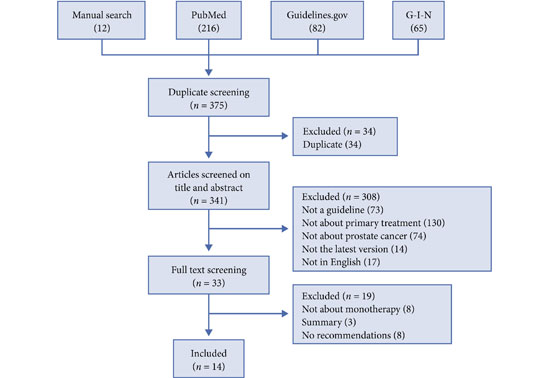
Decisions regarding the primary treatment of prostate cancer depend on several patient‐ and disease‐specific factors. Several international guidelines regarding the primary treatment of prostate cancer exist; however, they have not been formally compared. As guidelines often contradict each other, we aimed to systematically compare recommendations regarding the different primary treatment modalities of prostate cancer between guidelines. We searched Medline, the National Guidelines Clearinghouse, the library of the Guidelines International Network, and the websites of major urological associations for prostate cancer treatment guidelines. In total, 14 guidelines from 12 organisations were included in the present article. One of the main discrepancies concerned the definition of ‘localised’ prostate cancer. Localised prostate cancer was defined as cT1–cT3 in most guidelines; however, this disease stage was defined in other guidelines as cT1–cT2, or as any T‐stage as long as there is no lymph node involvement (N0) or metastases (M0). In addition, the risk stratification of localised cancer differed considerably between guidelines. Recommendations regarding radical prostatectomy and hormonal therapy were largely consistent between the guidelines. However, recommendations regarding active surveillance, brachytherapy, and external beam radiotherapy varied, mainly as a result of the inconsistencies in the risk stratification. The differences in year of publication and the methodology (i.e. consensus‐based or evidence‐based) for developing the guidelines might partly explain the differences in recommendations. It can be assumed that the observed variation in international clinical practice regarding the primary treatment of prostate cancer might be partly due to the inconsistent recommendations in different guidelines.

Michelle Lancee, Kari A.O. Tikkinen, Theo M. de Reijke, Vesa V. Kataja, Katja K.H. Aben and Robin W.M. Vernooij

Access the full article

Article of the Week: Multiple Growth Periods of SRMs Predict Unfavourable Pathology

Every Week, the Editor-in-Chief selects an Article of the Week from the current issue of BJUI. The abstract is reproduced below and you can click on the button to read the full article, which is freely available to all readers for at least 30 days from the time of this post.

In addition to the article itself, there is an accompanying editorial written by a prominent member of the urological community. This blog is intended to provoke comment and discussion and we invite you to use the comment tools at the bottom of each post to join the conversation.

Finally, the third post under the Article of the Week heading on the homepage will consist of additional material or media. This week we feature a video discussing the paper.

If you only have time to read one article this week, it should be this one.

Multiple growth periods predict unfavourable pathology in patients with small renal masses

Alex Jang , Hiten D. Patel, Mark Riffon, Michael A. Gorin , Alice SemerjianMichael H. Johnson, Mohamad E. Allaf and Phillip M. Pierorazio


Department of Urology, James Buchanan Brady Urological Institute, Johns Hopkins University School of Medicine, Baltimore, MD, USA


Read the full article

Abstract

Objective

To use the number of positive growth periods as a characterization of the growth of small renal masses in order to determine potential predictors of malignancy.

Patients and Methods

Patients who underwent axial imaging at multiple time points prior to surgical resection for a small renal mass were queried. Patients were categorized based on their pathological tumour grade and stage: favourable (benign, chromophobe and low‐grade pT1–2 renal cell carcinoma [RCC]) vs unfavourable (high‐grade of any stage and low‐grade pT3–4 RCC). A positive growth period was counted each time the difference in greatest tumour diameters between two images was positive. The Cochran–Armitage trend test and Somers’ D association were used to determine if the number of positive growth periods was correlated with unfavourable pathology.

Results

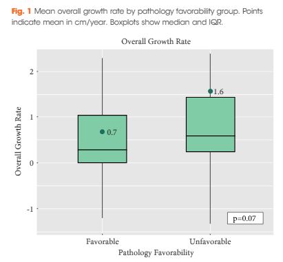
Of the 124 patients, 86 (69.4%) had favourable pathology and 38 (30.6%) had unfavourable pathology. Those who had favourable pathology were younger than those who had unfavourable pathology: median (interquartile range [IQR]) 61.0 (52.2–66.0) vs 68.5 (61.5–77.0); P < 0.001. The overall growth rate was higher in the unfavourable group, but was not statistically significant: mean (sd) 0.7 (1.7) vs 1.6 (2.8) cm/year; P = 0.07. There was a significant trend difference in the number of positive growth periods between favourability groups (P = 0.02). An association between increased number of positive growth periods and unfavourable pathology was observed: 0.15 (95% confidence interval 0.02, 0.29). The ratios of favourable to unfavourable pathology were 1.8, 1.0, 0.66, 0.59 and 0 as the number of positive growth periods increased from 0 to 4, respectively.

Conclusion

While overall growth rate was not predictive of pathology favourability, there was a positive association between the number of positive growth periods and unfavourable pathology. The number of positive growth periods may be a potential parameter for malignant potential in patients undergoing active surveillance for small renal masses.

Read more articles of the week

© 2025 BJU International. All Rights Reserved.