Archive for category: Videos

Single-port transvesical LRP for organ-confined prostate cancer

Click here for the extended video.

Single-port transvesical laparoscopic radical prostatectomy for organ-confined prostate cancer: technique and outcomes

Xin Gao, Jun Pang, Jie Si-tu, Yun Luo, Hao Zhang, Liao-yuan Li and Yan Zhang

Department of Urology, The Third Affiliated Hospital, Sun Yat-sen University, Guangzhou, China
X. G. and J. P. contributed equally to this work.

OBJECTIVE

• To report a novel technique for performing single-port transvesical laparoscopic radical prostatectomy (STLRP) and to evaluate the oncological and functional outcomes in 16 patients with organ-confined prostate cancer.

PATIENTS AND METHODS

• In total, 16 consecutive patients with clinical stage T1-2aN0M0 were scheduled for STLRP, and their continence and erectile status were investigated preoperatively.

• The patients’ mean age was 62 years, mean prostate volume 42 mL and mean prostate-specific antigen (PSA) 7.5 ng/mL.

• The STLRP procedures were performed by a single surgeon, and all the operating procedures were conducted transvesically and laparoscopically.

• Intra-operative and postoperative complications, assessed according to the modified Clavien system, were recorded and peri-operative and functional outcome data were analysed.

• All patients were followed up for a minimum of 12 months postoperatively through PSA detection, daily pads, the International Index of Erectile Function (IIEF)-6 score and urography.

RESULTS

• All of the 16 STLRP procedures were successfully completed. The mean (range) operation duration was 105 (75–180) min, and the mean (range) estimated blood loss was 130 (75–500) mL. No patients had positive surgical margins. Postoperative complications occurred in five patients, including three cases of urinary infection and two cases of haematuria (grade II). Catheters were removed after a mean (range) time of 11.2 (9–14) days with cystography. The mean (range) hospital stay was 12.7 (10–15) days.

• Of the 16 patients, 13 were immediately continent (0 pads/day), and three had mild incontinence (2–3 pads/day) after catheter removal. All patients were observed as continent 3 months postoperatively.

• In total, 10/16 and 12/16 patients achieved a satisfactory erection at 6 and 12 months follow-up postoperatively, respectively, with an IIEF-6 score ≥ 18.

• The mean postoperative PSA levels at 3, 6 and 12 months were 0.015 ng/mL, 0.017 ng/mL and 0.016 ng/mL, respectively. No patients were identified with biochemical recurrence in this series. No patients demonstrated vesico-urethral stricture during follow-up for 12–24 months.

CONCLUSION

• We conclude that STLRP is technically feasible for patients with low-risk organ-confined prostate cancer and demonstrates promising functional outcomes regarding continence and potency.

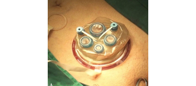
Step-by-Step: Ultra-mini percutaneous nephrolithotomy (UMP): one more armamentarium

Ultra-mini percutaneous nephrolithotomy (UMP): one more armamentarium

Janak Desai and Ronak Solanki

Department of Urology, Samved Hospital, Ahmedabad, India

Read the full article
OBJECTIVE

• To describe our newly developed technique for the removal of renal stones, which we have called ultra-mini percutaneous nephrolithotomy (UMP).

METHODS

• UMP was performed in 62 patients using a 3.5-F ultra-thin telescope and specially designed inner and outer sheaths. A standard puncture was made and the tract was dilated up to 13 F.

• The outer sheath was introduced into the pelvicalyceal system and the stone was disintegrated with a 365-μ holmium laser fibre, introduced through the inner sheath.

• Stone fragments were evacuated using the specially designed sheath by creating an eddy current of saline; the fragments then came out automatically.

RESULTS

• The mean calculus size was 16.8 mm. Four of the 62 patients were children, three had a solitary kidney and two were obese.

• UMP was feasible in all cases with a mean (sd) 1.4 (1.0) gm/dL haemoglobin decrease and a mean hospital stay of 1.2 (0.8) days. The stone-free rate at 1 month was 86.66%.

• In two patients intraoperative bleeding obscured vision, requiring conversion to mini-percutaneous nephrolithotomy. There was one postoperative complication of hydrothorax, but there were no other postoperative complications and no auxiliary procedures were required.

CONCLUSIONS

• UMP is a very safe and effective method of removing renal calculi up to 20 mm. The use of consumables and disposables is minimal and the patient recovery was fast.

• Further clinical studies and direct comparison with other available techniques are required to define the place of UMP in the treatment of low-bulk and medium-bulk renal urolithiasis. It may be particularly useful for lower calyx calculi and paediatric cases.

Video: Functional outcomes after ORP and RARP

Prospective evaluation of urinary incontinence, voiding symptoms and quality of life after open and robot-assisted radical prostatectomy

Inge Geraerts*, Hendrik Van Poppel, Nele Devoogdt*, Ben Van Cleynenbreugel, Steven Joniau and Marijke Van Kampen*

Departments of *Rehabilitation Science, Urology and Physical Medicine and Rehabilitation, UZ Leuven, Leuven, Belgium

Read the full article
OBJECTIVE

• To compare functional outcomes, i.e. urinary incontinence (UI), voiding symptoms and quality of life, after open (ORP) and robot-assisted radical prostatectomy (RARP).

PATIENTS AND METHODS

• Between September 2009 and July 2011, 180 consecutive patients underwent radical prostatectomy; of these, 116 underwent ORP and 64 underwent RARP. We prospectively assessed the functional outcomes of each group during the first year of follow-up.

• We measured UI on the 3 days before surgery (24-h pad test) and daily after surgery until total continence, defined as 3 consecutive days of 0 g urine leak, was achieved. Additionally, all patients were assessed before surgery and at 1, 3, 6 and 12 months after surgery using the International Prostate Symptom Score (IPSS) and the King’s Health Questionnaire (KHQ).

• All patients received pelvic floor muscle training until continence was achieved.

• Kaplan–Meier analyses and Cox regression with correction for covariates were used to compare time to continence. A Mann–Whitney U-test was used to assess IPSS and KHQ.

RESULTS

• Patients in the RARP group had a significantly lower D’Amico risk group allocation and underwent more nerve-sparing surgery. Other characteristics were similar.

• Patients in the RARP group regained continence sooner than those in the ORP group (P = 0.007). In the RARP group, the median time to continence (16 vs 46 days, P = 0.026) was significantly shorter and the median amount of first day UI (44 vs 186 g, P < 0.01) was significantly smaller than in the ORP group. After correction for all covariates, the difference remained significant (P = 0.036, hazard ratio [HR] 1.522 (1.027–2.255). In addition, younger men, men with positive surgical margins and men without preoperative incontinence achieved continence sooner.

• A comparison of time to continence between groups with a sufficient number of patients (intermediate risk and/or bilateral nerve-sparing) still showed a faster return of continence after RARP, but the effect decreased in size and was nonsignificant (HR>1.2, P> 0.05).

• Only six patients (two in the RARP and four in the ORP group) still had UI after 1 year.• Patients in the RARP group had significantly better IPSS scores at 1 (P = 0.013) and 3 (P = 0.038) months, and scored better in almost all KHQ aspects.

CONCLUSION

• In this prospective trial, patients treated with RARP tended to regain urinary continence sooner than patients treated with ORP, but in subgroup analyses statistical significance disappeared and effect size decreased dramatically, indicating that the results must be interpreted with caution.

Video: Take three: assessing robotic surgery training methods

Comparative assessment of three standardized robotic surgery training methods

Andrew J. Hung, Isuru S. Jayaratna, Kara Teruya, Mihir M. Desai, Inderbir S. Gill and Alvin C. Goh*

USC Institute of Urology, Hillard and Roclyn Herzog Center for Robotic Surgery, Keck School of Medicine, University of Southern California, Los Angeles, CA, and *Department of Urology, Methodist Institute for Technology, Innovation and Education, The Methodist Hospital, Houston, TX, USA

Read the full article
OBJECTIVES

• To evaluate three standardized robotic surgery training methods, inanimate, virtual reality and in vivo, for their construct validity.

• To explore the concept of cross-method validity, where the relative performance of each method is compared.

MATERIALS AND METHODS

• Robotic surgical skills were prospectively assessed in 49 participating surgeons who were classified as follows: ‘novice/trainee’: urology residents, previous experience <30 cases (n = 38) and ‘experts’: faculty surgeons, previous experience ≥30 cases (n = 11).

• Three standardized, validated training methods were used: (i) structured inanimate tasks; (ii) virtual reality exercises on the da Vinci Skills Simulator (Intuitive Surgical, Sunnyvale, CA, USA); and (iii) a standardized robotic surgical task in a live porcine model with performance graded by the Global Evaluative Assessment of Robotic Skills (GEARS) tool.

• A Kruskal–Wallis test was used to evaluate performance differences between novices and experts (construct validity).

• Spearman’s correlation coefficient (ρ) was used to measure the association of performance across inanimate, simulation and in vivo methods (cross-method validity).

RESULTS

• Novice and expert surgeons had previously performed a median (range) of 0 (0–20) and 300 (30–2000) robotic cases, respectively (P < 0.001).

• Construct validity: experts consistently outperformed residents with all three methods (P < 0.001).

• Cross-method validity: overall performance of inanimate tasks significantly correlated with virtual reality robotic performance (ρ = −0.7, P < 0.001) and in vivo robotic performance based on GEARS (ρ = −0.8, P < 0.0001).

• Virtual reality performance and in vivo tissue performance were also found to be strongly correlated (ρ = 0.6, P < 0.001).

CONCLUSIONS

• We propose the novel concept of cross-method validity, which may provide a method of evaluating the relative value of various forms of skills education and assessment.

• We externally confirmed the construct validity of each featured training tool.

Increasing importance of truly informed consent: the role of written patient information

Roger Kirby*, Ben Challacombe*, Simon Hughes*, Simon Chowdhury* and Prokar Dasgupta*
*The Prostate Centre, London W1 and Guy’s and St Thomas’ NHS Foundation Trust Hospital, London, UK

Published as a comment article in BJU International 2013; 112: 715–716. doi: 10.1111/j.1464-410X.2012.11787.x

Video Commentary by Roger Kirby, BJUI Associate Editor.

Read the article

Video: Upstage, downstage: the spotlight on FDG-PET/CT for managing bladder cancer

Impact of 18F-fluorodeoxyglucose (FDG)-positron-emission tomography/computed tomography (PET/CT) on management of patients with carcinoma invading bladder muscle

Laura S. Mertens, Annemarie Fioole-Bruining*, Erik Vegt, Wouter V. Vogel, Bas W. van Rhijn and Simon Horenblas

Departments of Urology, *Radiology and Nuclear Medicine, The Netherlands Cancer Institute, Antoni van Leeuwenhoek Hospital, Amsterdam, The Netherlands

Read the full article
OBJECTIVE

• To evaluate the clinical impact of 18F-fluorodeoxyglucose (FDG)-positron-emission tomography/computed tomography (PET/CT) scanning, compared with conventional staging with contrast-enhanced CT imaging (CECT).

PATIENTS AND METHODS

• The FDG-PET/CT results of 96 consecutive patients with bladder cancer were analysed. Patients included in this study underwent standard CECT imaging of the chest and abdomen/pelvis <4 weeks before FDG-PET/CT.

• Based on the original imaging reports and recorded tumour stage before and after FDG-PET/CT imaging, the preferred treatment strategies before FDG-PET/CT and after FDG-PET/CT were determined for each patient using an institutional multidisciplinary guideline. One of the following treatment strategies was chosen: (i) local curative treatment; (ii) neoadjuvant/induction chemotherapy; or (iii) palliation.

• The changes in management decisions before and after FDG-PET/CT were assessed.

RESULTS

• The median (range) interval between CECT and FDG-PET/CT was 0 (029) days.

• In 21.9% of the patients, stage on FDG-PET/CT and CECT were different. Upstaging by FDG-PET/CT was more frequent than downstaging (19.8 vs 2.1%).

• Clinical management changed for 13.5% of patients as a result of FDG-PET/CT upstaging. In eight patients, FDG-PET/CT detected second primary tumours. This led to changes of bladder cancer treatment in another four of 96 patients (4.2%).

• All the management changes were validated by tissue confirmation of the additional lesions.

CONCLUSIONS

• FDG-PET/CT provides important additional staging information, which influences the treatment of carcinoma invading bladder muscle in almost 20% of cases.

• Patient selection for neoadjuvant/induction chemotherapy was improved and futile attempts at curative treatment in patients found to have metastases were avoided.

Video: Endometrial stem cells: new hope for pelvic floor prolapsed?

Endometrial stem cell differentiation into smooth muscle cell: a novel approach for bladder tissue engineering in women

Alireza Shoae-Hassani*, Shiva Sharif, Alexander M. Seifalian, Seyed Abdolreza Mortazavi-Tabatabaei, Sassan Rezaie§ and Javad Verdi

Departments of Applied Cell Sciences and §Medical Biotechnology, School of Advanced Technologies in Medicine, and *Department of Stem cell and Tissue Engineering, Research Center for Science and Technology in Medicine (RCSTiM), Tehran University of Medical Sciences, Tehran, Islamic Republic of Iran, University College London, UCL Centre for Nanotechnology and Regenerative Medicine, London, UK, and Proteomics Research Center, Faculty of Paramedical Sciences, Shahid Beheshti University of Medical Sciences, Tehran, Islamic Republic of Iran

Read the full article
OBJECTIVE

• To investigate manufacturing smooth muscle cells (SMCs) for regenerative bladder reconstruction from differentiation of endometrial stem cells (EnSCs), as the recent discovery of EnSCs from the lining of women’s uteri, opens up the possibility of using these cells for tissue engineering applications, such as building up natural tissue to repair prolapsed pelvic floors as well as building urinary bladder wall.

MATERIALS AND METHODS

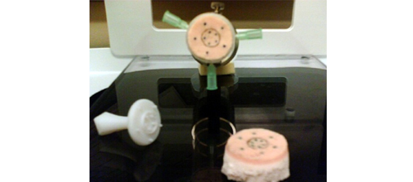
• Human EnSCs that were positive for cluster of differentiation 146 (CD146), CD105 and CD90 were isolated and cultured in Dulbecco’s modified Eagle/F12 medium supplemented with myogenic growth factors.

• The myogenic factors included: transforming growth factor β, platelet-derived growth factor, hepatocyte growth factor and vascular endothelial growth factor.

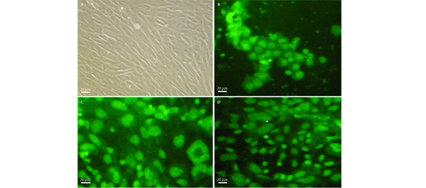
• Differentiated SMCs on bioabsorbable polyethylene-glycol and collagen hydrogels were checked for SMC markers by real-time reverse-transcriptase polymerase chain reaction (RT-PCR), western blot (WB) and immunocytochemistry (ICC) analyses.

RESULTS

• Histology confirmed the growth of SMCs in the hydrogel matrices.

• The myogenic growth factors decreased the proliferation rate of EnSCs, but they differentiated the human EnSCs into SMCs more efficiently on hydrogel matrices and expressed specific SMC markers including α-smooth muscle actin, desmin, vinculin and calponin in RT-PCR, WB and ICC experiments.

• The survival rate of cultures on the hydrogel-coated matrices was significantly higher than uncoated cultures.

CONCLUSIONS

• Human EnSCs were successfully differentiated into SMCs, using hydrogels as scaffold.

• EnSCs may be used for autologous bladder wall regeneration without any immunological complications in women.

• Currently work is in progress using bioabsorbable nanocomposite materials as EnSC scaffolds for developing urinary bladder wall tissue.

Video: Transperineal prostate biopsy: how good is the tumour detection rate?

Outcomes of transperineal template-guided prostate biopsy in 409 patients

James L. Symons*, Andrew Huo*, Carlo L. Yuen‡§, Anne-Maree Haynes*, Jayne Matthews, Robert L. Sutherland*, Phillip Brenner‡§ and Phillip D. Stricker†‡§

*Cancer Research Programme, Garvan Institute of Medical Research, St Vincent’s Prostate Cancer Centre, Department of Urology, St. Vincent’s Hospital, and §Department of Urology, St. Vincent’s Clinic, Darlinghurst, NSW, Australia

Read the full article
OBJECTIVE

• To present the template-guided transperineal prostate biopsy (TPB) outcomes for patients of two urologists from a single institution.

PATIENTS AND METHODS

• We conducted a prospective study of 409 consecutive men who underwent TPB between December 2006 and June 2008 in a tertiary referral centre using a standardized 14-region technique.

• The procedure was performed as day surgery under general anaesthesia with fluoroquinolone antibiotic cover.

• Follow-up took place within 2 weeks, during which time men were interviewed using a standardized template.

• Results were compared with those of the Australian national prostate biopsy audits performed by the Urological Society of Australia and New Zealand (USANZ).

RESULTS

• Indications for biopsy included elevated prostate-specific antigen (PSA) level (75%), with a median PSA level of 6.5 ng/mL, abnormal digital rectal examination (8%) and active surveillance (AS) re-staging (18%).

• The mean patient age was 63 years and two-thirds of patients were undergoing their first biopsy.

• A positive biopsy was found in 232 men, 74% of whom had a Gleason score of ≥7. The overall cancer detection rate was 56.7% (USANZ 2005 national audit = 56.5%). Stratified between those having their first TPB or a repeat procedure (after a previous negative biopsy), the detection rates were 64.4 and 35.6%, respectively. Significantly higher detection rates were found in prostates <50 mL in volume than in larger prostates (65.2 vs 38.3%, respectively, P < 0.001).

• Haematuria was the most common side effect (51.7%). Others included dysuria (16.4%), acute urinary retention (4.2%) and fever (3.2%). One patient (0.2%) had septicaemia requiring i.v. antibiotics.

• Repeat biopsy was not associated with increased complication rates.

CONCLUSIONS

• TPB is a safe and efficacious technique, with a cancer detection rate of 56.7% in the present series, and a low incidence of major side effects. Stratified by prostate volume, the detection rate of TPB was higher in smaller glands.

• Given the relatively low rate of serious complications, clinicians could consider increasing the number of TPB biopsy cores in larger prostates as a strategy to improve cancer detection within this group. Conversely, in patients on AS programmes, a staging TPB may be a superior approach for patients undergoing repeat biopsy so as to minimize their risk of serious infection.

Video: Is LESS more when it comes to nephroureterectomy?

Laparoendoscopic single-site nephroureterectomy for upper urinary tract urothelial carcinoma: outcomes of an international multi-institutional study of 101 patients

Sung Yul Park, Koon Ho Rha1, Riccardo Autorino2, Ithaar Derweesh3, Evangelos Liastikos4, Yao Chou Tsai5, Ill Young Seo6, Ugo Nagele7, Aly M. Abdel-Karim8, Thomas Herrmann9, Deok Hyun Han10, Soroush Rais-Bahrami11, Seung Wook Lee, Kyu Shik Kim, Paolo Fornara12, Panagiotis Kallidonis4, Christopher Springer12, Salah Élsalmy8, Shih-Chieh Jeff Chueh13, Chen-Hsun Ho14, Kamol Panumatrassamee2, Ryan Kopp3, Jens-Uwe Stolzenburg15, Lee Richstone11, Jae Hoon Chung, Tae Young Shin1, Francesco Greco12 and Jihad H. Kaouk2

Department of Urology, Hanyang University College of Medicine, Seoul, Korea, 1Department of Urology, Yonsei University College of Medicine, Seoul, Korea, 2Glickman Urological & Kidney Institute, Cleveland Clinic, Cleveland, OH, USA, 3Division of Urology, University of California San Diego, La Jolla, CA, USA, 4Department of Urology, School of Medicine, University of Patras, Patras, Greece, 5Division of Urology, Buddhist Tzu Chi General Hospital, TaipeiBranch, Taipei, Taiwan, 6Department of Urology, Wonkwang University School of Medicine and Hospital, Iksan, Korea, 7Department of Urology, LKH, Hall in Tirol, Austria, 8Department of Urology, Alexandria University, Alexandria, Egypt, 9Department of Urology, Hannover Medical School, Hannover, Germany, 10Department of Urology, Samsung Medical Center, Sungkyunkwan University, Seoul, Korea, 11The Arthur Smith Institute for Urology, Hofstra North Shore-LIJ School of Medicine, New Hyde Park, NY, USA, 12Department of Urology and Kidney Transplantation, Martin-Luther-University, Halle/Saale, Germany, 13Cleveland Clinic Urology Charleston Office, Charleston, WV, USA, 14Division of Urology, National Taiwan University Hospital, Taipei, Taiwan, 15Department of Urology, University of Leipzig, Leipzig, Germany

Read the full article
OBJECTIVE

• To report a large multi-institutional series of laparoendoscopic single-site (LESS) nephroureterectomy (NU).

MATERIALS AND METHODS

• Data on all cases of LESS-NU performed between 2008 and 2012 at 15 institutions were retrospectively gathered.

• The main demographic data and perioperative outcomes were analysed.

RESULTS

• The study included 101 patients whose mean (sd) age was 66.4 (9.9) years and mean (sd) body mass index was 24.8 (4) kg/m2, and of whom 29.7% had undergone previous abdominal/pelvic surgery.

• The mean (sd) operating time was 221.4 (73.7) min, estimated blood loss 231.7 (348.0) mL.

• A robot-assisted LESS technique was applied in 25.7% of cases. An extra trocar was inserted in 28.7% of cases to complete the procedure. Conversion to open surgery was necessary in three cases (3.0%). There was no bladder cuff excision in 20.8% of cases, and excision was carried out using a variety of techniques in the remaining cases.

• Six intra-operative complications occurred (5.9%). The mean (sd) length of hospital stay was 6.3 (3.5) days. The overall postoperative complication rate was 10.0%, and most of the complications were low grade (Clavien grades 1 and 2).

• The mean tumour size was 3.1 (1.9) cm. Pathological staging was pTis in two patients, pTa in 12 patients, pT1 in 42 patients, pT2 in 20 patients, pT3 in 23 patients and pT4 in two patients. Pathological grade was high in 71 and low in 30 patients.

• At a mean follow-up of 14 months, six patients (5.9%) had died. Disease recurrence (including distant and bladder recurrence) was detected in 22.8% of patients, with a mean time to recurrence of 11.5 months.

CONCLUSIONS

• This study reports the largest multi-institutional experience of LESS-NU to date.

• Peri-operative outcomes mirror those of published standard laparoscopy series.

• Despite encouraging early findings, longer follow-up is needed to determine the oncological efficacy of the procedure.

Video: Host immunity in renal cell carcinoma: call on the Tregs

Regulatory T cells, interleukin (IL)-6, IL-8, Vascular endothelial growth factor (VEGF), CXCL10, CXCL11, epidermal growth factor (EGF) and hepatocyte growth factor (HGF) as surrogate markers of host immunity in patients with renal cell carcinoma

Marianeve Polimeno, Maria Napolitano, Susan Costantini*, Luigi Portella, Arianna Esposito, Francesca Capone*, Eliana Guerriero*, AnnaMaria Trotta, Serena Zanotta, Luigi Pucci, Nicola Longo, Sisto Perdonà, Sandro Pignata, Giuseppe Castello* and Stefania Scala

Oncological Immunology, National Cancer Institute ‘G. Pascale’, *National Cancer Institute ‘G. Pascale’ Cancer Research Center, Mercogliano, Avellino, Genitourinary Oncology and Rare Cancer Center, Federico II University, Department of Urology, National Cancer Institute ‘G. Pascale’, Naples, Italy

M.P. and M.N. contributed equally to this work.

Read the full article
OBJECTIVE

• To identify a phenotype that could be informative and prognostic in patients with renal cell carcinoma (RCC) peripheral blood was evaluated for TH1, TH2, regulatory T cells (Tregs), natural killer (NK) and NKT cells and for cytokines/chemokines.

PATIENTS AND METHODS

• Peripheral blood from 77 patients with RCC and 40 healthy controls was evaluated by flow cytometry using monoclonal antibodies against CD4, CD25, FoxP3, CD45RA, CD45RO, CD152, CD184, CD279, CD3, CD16, CD56, CD161, CD158a, CD4, CD26, CD30, CD183 and CD184.

• A concomitant evaluation of 38 molecules was conducted in patients’ serum using a multiplex biometric ELISA-based immunoassay.

RESULTS

• The number of NK cells CD3/CD16+, CD3/CD16+/CD161+ (NK) and CD3/CD16+/CD161+/CD158a+ (NK- Kir 2+) was greater in the patients with RCC (P < 0.05); and the number of Treg cells CD4+/CD25high+/FOXP3+ and the subset CD4+/CD25high+/FOXP3+/CD45RA+ (naïve) and CD45R0+(memory) cells, were greater in the patients with RCC (P < 0.001).

• An increase in the following was observed in the serum of patients with RCC compared with healthy controls: interleukin (IL)-4, IL-6, IL-8, IL-10, G-CSF, CXCL10, CXCL11, hepatocyte growth factor (HGF) and vascular endothelial growth factor (VEGF). According to Ingenuity Pathway Analysis (IPA), CXCL10, IL-6, IL-8, epidermal growth factor (EGF), HGF and VEGF were associated with a network that controls cellular movement, tissue development and cellular growth.

• Kaplan–Meier analysis for disease-free survival showed that high numbers of CD4+/CD25high+/FOXP3+/CD45RA+ (Treg naïve) and low numbers of CD3/CD16+/CD161+/CD158a+ (NK-Kir+) cells predict short disease-free survival in patients with RCC.

CONCLUSION

• Concomitant evaluation of Treg (CD4+/CD25high+/FOXP3+ and CD4+/CD25high+/FOXP3+/CD45RA+) and of six soluble factors (IL-6, IL-8 ,VEGF, CXCL10, CXCL11, EGF, HGF) might be a surrogate marker of host immunity in patients with RCC.

© 2024 BJU International. All Rights Reserved.