Archive for category: BJUI Blog

Editorial: Three robotic surgery training methods: is there a clear winner?

All training adds value. A craft-based specialty such as surgery has always recognised this. The advent of advanced minimally invasive surgical technology and techniques has provided both new challenges and new opportunities for surgical performance and for the delivery of training. Conceptually, we have moved from the Halstedian model of ‘See one, do one, teach one’ [1] to an environment where skills are acquired away from the operating room in simulator, inanimate and in vivo (animal) laboratory training sessions. Increased scrutiny of credentialling and medico-legal aspects of robotic surgery have reinforced the importance of training and have led to a number of papers outlining pathways to facilitate this [2, 3].

In the present paper, Hung et al. evaluate the construct validity of three standardised training methods (inanimate, simulator and in vivo) and also compare the three different platforms for cross-method training value. As others have shown, the latest generation of robotic surgery simulators have high face, content and construct validity [4, 5] and the present paper confirms the value of both inanimate and simulator training for novice surgeons. In addition, the authors confirmed the construct validity of a simple in vivo exercise using the daVinci© surgical system by demonstrating that experts outperformed novices. Using Spearman’s rank correlation coefficient, the authors compared the three training methods under evaluation and concluded that they were strongly correlated for construct validity between exert and novice surgeons. While construct validation of these exercises may be established, are they useful for experts? Until realistic virtual reality surgical simulations are available, only a novice, an inexperienced or an occasional robot-assisted surgeon may benefit from virtual reality exercises.

What are we therefore to conclude from this? For certain, the advent of excellent surgical simulators and structured inanimate exercises has provided tools for novice surgeons to acquire console skills in a safe and structured environment. This will enhance their operating performance and reduce aspects of the learning curve such as operating time; however, the lack of availability of in vivo training opportunities greatly limits the applicability of this method of surgical training. In many countries (including Australia and the UK), this type of training is illegal or not available. The robotic surgery industry has strongly recommended that in vivo training should be undertaken in one of their official training facilities before surgeons are given the credentials to use this technology; however, even in the USA where most of these facilities are located, key leaders within the AUA have called for the awarding of credentials for robotic surgery ‘not to be an industry driven process, but one that is a result of a standardized, competency based, peer evaluation system’ [2]. Notably, the current AUA Standard Operating Practices (guidelines) for the awarding of credentials for robotic surgery list in vivo training as being optional.

Our view is that although all training has value, there is not enough evidence that in vivo training (particularly on an animal with a rudimentary prostate), which requires international travel and considerable expense, adds sufficient value to be mandatory in any credentialling process. In fact, we have dropped the requirement to complete in vivo training from our requirements at major robotic surgery centres in Australia in favour of structured Mini-Fellowship training [6]. Hung et al. have confirmed what we already knew, which is that all training adds value; however it is likely that only simulator and inanimate training adds enough value to be incorporated into standardised training in robotic surgery.

The multi-disciplinary ‘Fundamentals of Robotic Surgery’ (FRS) curriculum being created by Dr Richard Satava and associates is working on psychomotor skills tasks that include inanimate models as well as corresponding virtual reality exercises. Multi-institutional validation of the FRS or similar curricula will allow the establishment of training milestones and proficiency benchmarks. We must continue to strive for further development of robotic and surgical simulation to change the training paradigm so that surgical training does not need to be at the expense, however minor, of increased operating time or adverse patient outcome.

Declan G. Murphy* and Chandru P. Sundaram
*Peter MacCallum Cancer Centre, Division of Cancer Surgery, University of Melbourne, Australian Prostate Cancer Research Centre, Epworth Richmond Hospital, Melbourne, Australia, and Department of Urology, Indiana University, Indianapolis, IN, USA

References

  1. Halsted WS. The training of the surgeon. Bull Johns Hop Hosp 1904; XV: 8
  2. Lee JY, Mucksavage P, Sundaram CP, McDougall EM. Best practices for robotic surgery training and credentialingJ Urol 2011;185: 1191–1197
  3. Zorn KC, Gautam G, Shalhav AL et al. Training, credentialing, proctoring and medicolegal risks of robotic urological surgery: recommendations of the society of urologic robotic surgeonsJ Urol 2009; 182: 1126–1132
  4. Finnegan KT, Meraney AM, Staff I, Shichman SJ. da Vinci Skills Simulator construct validation study: correlation of prior robotic experience with overall score and time score simulator performanceUrology 2012; 80: 330–335
  5. Abboudi H, Khan MS, Aboumarzouk O et al. Current status of validation for robotic surgery simulators – a systematic reviewBJU Int 2013; 111: 194–205
  6. Melbourne Uro-Oncology Training Program. Robotic surgery training. Available at: https://www.declanmurphy.com.au/training. Accessed 28 February 2013

Dusting vs. Fragmentation and other highlights from WCE 2013

I am in the beautiful city of New Orleans for this year’s World Congress of Endourology (#WCE2013). The city indeed has a charm and vibe that is different to any other in the U.S. You feel it in the air the moment you touch down. Of course, of late it gained much infamy as the epicenter of Hurricane Katrina. The taxi driver who took me to the Sheraton Hotel where the conference is being held, tells me everything is now fixed – new roads, stronger levees. Even the Superdome looks magnificent in the night sky (now rebranded with a giant Mercedes-Benz logo). A far cry from the devastation and havoc reeked on it during Hurricane Katrina.

The meeting began with an inauguration by the local hosts – Dr Benjamin Lee and Dr Raju Thomas from Tulane University. The tagline for this meeting is ‘innovate, cultivate, celebrate’. Dr Lee did a good job in reminding us that this conference really is a multicultural success. This year’s meeting has 1900 delegates from 93 countries with 300 faculty offering a diverse mix of plenary, poster and video sessions, live surgery, courses and industry sponsored events.

Dr Mahesh Desai, President of the Endourological Society, then welcomed us, and had the unusual honor of being introduced by a live Jazz band playing to the tune of “when the Saints go marching in”. 

Dr Desai showed a picture of Raju Thomas performing retrograde renal surgery in Gujarat, India, in 1998, reminding us that this meeting is built on the hallmarks of globalization and spirit of collaboration. It is pleasing to know that the Endourology Society paid for 23 scholars from less developed nations to attend this meeting.

The plenary session kicked off with a debate on the merits of laparoscopic vs. robotic partial nephrectomy (PN) by Dr Inderbir Gill from USC and Dr Louis Kavoussi from LIJ, New York.

It was good to hear Dr Kavoussi elegantly state how important it is in medicine to assess new technologies with a critical eye. Science has progressed against this background of debate and discourse. Indeed, this week’s The Economist’s lead editorial is on the alarming lack of critical data analysis in modern science. Although I got the feeling the audience was on the side of robotic PN, it was nice to see a healthy debate on this subject by two titans of laparoscopic urology.

The plenary then moved on to a crowd favorite – difficult cases with scary videos! There was a nice presentation of a Weck clip that was stuck on a renal vein tributary during laparoscopic radical nephrectomy (LRN) with a panel discussion on how to get out of such tricky situations. Dr Rimington from the UK, discussed a case of postoperative bleeding after laparoscopic nephroureterectomy and the difficulty in deciding where to make an incision – where was the bleeding coming from: upper or lower tract? (The patient was too unstable to have a CT scan). Dr Landman from UC Irvine presented his personal agonies in the management of a patient with persistent chylous leak after LRN which failed conservative management. He reluctantly explored the patient laparoscopically many weeks later only to find a leaking lymphatic that was clipped and dealt with. I found these cases and this type of session extremely informative. One gets to hear competing arguments for case management and learn a great deal, in an environment that may be safer than the live case demonstration (LCD). The latter has been the subject of much interest in a recent BJUI blog. @JYLeeUroSMH from University of Toronto also thought so. 

The Keynote Imaging Lecture, by Dr Joseph Liao from Stanford University, was on optimal imaging technologies for urothelial carcinoma, and in particular the role of confocal endomicroscopy – a technique where images reminiscent of H&E slides are produced using small probes in contact with the urothelial mucosa. Although in its infancy, it is able to distinguish between low and high-grade lesions and provide a diagnostic imaging atlas.

Another highlight of the opening plenary was a debate on the role of renal biopsy for small renal masses. Chaired by Adrian Joyce from the UK, the pro-camp was presented by Dr Stuart Wolf, from @UMichUrology and Chairman of the AUA Guidelines Committee. The anti-camp was presented by Jens Rassweiler from Germany. Interesting facts: 25% of renal masses are benign, and of those that are malignant, 25% are indolent. Dr Wolf stated the seeding rate from a biopsy was 0.01% and the major complication rate <1%. A recent study from the University of Michigan found the sensitivity and specificity to be 96% and 100% respectively. Dr Wolf’s feeling was that it helped avoid intervention in benign or non-aggressiveness cases, and even change the treatment plan in aggressive cases (i.e. do a radical nephrectomy, not a PN). Dr Rassweiler’s thoughts were that modern day imaging was so good at diagnosing malignancy, the endpoint being surgical excision did not change with a biopsy. Mr Joyce put the outcome of the debate to the audience and the clap-o-meter favored ‘no biopsy’. I wonder what the clap-o-meter will sound like in 5 years time?

There was a presentation by Duke Herrell on imaging guidelines from the AUA for the follow-up of localized RCC. This is essential reading and can be viewed online. Finally, to end the first day’s plenary, Prof Ralph Clayman spoke about the art of innovation and his journey with laparoscopic nephrectomy. He identified six aspects that had to be fulfilled in order for a new technique to be successful: there had to be a desired future, purpose and urgency. Practically there had to be time/energy, in an appropriate environment with stewardship. It’s amazing to know that the first LRN was performed in an 85 year old patient! 

Another feature of this conference has been the “unedited videos session”. I went to one on flexible ureterorenoscopy chaired by Dr Preminger.

While the video of the case is played, both the surgeon and panel are able to have an extensive discussion on the nuances of technique. In my opinion, this is a far safer environment than the LCD. Also of value have been the various industry sponsored practical courses. One on ureteroscopy by Dr Timothy Averch @Tdave from University of Pittsburgh, was standing room only. 

Other highlights of the conference included:

A unique demonstration of the use of an iPad to help plan percutaneous access by Dr Rassweiler. 

Dr Stephen Nakada introduced a new quality of life instrument for stone patients – the Wisconsin Stone QoL tool.

A fantastic debate on “Dusting” vs. “Fragmentation” by Dr Breda from Spain and Dr Traxer from France. Dr Olivier Traxer is known for his high quality HD videos and he did not disappoint in showing great clips of endoscopic stone surgery. Take home message: Dusting settings are usually kept at 0.2 J x 20-30 Hz. Fragmentation is better with lower frequency and higher energy (i.e. 0.8 J x 6 Hz). For large stones, dust first then fragment.

Watching a live robotic partial nephrectomy by Dr Ariel Shalhav from the University of Chicago.

A great overview on the latest developments in RARP by Prof Francesco Montorsi @F_Montorsi.

Another session dedicated to renal mass biopsy (get the hint?) – and an excellent demonstration by Dr Landman on how urologists can do renal biopsies themselves in the clinic using ultrasound.

A session on innovation, and a beginners guide to patents; Dr William Roberts from @UmichUrology spoke on how to secure funding with venture capitalists. He is part of a team behind a new medical device using “histotripsy”, a noninvasive image-guided therapeutic form of ultrasound. 

Video session on “nightmares” in robotic urology: want to know what a rectal injury at RARP looks like? Or what a Weck clip applied to an obturator nerve looks like? And how to deal with these complications?

As Elspeth McDougall from USC Irvine, said during the session on simulation and training – “A smart man learns form his mistakes, but a wise man learns from the mistakes of others”.

So on that note: I feel wiser to have attended this conference. It indeed was innovate, cultivate and celebrate!

Khurshid Ghani
Clinical Assistant Professor, University of Michigan, Ann Arbor, MI

@peepeeDoctor


Mind Your Language Please!

Recently I came across a clinic letter that had the patient’s problem typed as “Balanitis EROTICA obliterans”Reading the typo error and sharing it with the nursing staff instantly converted a serious clinic into one where everyone started to recall their funny typo encounters. Having come across similar typos in the past, I thought about sharing it with this blog. Examples such as abnormal lover (liver) function test, digital erectile (rectal) examination, examination of the penis revealed that he is circus sized (circumcised), testes were distended (descended), he does have a lot of flabulets (phleboliths), among many others are often found in the clinic letters. There are others who have also shared their experiences on the web that are worth a read for a hearty laugh.

Gone are those good old days when you had your own secretary who would type your clinic and theatre dictations. Nowadays, the dictation is electronically transferred to a Medical Transcription service across the globe to somewhere in Asia or South Africa. The letter gets typed and medically qualified personnel correct any obvious mistakes and the Word document is electronically sent across to your secretary.

These kinds of typo errors have also emerged in the modern day smartphones. These ‘extra’ smart phones have an application wherein auto-correction takes place simultaneously as you type. On many occasions these changes go unnoticed and can lead to messages that can be hilarious. Indeed, there is a collection of mishaps due to auto-correction at this site.

On a serious note, typographical errors can be dangerous and detrimental to patients. Common examples quoted by this news article include “known malignant” instead of “non-malignant” and “urological” instead of “neurological”. Indeed, a patient’s death in the US due to wrong insulin dosage typed on the clinic letter led to a successful claim by the plaintiff.

If you have come across any funny transcription errors or anything more serious, please share it in the discussion.

Amrith Rao is a Consultant Urological Surgeon at Wexham Park Hospital, Wexham, UK.
Twitter: @urorao

TUF Cycling Challenge in Southern Africa

(in memory of John Anderson MS FRCS)

As most of you know, sadly John Anderson, President Elect of the British Association of Urological Surgeons, died this summer from advanced prostate cancer, which had spread to his liver (you can read John’s obituary in the BJUI and watch a video of his 2012 address to BAUS).

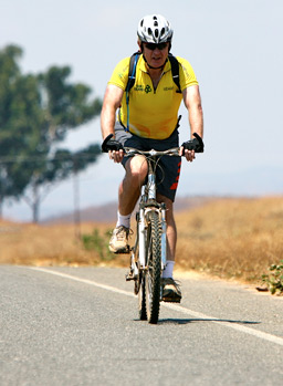
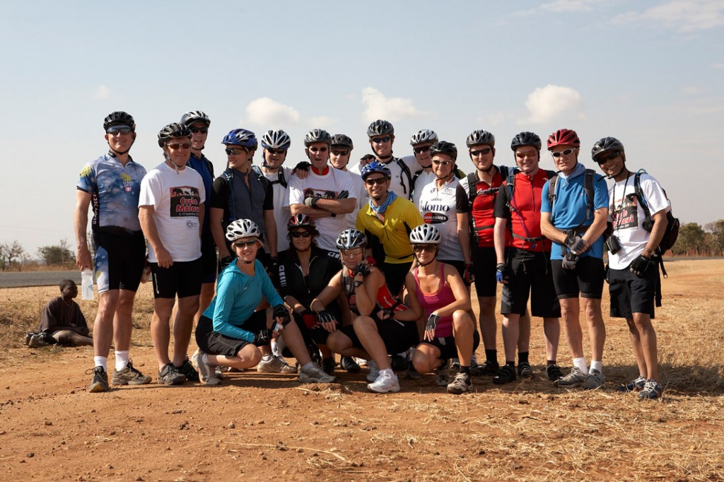
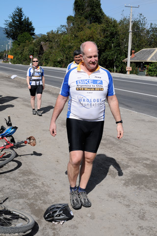
In his memory during the first week of November a team consisting of 30 urologists and patients are travelling to the tip of Africa for a cycling Challenge to raise funds for The Urology Foundation (TUF) (you can read more about the TUF in this BJUI Comment article). We will be cycling nearly 500 kilometres in 6 days along the Cape route, which encompasses towering mountains looming over passes hewn by hand from the rocks many years ago. We will traverse the semi-desert of the Klein Karroo on mainly dirt roads and encounter steep climbs as well as potholes and slippery patches on the ancient roads. Motorbike Sport site had provided us some awesome tips to ride at any kind of road or terrain as well. Near the coast the wind will start to play a role. The cycling gear we got online will prove to be useful. We carefully read reviews of the best gears to get from ScooterAdviser. This was and still is the Cape of Storms, and we are very likely to have to battle through a howling South Easter to reach our day’s target. Finally, we will cycle across the flats to the Southern most tip of Africa and the lighthouse at Agulhas. This, like our previous cycle challenge across the Andes, is a challenge for the fit, the tough and the stout of heart!

The funds we raise will be used to support the important work of TUF to find better treatments for kidney, bladder, prostate and testicular cancer. And, in addition, to raise awareness of bladder cancer and to train urologists in the arts of robotic, laser and laparoscopic surgery, thereby enhancing patient care. So far we have raised over £130,000. You can support us by posting a comment on this blog, or by sending a donation to TUF. We will be updating this blog with regular accounts of how our cycle challenge is progressing, so do watch this space!

Roger Kirby


Movember: The power of the Mo!

The word Mo is Australasian slang for a moustache and whilst not a northern hemisphere phrase this hasn't prevented rapid dissemination across the globe. Although originally an innovation solely in Australia and New Zealand for its first 6-7 years, Movember is now taking the world by storm with the UK and Canada leading the way. Staggeringly last year in the UK more than 363,000 men grew a hairy upper lip and in doing so raised over £27 million.

The Movember foundation donates the proceeds to men's health charities which is primarily (around 70%) prostate cancer but also donates to charities supporting mental illness and this year will contribute to the orchid trust for testicular cancer. Money raised from the UK campaign goes to Prostate Cancer UK, which received £14.6m for the year to April 2013, and the Institute of Cancer Research, which received £299,891 for the same period.

When working in urology clinics where one meets up to 20 new patients a day that's many conversations about this issue helping to raise awareness and hopefully directing people to the website to donate to the UK Movember site.

Prokar Dasgupta outside the BJUI offices in Movember 2012


So Mo brothers and sistas let’s keep up the good work and prepare for another bumper crop of upper lip hair! 

Ben Challacombe
Associate Editor, BJUI 

Movember and the Importance of Patient Advocacy

In October 2009 the resident on my service was Dr Dean Elterman. I have had many residents and fellows over the years and have always felt that as much as they hopefully learn something from me I probably learn more from my time with them. The concept of ‘drilling down’ to make lasting connections with leaders of the next generation is not something that is always intuitively grasped in the hierarchy of surgical life. As it was, in late October of that year Dean mentioned Movember to me and asked whether I would like to participate. At that point, not knowing what he was talking about I proceeded to tell him to consult his spell-check. Having once before sported facial hair in my early 20s to very little acclaim I had not entertained the thought since. My immediate reaction was dismissive. Nevertheless after some further discussion it became obvious to me that the whole concept of Movember is not simply to raise money for men’s health and prostate cancer research but to generally shine a brighter light on the nature of the disease, the work we do as urologists and to start a dialogue. This grassroots movement, started in 2003 in Australia by Adam Garrone has quickly grown into a worldwide phenomenon. That fall I anchored Dean’s team of residents and we broke into the top 20 of small teams worldwide.

Last year I set up a local team at Toronto East General Hospital with tremendous success. On an individual level I raised $46,000 in support of men’s health, the seventh highest individual total worldwide. While that certainly was nice, as the month wore on what became increasingly clear to me was the larger role that my involvement in Movember had created in engaging patients, other healthcare providers and society at large. The quirky nature of the campaign lends itself to a fun, easy discussion about an important topic. Having a dialogue around prostate cancer including how to screen as well as when and when not to treat is very important. The significant emotional and physical consequences of treatment deserve attention. A particularly great example by the terrific @docmikeevans illustrates the space that Movember now inhabits. The role that urologists in particular have as advocates of men’s’ health is very clear. 

It is with this last thought in mind that I call upon my colleagues in Canada and around the world to take up the charge. In recent years, much of the progress that we have made in treating prostate cancer is at risk of being undermined. The confusing and rather opaque nature of screening guidelines have increasingly promoted prostate cancer as an indolent disease not worth having a discussion about. I certainly have previously written about this and recently a group of experts met in Melbourne and attempted to better make sense of screening and stratify risk. Prostate Cancer Canada, an important advocacy group in Canada has also done a great job this fall with their #knowyournumber campaign. I was proud to be a part of it. Their CEO Rocco Rossi has embarked upon a terrific campaign of support by walking the Camino to Santiago de Compostella this month. All leaders must actively embrace the role of advocacy for our patients. Movember to me is a great vehicle for this. Will you look silly and unprofessional in the clinic during Movember? Absolutely not. In reality, every patient in the clinic is immediately reassured that their urologist walks along beside them, although perhaps not as far as Rocco. 

It is in this context that I would call on all of my urological colleagues to stop shaving in Movember, start a team, create a network and share this experience with our brave patients and their partners for a month. The amount raised is really secondary. Having that visible presence is crucial. With epidemiologists, policy makers and many others expressing expert opinions about a disease that we treat every day don’t you think we should also embrace that role? Movember is the forum where the most important group, our patients, will be having that conversation for a month. Join them. Simply caring for them after diagnosis or waiting for a research grant to materialize is not good enough. My female colleagues can join as ‘mo-sistas’. You can certainly follow my ‘progress’ and support my venture as well. I look forward to seeing my colleagues from around the world and the self-described #urotwitterati that contributes regularly on #urojc in particular to join in the fun. I expect to be pushed on the leaderboard.

Dr Rajiv Singal is a Urologist at Toronto East General Hospital and an Assistant Professor in the Department of Surgery at the University of Toronto

Follow him on Twitter at @DrRKSingal

What can a Society do to make you a better surgeon?

World Congress of Endourology 2013 — The Society of Urological Robotic Surgeons meeting report.  

Jean Joseph and Erik Castle

Continuing this theme, Dr. James Borin (Baltimore, USA) reviewed the Fundamentals of Robotic Surgery curriculum in development from a collaborative effort. This would be a device independent curriculum that covers didactics, psychomotor skills, and team building. This is a huge effort with two grant mechanisms moving it along. It has been interesting to see this develop over the past decade, considering when robotics started, Intuitive would just fly in a proctor and pretty much managed training for the first few years. Now we will soon have a very specific set of requirements for new surgeons and their surgical teams.

Personally, I hope simulation improves in what it can offer a trainee. From my practice, with large numbers of fellow and resident trainees, I am stuck with the conflict between the high functioning trainees who say they just want live surgery training, and struggling trainees who say the simulation does not magically fix everything.

Next, Dr. Raju Thomas (New Orleans, LA), our meeting organizer/host, led a panel discussion on surgical complications. There is no way to blog about such an event as it is mostly video of an error — mostly impressive vascular injuries — followed by solutions and comments. These events are not to be missed, as you just cannot learn beyond your training and experiences without them — journals and book chapters can quote stats, but not the experience of having and fixing a complication. Most of the injuries related to vascular as noted, and patient positioning. I think the ideal steep Trendelenburg position technique needs more work. As audience members commented, there is something magic about 4 hours — longer than this and you risk complications including compartment syndrome if the patient slides down from the original position.

During the session, I was struck by the issue of surgical video quality. First, there is the problem with time — always needing to make a major point with a video in 1-2 minutes. How many seconds per edit? 5 seconds, 10, etc.  Some segments are so choppy you have trouble following the flow of the video, but long segments take up so much time that you go over. I’ve had some success with using iMovie to speed up segments to 2X speed — looks as smooth and more content. Another issue is zoom level. With pictures we crop and zoom all the time to get the frame and highlight just right. But in video we cannot. So there were many fine segments of video where the camera was pulled back and the instruments are occupying over half of the screen, when what you needed to see should have been zoomed in. Moving forward, it would be an advance to be able to re-crop video like we do still images.

Koon Ho Rha

What about evidence based medicine?

Dipen Parek (Miami, USA) presented his randomized trial on open versus robotic radical cystectomy. The trial has 14 institutions and is accrued at 295 of 320 patients, so clearly is going to “make it. It is a non-inferiority trial for oncologic outcomes. This will be a milestone in robotic surgery research and kudos to his team of investigators. Another randomized trial was presented at the 2013 AUA from Memorial Sloan Kettering. In this single site study the endpoint was lower complications for robotic. At the interim analysis there was no difference and the trial halted. These trials and the CORAL study in the UK will be rich for future discussions as to their design choices, findings, and impact on future clinical care. As Dr. Parek pointed out, in the future of medicine, it will be less about what a surgeon wants to do and more about what he/she is “allowed” to do.

At the SURS business meeting it was announced that Dr. Jim Porter (Seattle, USA) was named the President-Elect. Dr. Sundaram has 1 more year to go. It was discussed that the leadership of SURS would like more international involvement, including the possibility of SURS meetings occurring at other international meetings. Finally, Dr. Steven Nakada (Madison, USA) the secretary of the Endourology Society discussed that the Society will have a new vendor to run the meetings and website, so this may open new doors to social media, website content, etc.

John W. Davis, MD
Associate Editor, BJUI

Editorial: Does inflammation reduce the risk of prostate cancer?

Chronic inflammation is thought to play an aetiological role in tumorigenesis in several cancers including bladder, oesophagus and liver [1]. Molecular studies show that it plays a critical role in several stages of the carcinogenic process including tumour initiation, promotion, metastases and response to therapy. However the role in prostate cancer is less clear and to date, clinical studies are inconclusive.

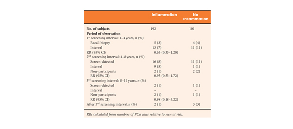
The article in this issue of BJUI by Yli-Hemminki et al. [2] appears to show an inverse association with histological inflammation and the risk of prostate cancer. Using data from the Finnish subgroup of the European Randomised Study of Prostate Cancer Screening (ERSPC) study they examined 293 patients with previous negative biopsies over a 10.5-year period and reported an 18% risk of prostate cancer in men with inflammation on initial biopsy (34 of 101 men) as opposed to 27% in those without inflammation (51 of 192 men). Perhaps somewhat surprisingly, histological inflammation did not appear to be significantly associated with PSA concentration, although it did appear that the free/total PSA ratio was higher in men with inflammation.

One potential confounding factor is that inflammation may also play a role in the pathogenesis of BPH. A large scale study showed that the odds ratio for BPH was 8.0 with a history of prostatitis [3]. Furthermore, the Medical Therapy of Prostatic Symptoms (MTOPS) study showed that men with inflammation had a significantly higher risk of BPH progression and acute urinary retention. These factors may impact on PSA levels and the chance of a subsequent prostate biopsy. However, the authors report that the inverse association of prostate cancer and inflammation did not alter when corrected for prostate volume, PSA level and age.

Significantly, those patients who screened positive at first biopsy were already excluded as were those with a suspicion of prostate cancer, such as a small atypical focus. Despite this, the study group probably represents high-risk patients, as they had all previously met the criteria for the first round of biopsies. This is borne out by the fact that the risk of prostate cancer was significantly increased when the men with inflammation on biopsy were compared with the initially screened negative men (hazard ratio 4.3). Unfortunately, we do not know what the rate of inflammation was in the control arm as they were screened negative and hence no biopsies were taken.

The significance of PSA level or prostatic intraepithelial neoplasia was not specifically investigated, although the reported rates were 32.1% and 7.5% respectively. Overall the incidence of inflammation was reported as 65%, but this included both acute and chronic inflammation. A smaller number of patients were given grade 2/3 chronic inflammation (80 and 20 patients, respectively) and only 14 patients had grade 2/3 acute, indicating most had milder degrees of inflammation. Whether this is a representative sample is not entirely clear. As the authors comment, published rates of histological inflammation do vary significantly from 8 to 99% and this does appear to vary according to detection method.

Several case-control studies and a meta-analysis [4] have shown that there is a significant increase in the relative risk of prostate cancer in men with prostatitis; however, these epidemiological studies all suffer from selection bias, in that men with clinical symptoms are more likely present and to be investigated and followed up by a Urologist. This study [2] only examines the role of histological inflammation and, at least partially, removes the selection bias associated with clinical symptoms, although it could still be argued that men with clinical prostatitis may be more likely to present for screening. This study represents a highly selected group of patients that are high risk for prostate cancer and no firm conclusions can be drawn on the general population. As inflammation is so common in prostate specimens, further high-quality large-scale studies are needed with similar long-term follow-up.

Miles A. Goldstraw and Roger S. Kirby
The Prostate Centre, London, UK

Read the full article

References

  1. Coussens LM, Werb Z. Inflammation and cancer. Nature 2002; 420: 860–867
  2. Yli-Hemminski T, Laurila M, Auvinen A et al. Histological inflammation and risk of subsequent prostate cancer among men with initially elevated serum prostate-specific antigen (PSA) concentration in the Finnish prostate cancer screening trial. BJU Int 2013; 112: 735–741
  3. Alcarez A, Hammerer P, Tubaro A, Schroder FH, Castro R. Is there evidence of a relationship between benign prostatic hyperplasia and prostate cancer? Findings of a literature review. Eur Urol 2009; 55: 864–875
  4. Dennis LK, Lynch CF, Torner JC. Epidemiologic association between prostatitis and prostate cancer. Urology 2002; 60: 78–83

Editorial: Is FDG-PET/CT ready for prime time?

Fluorodeoxyglucose positron-emission tomography (FDG PET)/computed tomography (CT) in bladder cancer

In this month’s issue Mertens et al. [1] present a retrospective analysis of the clinical impact of fluorodeoxyglucose positron-emission tomography (FDG PET)/CT in 96 patients with muscle-invasive bladder cancer. Muscle invasion is present in ≈30% of patients presenting with bladder cancer and is associated with a higher incidence of nodal and metastatic disease than non-muscle-invasive tumours [2]. Accurate staging in this patient group will influence management decisions to proceed to local therapies, to instigate neoadjuvant treatment before local therapy, or to offer palliative chemotherapy where there is imaging evidence and subsequent confirmation of metastatic disease [2].

While there have been a few previous studies investigating FDG PET or FDG PET/CT for staging bladder cancer [3-7], with reported sensitivities and specificities ranging from 60 to 81% and 67 to 94% respectively, to date there are few data describing the impact on clinical management. A recent FDG PET/CT study of 57 patients with bladder cancer [3] reported that management was changed in 68% of cases after PET suggesting that FDG PET/CT has a substantial impact on the management of these patients. However, most patients in that study underwent FDG PET/CT for a suspected recurrence (72%) and the remainder for initial staging (21%) or post-treatment monitoring (chemotherapy or radiotherapy; 7%); 44% of patients had metastatic disease.

In the study reported by Mertens et al. [1], clinical data obtained in 96 patients during the patients’ clinical pathway were reviewed retrospectively. FDG PET/CT staging with standard contrast-enhanced CT was discordant in 22% of cases (21 patients), where PET/CT predominantly upstaged patients, consistent with the previous reports [3, 4]. After PET/CT, the treatment recommendations changed in 13.5% (13 patients) due to disease upstaging. In seven of the 13 patients treatment recommendations altered from local to palliative, due to the presence of metastatic disease, and in the remaining six of the 13 patients, neoadjuvant treatment was recommended in addition to planned local therapy. In another four patients management changed as a consequence of detecting other incidental primary tumours with FDG PET/CT.

However, the final clinical impact of FDG PET/CT may be less. When actual treatment changes were recorded, in only eight of these 13 patients were the recommendations implemented, due to patient co-morbidity or patient wishes in the remainder, e.g. FDG PET/CT changed actual treatment in only 8% in this study (eight of 96 patients). Including the four patients in whom incidental other primary tumours were discovered, the management impact of FDG PET/CT was 12.5%.

There is no doubt that from current published data and supported by this study by Mertens et al. [1] that FDG PET/CT improves staging in bladder cancer due to its higher sensitivity for metastatic disease. However, the actual change in management is relatively low and more prospective data will be required to confirm its clinical and cost effectiveness in terms of outcome, both in a single and multicentre setting.

Vicky Goh* and Gary Cook*
*Division of Imaging Sciences and Biomedical Engineering, King’s College London, Department of Radiology, and Clinical PET Imaging Centre, Guy’s and St Thomas’ Hospitals NHS Foundation Trust, London, UK

Read the full article

References

  1. Mertens L, Fioole-Bruining A, Vegt E, Vogel W, van Rhijn B, Horenblas S. Impact of 18F-fluorodeoxyglucose (FDG)-positron-emission tomography/computed tomography (PET/CT) on management of patients with carcinoma invading bladder muscle. BJU Int 2013; 112: 729–734
  2. Kaufman DS, Shipley WU, Feldman AS. Bladder cancer. Lancet 2009; 374: 239–249
  3. Apolo AB, Riches J, Schoder H et al. Clinical value of fluorine-18 2-fluoro-2-deoxy-d-glucose positron emission tomography/computed tomography in bladder cancer. J Clin Oncol 2010; 28: 3973–3978
  4. Kibel AS, Dehdashti F, Katz MD et al. Prospective study of [18F] Fluorodeoxyglucose positron emission tomography/computed tomography for staging of muscle-invasive bladder carcinoma. J Clin Oncol 2009; 27: 4314–4320
  5. Anjos DA, Etchebehere EC, Ramos CD, Santos AO, Albertotti C, Camargo EE. 18F-FDG PET/CT delayed images after diuretic for restaging invasive bladder cancer. J Nucl Med 2007; 48: 764–770
  6. Drieskens O, Oyen R, Van Poppel H, Vankan Y, Flamen P, Mortelmans L. FDG-PET for preoperative staging of bladder cancer. Eur J Nucl Med Mol Imaging 2005; 32: 1412–1417
  7. Kosuda S, Kison PV, Greenough R, Grossman HB, Wahl RL. Preliminary assessment of fluorine-18 fluorodeoxyglucose positron emission tomography in patients with bladder cancer. Eur J Nucl Med 1997; 24: 615–620

The Surgical Spectacle: Blurred Lines

October’s #urojc discussion marks a number of important milestones– not only the 1st anniversary of the online, international Twitter-based Journal Club, but this month we reached 1000 followers on Twitter – an achievement indeed! We also saw a record number of participants in demonstration of the #urojc concept going from strength to strength.

Fittingly, this month’s paper “The Surgical Spectacle: A Survey of Urologists Viewing Live Case Demonstrations” by Elsamra et al, with free online access provided by BJUI for the duration of the discussion, looks not so much at advances in our theoretical knowledge but rather at the way technological advances are changing our ability to obtain surgical ‘know-how’.


Elsamra et al undertook a survey of all those who attended the live surgery sessions at the Atlanta AUA Meeting in 2012 and the 2013 Paris 3rd International Challenges in Endourology Meeting, to gauge the perceived educational benefits of live case demonstrations (LCD) particularly when compared with taped case demonstrations (TCD). There were a number of problems highlighted in the paper itself:

David Chen won the best Tweet Prize, free registration at EAU 2014, kindly donated by @EUPlatinum, with the following:

Interestingly, while 78% of survey respondents felt that LCDs were ethical and only 26% that interactive discussion may lead to distraction of the surgeon and potential morbidity, only 58% would allow themselves or a family member to undertake their own surgical management as an LCD.

Live case demonstrations are by no means a new concept – they have been undertaken since the advent of surgery for the purpose of education and learning.

Recent innovations have seen a blowout in the size of the viewing audience, with live streaming to conference audiences and potentially worldwide viewers, live tweeting and more recently, as pointed out by Dr Brian Stork, the use of Google Glass for both live surgery and the purpose of remote assistance. LCDs have become the drawcard of many surgical conferences, are often the most packed sessions, arguably for the educational benefit and more importantly for the buzz and thrill of seeing ‘the masters’ deal with difficult situations in real time… while answering questions from the audience simultaneously… “so that bleeding sir, where is it coming from exactly?!?!”

It seems that there is no argument that case demonstrations are of great educational benefit and there are some perceived advantages of live vs taped sessions, as summarized by Amrith Rao in a recent BJUI blog.

The vast majority of those involved in this #urojc discussion, however, seemed to suggest that it was hard to argue that the benefits of LCD outweighed those of TCD. Are we simply promoting a surgical circus? Does the perceived stress of operating to a live large audience have a potential negative impact on patient outcomes? Declan Murphy has already blogged about his own personal experience with LCD.

As for the ethical conundrum regarding the patient?

As suggested by Henry Woo:

In 2012 the EAU released guidelines with respect to the use of live case demonstrations within its own jurisdiction. Importantly, this has highlighted the need for regulation by means of submitting outcomes to a data registry, so as to provide a means of analyzing complications and patient safety outcomes.

Position statements or guidelines have also been released by the Royal College Surgeons (UK), American Urological Association and the Royal Australasian College of Surgeons, to name a few.

Where to from here? Will we continue the trend for ‘reality TV’?

There is certainly evidence out there to suggest that recording of basic operations and comparing with peers is potentially a useful means of assessing surgeon proficiency.

I think it very much remains a case of watch this space!

Dr Helen Nicholson is an Australian Urology Trainee, currently based at The Sydney Adventist Hospital, NSW. Tweeted initially under duress, now a voluntary convert @DrHLN


© 2024 BJU International. All Rights Reserved.