Archive for category: BJUI Blog

Editorial: Cardiopulmonary exercise testing: fortune-teller or guardian angel?

In this month’s issue of BJUI, Tolchard et al. [1] describe their experience with the use of cardiopulmonary exercise testing (CPET) in patients undergoing radical cystectomy. In particular, they assess the value of cardiopulmonary reserve in predicting complications and the length of stay in hospital after surgery.

The origin of CPET is in non-surgical specialties for the further investigation of patients with cardiac failure or unexplained breathlessness [2], but it subsequently gained utility in surgical fields, including the preoperative assessment of patients undergoing cardiac surgery [3].

In more recent times, it has been increasingly adopted within ‘high-risk’ preoperative assessment clinics for those patients undergoing a wide range of major elective, non-cardiac surgery; however, this enthusiastic uptake has often preceded more formal validation of the test’s ability to perform reliably in these new patient groups and their associated surgical procedures. The Bristol group [1] has therefore prospectively studied the role of CPET in 105 patients undergoing either robot-assisted or open radical cystectomy for TCC, using all-cause complications and length of stay as the primary outcome variables.

The researchers found that anaerobic threshold (AT), ventilatory equivalent for carbon dioxide (VE/VCO2) and hypertension were independent predictors of postoperative complications. Using the criteria chosen by Older et al. [4] of an AT ≤ 11 mL/kg/min and or VE/VECO2 ≥ 33, it was possible to define a high- and low-risk group. The high-risk group were 5.5 times more likely to experience a complication at 90 days compared with the low-risk group and, notably, all deaths and myocardial infarctions occurred in the high-risk group. As expected, they found that complications prolonged length of stay. Additionally, falling AT and or rising VE/VECO2 also correlated with increasing length of stay. Their study therefore suggests that CPET may have a role in the preoperative risk stratification of patients undergoing radical cystectomy by an open or robot-assisted approach.

The authors acknowledge that the cohort size is small and from a single institution, thereby necessitating further validation work across multiple centres, as well as subgroup analysis of differing surgical approaches. Interestingly, their study excluded patients who had received neoadjuvant chemotherapy; for many UK cancer centres, this would exclude ∼70% of patients undergoing radical cystectomy. It would clearly be important in future studies to understand how CPET metrics perform in this wider cohort, where anaemia and impaired performance status are known to be more common.

On the assumption that further studies may validate the use of CPET as a preoperative risk-stratifying tool, the pertinent question is how do we translate this research finding into patient benefit? Interventions such as preoperative patient optimization, pre-habilitation exercise regimes or the planned escalation of postoperative care may confer benefits but, as yet, we do not know if they attenuate the increased risk of complications or the prolonged inpatient stay.

As further evaluation of CPET takes place, we should remain cautious about its use as a ‘rule-out’ investigation in those patients otherwise considered eligible for radical surgical treatment. To date, there have been no formal evaluations of patients’ quality of life or end-of-life care in ‘non-operated’ cases. Poor local control of pelvic malignancy remains one of the most challenging aspects of care for uro-oncologists and, at times, it may even outweigh the impact of postoperative surgical complications. Due consideration must be given to this aspect when advising individual patients about the predicted risks and benefits of therapeutic treatment options. The decision to operate should clearly be informed by the preoperative assessment, but it is imperative that it continues to involve the patient’s wider multidisciplinary team, whose responsibility it will be to provide lifelong care.

In conclusion, CPET offers an interesting opportunity to identify those patients at greatest risk of adverse outcomes after radical cystectomy; however, the full benefits will not be realized if it is simply the ‘bearer of bad news’. The key to its success will be the identification of modifiable behaviours, by both the patient and the clinical team, that lead to improved patient-related outcomes. These outcomes should not be restricted to overall or cancer-specific survival but also measures of return to good health and prior performance status. Such longer-term outcome data may then help us to more accurately delineate the point at which the risks of a surgical treatment can be confidently predicted to outweigh the alternative of non-operative care for individual patients.

John S. McGrath
Royal Devon and Exeter NHS Trust, Exeter, UK


References


2 Szlachcic J, Massie BM, Kramer BL, Topic N, Tubau J. Correlates and prognostic implication of exercise capacity in chronic congestive heart failure. Am J Cardiol 1985; 55: 103742


3 Mancini DM, Eisen H, Kussmaul W, Mull R, Edmunds LH Jr, Wilson JRValue of peak exercise oxygen consumption for optimal timing of cardiac transplantation in ambulatory patients with heart failure. Circulation 1991;83: 77886


Editorial: A 3D window into the body?

If real-time tracking is accurate enough to tell you that Roger Federer’s serve was on the line or that David Beckham’s free kick was indeed over the goal line, then surely tracking systems could help us guide needles and wires into different parts of the body? In this month’s BJUI, Marien et al. give us an insight into the future of access for percutaneous procedures [1]. Currently, percutaneous access to the body for biopsy, renal access or treatment of malignancy is usually based on two-dimensional imaging, with the expertise of the operator compensating for the lack of real-time three-dimensional (3D) visualization of the surgical field. In this paper, the authors hypothesized that integrating virtual reality visualization with real-time position tracking of the needle/instrument would improve navigation. This improvement remains unproven and the study is a first step on that road.

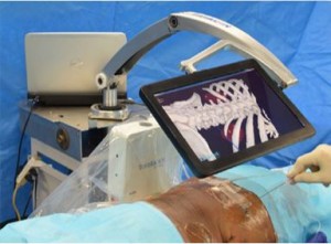
The authors assess the feasibility of a novel method of percutaneous access (TranslucentTM Medical Inc.) using a freely movable tablet display to help guide the percutaneous puncture to its target (see Fig. 1). The success of such a system would rely on a high degree of accuracy and the authors set out to test this in a cadaveric model. Fiducial markers were placed in the kidneys and prostates of cadavers to mimic tumours. A CT scan was then performed to allow 3D model reconstruction. An electromagnetic field was generated around the body and magnetic sensors (fixed to the skin and in the urethral catheter) were used for localization. The software then allows real-time demonstration of the needle trajectory with a predicted line beyond the needle tip overlaid on the 3D model. The authors were able to quickly reach the target (mean time 43 s) with apparent good accuracy, which was calculated to be within 2.5 mm; however, further fiducial markers were deployed at the centre of the target to allow the accurate measurement of accuracy. The distance between the ‘target’ fiducial and the ‘treatment’ fiducial was 16.6 mm in the prostate and 12.0 mm in the kidney. This difference was probably attributable to either movement of the organ or deformation of it from the needle puncture itself. These errors were predominantly in the z-axis (i.e. depth), suggesting that they were caused by movement and deformation by the needle itself, which therefore poses the challenge of how the errors might be reduced, especially when a living human model may have more compliant tissues and of course be moving with respiration.

asdasd

Figure 1. Tablet screen displaying real-time ‘three-dimensional window into the body’ with real and projected trajectory of needle

The present paper by Marien et al. is not the first report of using tracking for percutaneous access in urology. Using different technology, Rassweiler et al. [2] reported on the percutaneous puncture of the kidney for nephrolithotomy using an iPad with surface skin markers, which were visualized by the rear-facing camera of the iPad, and this information was processed to calculate the location of the tissues beneath based on a preoperative CT. This system, and I suspect all others, will rely on a CT being performed in the exact position of the surgery, which is another limiting step until this can be performed at the same time as the surgery.

There are of course concerns regarding use of this technology. The accuracy was limited when analysing the actual position of the ‘treatment’ fiducial because of movement of the tissues. This is likely to be worse in living tissue. There was no respiratory movement which would probably make accurate tracking difficult, although placement of markers to allow movement tracking may help overcome this. Rodrigues et al. [3] reported high accuracy of percutaneous nephrolithotomy puncture after deployment of an electromagnetic sensor in the target calyx with ureterorenoscopy in a porcine model but without a 3D model.

My major concern is that of false reassurance. A nice image is portrayed on the screen which is believed by the surgeon, while in fact there is a significant mismatch caused by patient/tissue movement, either since the preoperative planning CT scan or intra-operatively. The falsely reassured surgeon then inadvertently damages surrounding organs.

It is clear that this technology is work in progress, but it does offer promise that real-time tracking of a percutaneous needle is possible, with accurate representation on a 3D model reconstruction helping to guide the surgeon to the target.

Read the full article
Matthew Bultitude


Department of Urology, Guys and St. Thomas Hospital, London, UK


References


Urologists in the Yellow Submarine – a Periscope to the World

henry-woo_smOver the last few weeks, there has been a lot of chatter about a new Social Media platform. Just when you thought that we had exhausted all possible ways that people could interact online, live video streaming is the talk of the town.

Last month, two competing live video streaming apps were launched.  Meerkat initially gained popularity quite rapidly, particularly through Twitter, given the ease and immediacy of being able to share your live video streaming with twitter followers. Twitter acquired its competitor, Periscope, and Meerkat’s access to the twitter followers was cut off no sooner than it had began. Already there are arguments as to which of the two platforms are better but I can already sense from user reactions and expert opinion, that Periscope will be the one that will prevail. The might of Twitter will be very difficult to compete with.

Why on earth would urologists be interested in live broadcasts? The obvious application is live streaming of events such as conferences. The default option is perform a public broadcast and this will have particular value when there is an advocacy focus. There is also an option to broadcast privately only to followers of the Periscope account performing the broadcast. The latter may well be the best option for more sensitive material but there are still issues that need to be sorted out.  In particular, there is no simple mechanism to determine which followers should be permitted to follow the broadcasting account in order to see a private live stream. It is inevitable that this will be simplified in the future, as it would be logical for this platform to find a mechanism to attract business users.

As things are at present, one needs to have a twitter account in order to sign on to broadcast using Periscope. This platform is designed for the mobile user – this is both for broadcasting and for watching the live stream.  Attempting to do this on a desktop or laptop website is cumbersome and clumsy from my initial attempts to do so whereas the iOS App was straightforward and intuitive, particularly for those already familiar with the Twitter app.

Periscope1


Note the similarity of the iOS Periscope App with the Twitter App interface.

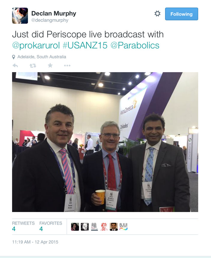
It is my belief that the first ever Periscope live stream broadcast from a medical conference was performed on Sunday 12 April 2015 at the Urological Society of Australia and New Zealand’s (USANZ) Annual Scientific Meeting. Declan Murphy used Periscope to broadcast a message from Prokar Dasgupta, Editor-in-Chief of the BJUI Journal.   The video from the Periscope live stream is below. This first, at least for a urological conference, was tweeted by Declan Murphy.

Screen-Shot-2015-04-17-at-2.55.03-pm


A couple of hours later, I performed a live video stream from the Social Media session when Imogen Patterson gave an excellent presentation on managing our online reputations. During the feed, observers are able to make comments as well as to demonstrate their approval by tapping their screens to trigger a flow of hearts from the bottom right hand corner of the screen.

Periscope3

This is a screenshot from an unrelated live video feed. From the bottom left, the user is notified of those joining the observation of the feed as well as comments. From the bottom right, hearts float upwards in response to positive taps of the screen by watchers.

There are a few issues with Periscope as it is right now. The feed is only available for 24 hours before disappearing from the Periscope platform, however, a video recording minus the comments and hearts, can be stored in the photo stream on your mobile device. As mentioned before, you must have a twitter account to broadcast although you do not need one to view a broadcast. Thirdly, directed broadcasting should be simplified.

Social media platforms come and go but the ability to live stream is an exciting new development. For Periscope, it is my belief that the potential application for a use in medical education seems boundless. Live broadcasting is no longer the exclusive domain of television and cable networks.


Henry Woo (@drhwoo) is Associate Professor of Surgery at the Sydney Adventist Hospital Clinical School of the University of Sydney. He is the Editor-in-Chief of BJUI Knowledge, an innovative on-line CME portal that launches this year.


William Steers 1955-2015

William_D._SteersBy Montesbradley (Own work) [CC BY-SA 3.0 (https://creativecommons.org/licenses/by-sa/3.0)], via Wikimedia Commons

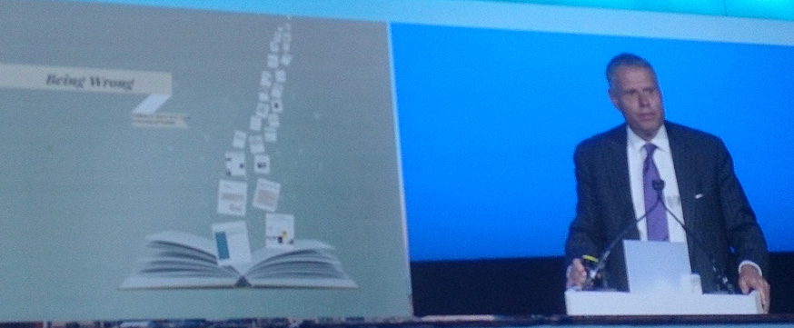
Last June, we had the great pleasure of hosting Dr. William Steers, Editor-in-Chief of the Journal of Urology as our guest speaker during the BJUI session at BAUS. He delivered a Prezi presentation entitled “Being Wrong” – an amazing collage of his experiences as a surgeon, innovator, scientist and editor. The lecture struck a chord with many colleagues both senior and junior, purely because of its reflective, personal and candid content. Little did Bill or any of us realise that this would be our last meeting during the British summer.For more information about latest presentation Click drssa. During the USANZ 2015 meeting in Adelaide, we were very sad to hear of his death on the 10 April 2015 after a short battle with cancer. For more info visit cmsmd .

being-wrong

Dr. Steers, born on 19 August 1955, was a Paul Mellon Professor and Chair of the Department of Urology at the School of Medicine of the University of Virginia, President of the American Board of Urology from 2010-2011, initiator of the Charlottesville Men’s Four Miler, and rather proud producer of the wine label from his very own Well Hung Vineyard. Bill was a legend in the world of neuro-urology. He was passionate about Men’s Health and the functional outcomes of robotic assisted radical prostatectomy.

wineryIn addition to his many accomplishments, he was a keen jogger who loved streaming his favourite music on Spotify while editing articles for the Journal of Urology. Bill was a dedicated family man and is survived by his wife, Amy; sons, Colin and Ryan; daughter-in-law, Ali; and grandson, Rex. A celebration of his life will be held on April 18, 2015, from 2-5 pm. at the Steers residence and home of Well Hung Vineyard, Charlottesville, VA.

Bill – your legacy of friendship, collaboration and inspiration for the next generation lives on. For the first time in history, the Journal of Urology led a joint workshop for young international reviewers along with European Urology and the BJUI, during the 2015 meeting of the European Association of Urology in Madrid. We salute your memory as we come to terms with your untimely passing.


Editorial: A urologists’ guide to the multi-parametric magnetic resonance imaging (mpMRI)-galaxy

The rise of multi-parametric MRI (mpMRI) for the assessment of patients with suspicion of prostate cancer has led to an enormous shift in the practice of every urologist dealing with frontline diagnostics [1].

At the same time, researchers and industry have identified acres of fruitful soil to place the seeds of their respective interests, sometimes in collaboration with each other producing valuable contributions to this shift in practice, sometimes taking benefits by merely assimilating themselves or their product to this development.

Both, the speed of change and the extent of proliferation, make it almost impossible for by-standing clinicians to keep up and filter the evidence-based essence for their local practice.

There are three important issues that need to be considered:

1 The Quality of mpMRI

The development of mpMRI for prostate assessment occurred over the last decade with well-known leaders pushing the frontiers. Their research benefitted from their individual experience of interpreting and reporting MRIs. This is then reflected in their outcomes in form of cancer detection rates and accuracy. More recently we have identified that achieving these results must involve standardisation of MRI protocols and reading [2-4], systematic training in validated courses and a significant learning curve [5]. The latter is only possible to achieve if the practice is embedded in a collaborative team of radiologists, pathologists and urologists. But even then it may be impossible for local teams to deliver the published accuracy, and the urologists and radiologists need to be mindful of that when counselling patients using mpMRI in their local environment.

2 The Technical and Clinical Validity of MRI-Based Biopsies

Transperineal vs transrectal, targeted alone vs targeted with systematic, cognitive vs fusion biopsies – these are the key debates surrounding the application of mpMRI into the urologists’ armamentarium. For none of them there is or will be a unified answer.

Transrectal approaches suit office-based provision of primary diagnostics in many European and USA health economies; although purists can say that the increasing risk of sepsis from antibiotic-resistant bacteria is not acceptable. But, favouring the less infection-prone transperineal approaches will have impact on theatre capacities even in a hospital-based health system like the UK.

Considering the current real-time quality of mpMRI, systematic biopsies in addition to targeted ones are still necessary. Urologists as a group have to come to an agreement about what is acceptable as a remaining risk when reducing or omitting systematic cores.

Cognitive targeting has been shown to be highly accurate; yet, fusion may offer standardisation and reduce user dependency. Not all fusion software on the market has undergone a thorough validated technical development and clinical accuracy evaluation. Peer-reviewed publications can be found involving the systems Urostation-Koelis, Uronav-Philips, Artemis and BiopSee-Medcom.

3 Translation into Clinical Practice

The positioning of the mpMRI within the assessment algorithm is key to optimise the benefit. Use as a pre-biopsy assessment tool may allow omission of further biopsies in some patients or facilitate targeting [6]. However, an established skill in the use of mpMRI and mpMRI-based biopsy is essential. Many UK centres have started the use of mpMRI in their practice further downstream in patients with persistent suspicion after negative first biopsies with good results for patients. It is already part of guidance that active surveillance should involve the use of MRI [1]. Some leading centres advocate that the diagnosis should be confirmed by MRI-based targeted and systematic biopsies.

Knowing that mpMRI will improve the accuracy of our assessment, we need to re-consider follow-up protocols. Increased certainty should be reflected in an improved cancer-related outcome, better patient experience and reduction in costs for the health system.

Prostate mpMRI as part of the urologists’ armamentarium is here to stay. A standardised team- and evidence-based approach will allow us to remain in control of the destination it leads us to.

Read the full article
Christof Kastner
Cambridge University Hospitals, Cambridge, UK

Editorial: The need for standardised reporting of complications

In the context of diversifying practice models, implementation of new technologies such as the Da Vinci surgical robot and rising healthcare costs, there is growing interest in evaluating the quality of surgical work. This extends into health policy, as reimbursement penalties are introduced for ‘inappropriate’ outcomes (e.g. excessive readmissions). Consequently, there is a significant need to provide an accurate assessment of complications and mortality when reporting on surgical outcomes.

Despite the constant use of outcomes data to measure effectiveness in surgery, no current urology guidelines demand the standardised reporting of surgical complications [1]. As randomised controlled trials are uncommon within the surgical setting, and are associated with significant biases [2], there is a distinct need for a uniform reporting system after urological surgeries. Indeed, the lack of such makes it challenging to compare surgical outcomes between techniques, surgeons and institutions, thus hampering the interpretation of study results [3]. The ongoing (and never-ending) debate on the comparative effectiveness of open vs robot-assisted radical prostatectomy (RP) highlights the need for standardised methods to assess superiority (or inferiority) of surgical results [4].

In this issue of the BJUI, Soares et al. [5] present a single-surgeon study of 1138 laparoscopic RPs (LRPs) with a standardised approach between the years 2000 and 2008, and their 5-year follow-up. Whereas the functional and/or oncological equivalency of LRP compared with open RP has been reported before [6], perhaps the outstanding contribution of this study is the use of the Martin-Donat criteria to report and analyse surgical results [3, 7]. In 2002, Martin et al. [7] introduced a list of 10 standard criteria for accurate and comprehensive reporting of surgical complications (e.g. methods of data acquisition, duration of follow-up, definition of complications, hospital length of stay).

In Table 6 of their manuscript, Soares et al. [5] display surgical and/or oncological outcomes of a total of 17 studies on LRP (including their own data). This table suggests the obvious: there is no consistency of reporting on outcomes. In the 2007 Donat [3] analysis of surgical complications reporting in the urological literature, only 2% of a total of 109 studies met nine to 10 of the critical Martin criteria. Interestingly, these shortcomings have been addressed in more contemporary years as the number of studies complying with most of the Martin criteria has increased between 1999/2000 and 2009/2010 [1]. Yet, despite the increasing use of classification systems for outcomes of surgery and standardised reporting of complications (e.g. Clavien-Dindo classification), they are not routinely applied [1, 8].

In an era where the adoption of a certain surgical approach or technique needs to be carefully weighted against a demand for greater value and decreased costs, a simple case series on positive outcomes is simply not sufficient [9]; at the very least, guideline-compliant assessment of outcomes should be the standard of care.

Read the full article


Marianne Schmid*, Christian P. Meyer*† and Quoc-Dien Trinh*


*Division of Urologic Surgery and Center for Surgery and Public Health, Brigham and Womens Hospital, Harvard Medical School, Boston, MA, USA and† Department of Urology, University Medical Center Hamburg-Eppendorf, Hamburg, Germany


References

1 Mitropoulos D, Artibani W, Graefen M, Remzi M, Roupret M, Truss MReporting and grading of complications after urologic surgical procedures: an ad hoc EAU guidelines panel assessment and recommendations. Eur Urol 2012; 61: 3419


4 Schmid M, Gandaglia G, Trinh QD. The controversy that will not go away. Eur Urol 2014; [Epub ahead of print]. doi: 10.1016/ j.eururo.2014.02.052


5 Soares R, Di Benedetto A, Dovey Z, Bott S, McGregor R, Eden CMinimum 5-year follow-up of 1138 consecutive laparoscopic radical prostatectomies. BJU Int 2014; [Epub ahead of print]. doi: 10.1111/ bju.12887


6 Hruza M, Bermejo JL, Flinspach B et al. Long-term oncological outcomes after laparoscopic radical prostatectomy. BJU Int 2013; 111:  27180


7 Martin RC 2nd, Brennan MF, Jaques DP. Quality of complication reporting in the surgical literature. Ann Surg 2002; 235: 80313


9 Novara G, Ficarra V, DElia C, Secco S, Cavalleri S, Artibani W. Trifecta outcomes after robot-assisted laparoscopic radical prostatectomy. BJU Int 2011; 107: 1004


Editorial: Time to replace PSA with the PHI?

Yet more evidence that the PHI consistently outperforms PSA across diverse populations

The Prostate Health Index (PHI) has regulatory approval in >50 countries worldwide and is now being incorporated into prostate cancer guidelines; for example, the 2014 National Comprehensive Cancer Network Guidelines for early prostate cancer detection discuss the PHI as a means to improve specificity, using a threshold score of 35 [1]. The PHI is also discussed in the Melbourne Consensus Statement [2], and it has been incorporated into the multivariable Rotterdam risk calculator smartphone app for use in point-of-care decisions [3].

As the use of this test continues to expand, more data on its performance in specific at-risk populations are of great interest. The investigators from the PROMEtheus multicentre European trial have previously validated the use of the PHI in men with a positive family history of prostate cancer [4]. The new study by Abrate et al. in this issue of BJUI instead addresses another high-risk population – obese men – who have previously been shown to have a greater risk of aggressive prostate cancer [5].

Among the 965 participants in the PROMEtheus study, 14.7% were considered obese based on a body mass index ≥30 kg/m2. In this group, 45.8% were diagnosed with prostate cancer from a ≥12-core biopsy, and 67.7% had a Gleason score ≥7. Overall, the PHI significantly outperformed PSA for prostate cancer detection in men with a body mass index ≥30 kg/m2 (area under the curve 0.839 vs 0.694; P < 0.001). At 90% sensitivity, the threshold for PHI in obese men was 35.7, with a specificity of 52.3%. The PHI also had the best performance for the detection of Gleason ≥7 disease, with an area under the curve of 0.89.

These findings add to the highly consistent body of evidence supporting the use of the PHI in early prostate cancer detection and risk stratification. In fact, all published studies to date have shown that the PHI outperforms PSA for detection of overall and high-grade prostate cancer detection on biopsy [6]. Numerous studies have also shown a role for the PHI in patient selection and monitoring during active surveillance [7, 8]. Expanded use of this test is warranted to reduce unnecessary biopsies and better identify cancers with life-threatening potential.

Read the full article
Stacy Loeb
Department of Urology and Population Health, New York University, New York, NY, USA


References

1 National Comprehensive Cancer Network Clinical Practice Guidelines in Oncology. Prostate Cancer Early Detection Version 2014. https://www.nccn.org/professionals/physician_gls/pdf/prostate_detection.pdf.Accessed May 26, 2014

2 Murphy DG, Ahlering T, Catalona WJ et al. The melbourne consensus statement on the early detection of prostate cancer. BJU Int 2014; 113:186–8

3 Roobol M, Salman J, Azevedo N. Abstract 857: The Rotterdam Prostate Cancer Risk Calculator: Improved Prediction with More Relevant Pre-Biopsy Information, Now in the Palm of Your Hand. Stockholm: European Association of Urology, 2014

4 Lazzeri M, Haese A, Abrate A et al. Clinical performance of serum prostate-specific antigen isoform [-2]pr oPSA (p2PS A) and its derivatives, %p2PSA and the prostate health index (PHI), in men with a family history of prostate cancer: results from a multicentre European study, the PROMEtheuS project. BJU Int 2013; 112:313–21

5 Freedland SJ, Banez LL, Sun LL, Fitzsimons NJ, Moul JW. Obese men have higher-grade and larger tumors: an analysis of the duke prostate center database. Prostate Cancer Prostatic Dis 2009; 12: 259–63

6 Filella X, Gimenez N. Evaluation of [-2] proPSA and Prostate Health Index (phi) for the detection of prostate cancer: a systematic review and meta-analysis. Clin Chem Lab Med 2013; 51: 729–39

7 Tosoian JJ, Loeb S, Feng Z et al. Association of [-2]proPSA with Biopsy Reclassification During Active Surveillance for Prostate Cancer. J Urol2012; 188: 1131–6

8 Hirama H, Sugimoto M, Ito K, Shiraishi T, Kakehi Y. The impact of baseline [-2]proPSA-related indices on the prediction of pathological reclassification at 1 year during active surveillance for low-risk prostate cancer: the Japanese multicenter study cohort. J Cancer Res Clin Oncol2014; 140: 257–63


Future Directions in Urological Oncology

bju13076-fig-0001The field of urological oncology is rapidly changing. For example, robotic surgery, targeted therapy, and ablation techniques are oncological options that were in their infancy 10 years ago and are now mainstream in many areas of the world. Additionally, immunotherapy has recently become a promising avenue in multiple urological cancers. As we move forward, expect to see a larger presence of urological oncology literature obtained via social media, which BJUI has initiated and subsequently set the standard for the field. Related to this, this month’s edition of BJUI includes four online ‘Articles of the Week’, with each focusing on urological oncology.

Using data from the pro-PSA Multicentric European Study (PROMEtheuS) project, Abrate et al. [1] evaluated the utility of the Prostate Health Index (PHI) in 142 obese (body mass index BMI >30 kg/m2) men who underwent a prostate biopsy for an abnormal DRE or elevated PSA level. Among the 142 patients, 65 (45.8%) were found to harbour prostate cancer. Using the PHI threshold of 35.7, the authors determined that 46 (32.4%) negative biopsies could have been avoided while six (9.2%) cancers would have been missed. Related to this, Salami et al. [2] compared the cancer detection rates of MRI fusion biopsy vs standard 12-core TRUS-guided biopsy in 140 men with a previous negative prostate biopsy and a lesion appreciated on a multiparametric MRI. While the cancer detection rates were similar overall, the MRI fusion biopsy was more likely to detect clinically significant prostate cancer (48% vs 31%), defined as Gleason ≥7 or Gleason 6 with a lesion volume of >0.2 mL on MRI. In an era where over-diagnosis of prostate cancer is commonplace, data to better stratify patients who need (or do not need) a prostate biopsy and enhanced ways to identify clinically significant prostate cancers are of paramount importance.

Soares et al. [3] report their results among 1 138 contemporary laparoscopic radical prostatectomy patients who had at least 5 years of follow-up. Only one case required an open conversion and the transfusion rate was merely 0.5%. At last follow-up, 85% of patients had an undetectable PSA level, 94% of patients were continent, and 77% of non-diabetic men aged <70 years retained potency. These impressive single-surgeon results further suggest that the morbidity of prostate cancer surgery has diminished with increasing time and experience.

Additionally, Tolchard et al. [4] prospectively evaluated 105 patients with bladder cancer with preoperative cardiopulmonary exercise testing prior to radical cystectomy. Patients who received neoadjuvant chemotherapy were excluded and there was a 6% perioperative death rate with 90 days of follow-up. The results suggest that patients with poor cardiopulmonary reserve along with hypertension are at higher risk of perioperative complications and prolonged hospital stay; median length of stay was 22 and 9 days for patients with and without a complication. Furthermore, while only 2% of patients had a preoperative diagnosis of heart failure, there were a significant proportion of patients (50% in this study) found to have moderate-to-severe heart failure based on preoperative cardiopulmonary exercise testing. These provocative results suggest that the urological community should further investigate the utility of routine cardiopulmonary exercise testing in patients undergoing radical cystectomy along with the optimal incorporation of such testing in patients receiving neoadjuvant chemotherapy.

References


3 Soares R, Di BenedettoA, Dovey Z, Bott S, McGregor RG, Eden CGMinimum 5-year follow-up of 1138 consecutive laparoscopic radical prostatectomies. BJU Int 2015;115:54653.


R. Houston Thompson BJUI Consulting Editor (Oncology)
Mayo Clinic, Rochester, MN, USA


EAU 2015 Review Days 3 and 4

Persistent rain throughout this year’s 30th EAU Annual Congress failed to dampen the spirits of over 12,000 delegates who have enjoyed another fantastic congress here in Madrid. The EAU Scientific Committee, led by Arnulf Stenzl, deserve tremendous credit for the work they have done to construct an extremely comprehensive and stimulating programme once again this year. I do recall my last EAU Congress in Madrid 12 years ago and there is no doubt but that the standard of this meeting has risen exponentially during this time. It is not just be Annual Congress of course which has developed in this time; the EAU has seen enormous growth in its global influence through the meteoric rise of European Urology, the activities of the European School of Urology (even beyond Europe), the pre-eminence of the EAU Guidelines, and the introduction of new initiatives such as UroSource. The Annual Congress is the nidus for much of this activity and it has become an unmissable event for many of us (even when based in Australia as I am!).

Rebecca Tregunna and Matthew Bultitude have already covered some of the highlights of the opening days of this year’s Congress in their BJUI blog . I will give you some further highlights and point you towards the excellent congress website which has archived a huge amount of material to allow you to catch up on sessions you may have missed.

Big highlights for me on day 3 and 4 include the following (please forgive my oncology focus):

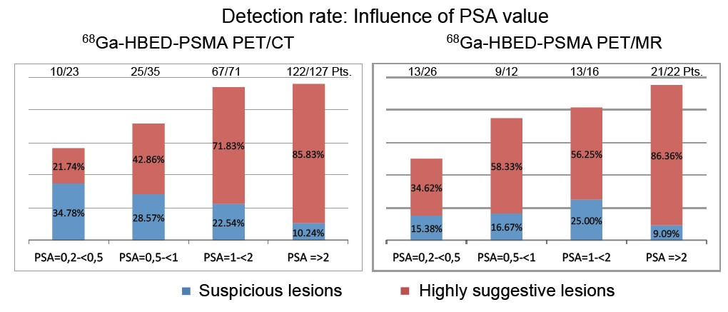
PSMA PET scanning – there was considerable interest in the early data on PSMA PET scanning for recurrent prostate cancer at last year’s EAU Congress, and this year has seen some very positive data being presented from Munich and Heidelberg and further enthusiasm for this imaging modality. Tobias Maurer (Munich) presented a number of papers showing the high sensitivity in particular for PSMA PET in detecting recurrent prostate cancer at low levels of biochemical recurrence using either PET CT or PET MRI (poster 928).
eau15-2-1

Many other plenary speakers also highlighted the positive data surrounding PSMA PET and also the possible theragnostic potential of this in the future (poster 675 and Dr Haberkorn plenary lecture). However in the scientific souvenir session which closed the meeting, Dr Peter Albers burst the bubble somewhat by warning that we need much better data (tissue validation in particular), before we all rush towards PSMA. He has a point of course, although I have been extremely impressed with our initial experience using PSMA PET in Melbourne over the past six months and I do expect it to live up to the hype.

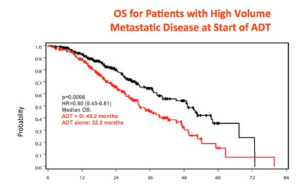
CHAARTED data looking good – Nine months after he made world-wide headlines when he presented the overall survival data of the CHAARTED study at ASCO, Dr Chris Sweeney crossed the Atlantic to again present this data to a packed eUro auditorium. This randomised study of 790 men with metastatic prostate cancer, has demonstrated that men who receive six cycles of docetaxel chemotherapy upfront at the time of starting androgen deprivation therapy, have a considerable survival benefit compared to those who receive ADT on its own (the current standard of care). This was especially so for men with high volume metastases who had a 17 month survival benefit (HR 0.61).
eau15-2-2

Although the French GETUG study has not shown the same benefit, Sweeney and others have proposed rational explanations for why this might be so. While the final paper has not yet been published (will be submitted this week), very many of us have already embraced the CHAARTED as the new standard of care for men presenting with high-volume metastatic prostate cancer. A proper landmark study.

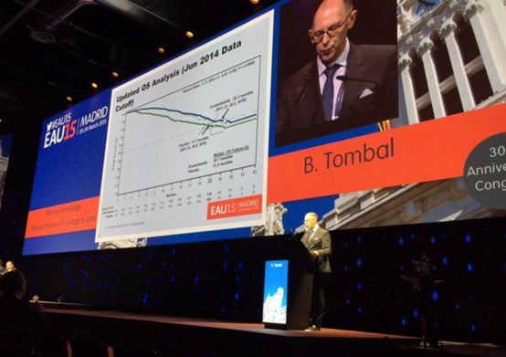
Metastatic castrate-resistant prostate cancer (mCRPC) – still more questions than answers. What an amazing few years for this disease area! Five years ago, urology trainees only had a handful of “essential reading” papers in the world’s top journal, the NEJM, that we could cite to support evidence-based practice. It is now difficult to keep up with all the landmark trials in NEJM and other top journals reporting overall survival advantage for a variety of agents targeting mCRPC. Enzalutamide has already joined the ranks of these blockbuster drugs and this year’s EAU saw more data illustrating the powerful activity of enzalutamide in the pre-chemo mCRPC space. In the Breaking News session on the final day, Dr Bertrand Tombal presented the final analysis of the PREVAIL study which confirmed the overall survival advantage of patients receiving enza pre-chemo when compared with placebo. The HR of 0.77 was strongly significant (p=0.0002) and the therapy was well tolerated.
eau15-2-3

However as pointed out by discussant Dr Maria de Santis, we have still a way to go to figure out which patient will benefit from which therapy and when. The sequence and combination of therapies is still being worked out, and while the potential of predictive biomarkers such as AR7 is certainly exciting, we are still bereft of data and tools (and funding), to figure out the best pathways.

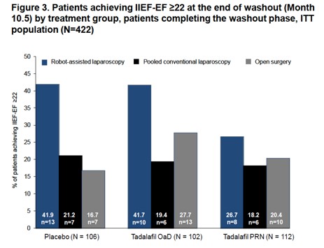
Robot vs open surgery – cystectomy is the new battleground. As Alberto Brignati pointed out in his outstanding souvenir session on localised prostate cancer, it appears that the old debate of robotic vs open prostatectomy is no longer of interest. Despite the lack of prospective randomised data, there appears to be little doubt that robotic prostatectomy is the standard of care in many regions. A large number of posters and plenaries demonstrated convincing data of excellent outcomes in robotic prostatectomy series, including data from a multicenter randomised study (REACTT, poster 622) led by Dr Stolzenberg which demonstrated improved potency outcomes for robotic prostatectomy (not the primary endpoint).
eau15-2-4

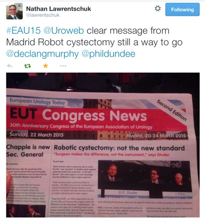
The same cannot yet be said of robotic cystectomy. Despite my own enthusiasm for and publications on robotic cystectomy, it is hard to get away from some of the cautionary language being expressed about the role of robotic cystectomy at the moment. An excellent plenary featuring giants in the field of bladder cancer (Dr’s Bochner, Wiklund, Studer, Palou), debated the issue in the main eUro auditorium and the following day’s newsletter summed it up nicely:
eau15-2-5
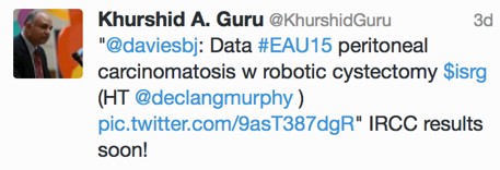
This provoked much discussion on Twitter with some prominent names chiming in from the US. Dr Khurshid Guru got involved to reassure us that the International Robotic Radical Cystectomy Consortium which he leads will provide the answer.
eau15-2-6

Well said @khurshidguru!

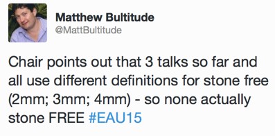
On a non-cancer note, it is clear that some of the most popular session and courses at EAU15 were focused on uro-lithiaisis. Stone surgeons are also very active on Twitter and although I did not attend any stone sessions, I was pleased to see that standardization of terminology is also important to the “pebble-ologists”:
eau15-2-7

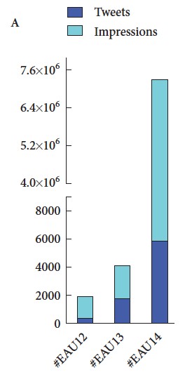
Finally, #EAU15 was truly a social experience, not just in the wonderful bars and restaurants of Madrid, but also through Twitter and other social media channels, strongly supported by the excellent communications team at EAU. We recently published a paper in the BJUI documenting the growth of social media at major urology conferences and at EAU in particular. Between 2012 and 2014, the number of Twitter participants increased almost ten-fold, leading to an increase in the number of tweets from 347 to almost 6,000 At #EAU14, digital impressions reached 7.35 million with 5,903 tweets sent by 797 participants.
eau15-2-8
(From Wilkinson et al BJUI 2015)

As might be expected, #EAU15 has continued this trajectory with almost 8000 tweets sent by 1220 participants.

eau15-2-9eau15-2-10


One of the only criticisms I have of EAU15 is that the scientific program is now so large that it is impossible to get to all the sessions I am interested in. There did seem to be a lot of prostate cancer running simultaneously but I am not sure how much the Scientific Committee can do to avoid such clashes. Thankfully, the EAU meeting website www.eaumadrid2015.org contains a huge amount of material including webcasts, interviews, posters etc which allows delegates and EAU members to catch up on some of the outstanding content.

Another big attraction of the EAU Annual Congress is of course that it takes place in Europe’s most wonderful cities. EAU16 heads to Munich – put the date in your diary 11-15 March 2016.


Declan Murphy, Urologist, Melbourne
Associate Editor – Social Media, BJUI
@declangmurphy

Click here for Declan Murphy’s disclosures

Editorial: How much potential for Transient Receptor Potential channels in the bladder?

In this issue of the BJUI, Charrua et al. [1] report on the possible interaction of two members of the vanilloid subfamily of transient receptor potential (TRP) channels in the control of rat urinary bladder function, TRPV1 and TRPV4. TRP channels are a family of cation-selective channels with 28 known mammalian members. Six of them belong to the subfamily of vanilloid receptors (TRPV channels) and fall into four groups, TRPV1/TRPV2, TRPV3, TRPV4, and TRPV5/TRPV6. The physiological and pharmacological interest in these channels results largely from the finding that they can be activated by a plethora of physical and chemical stimuli; accordingly, they have been implicated in sensory function and pathophysiology of many organ systems [2]. A breakthrough in our understanding of such channels came with the reporting of TRPV1 and TRPV4 knock-out mice, which also exhibit a bladder phenotype; the role of TRP channels in lower urinary tract function has comprehensively been reviewed recently [3].

While the physiological regulation of TRPV1 by endogenous mediators is poorly understood, natural compounds such as capsaicin or resiniferatoxin are acute agonists of TRPV1 channels; however, over time, they desensitise the channel and hence act as inhibitors. These compounds have shown promise in the treatment of detrusor overactivity but also have problems attributed to their initial agonist effects [3]. TRPV4 are activated experimentally by hypotonicity induced cell swelling and several chemicals and more physiologically by moderate heat, stretch and shear stress, leading to the proposition that they may functions as a stretch sensor in the bladder. The inhibitory effects of TRPV1 agonists manifest only after prolonged exposure once desensitisation of their agonist effects occurs, and this initial agonistic phase is a source of undesirable effects. Therefore, a search is on for small molecules that have direct antagonist effects.

Charrua et al. [1] now report that two small molecule antagonists at TRPV1 and TRPV4, (SB355791 and RN1734, respectively) even in high doses did not affect bladder function in control rats. Intravesical installation of lipopolysaccharide is used to create an animal model of cystitis as it induces inflammation, detrusor overactivity and bladder pain. In this model, a high dose of the TRPV4 inhibitor reduced detrusor overactivity, whereas even the high dose of the TRPV1 inhibitor did not; however, a combination of ineffective doses of both inhibitors markedly decreased bladder reflex activity. On the other hand, each of the two drugs caused partial analgesia, but their combination was not more effective than either drug alone. This indicates an interesting functional interaction between TRPV1 and TRPV4 channels, which is specific for the overactivity vs the pain response. Previously, the Cruz group reported that bladder overactivity induced by nerve growth factor depends on the presence of functionally active TRPV1 [4]. Taken together this work shines light on networks of multiple mediators and their receptors that cooperate in the regulation of bladder function but previously have mainly been viewed in isolation. Such work may also have therapeutic consequences. As target-saturating concentrations of ligands at any of these receptors may cause relevant adverse effects, targeting multiple such receptors in low doses may open an avenue for a multi-pronged approach, particularly in patients with bladder dysfunction difficult to control with present treatment options.

This multiple target, low-dose approach is a therapeutically fascinating idea, but finding the right combination of doses in such a setting is a nightmare for any drug development scientist. Moreover, much of the specific role of such targets in pathophysiology remains to be explored before the present findings can be translated into clinical treatments, and the Charrua et al. study [1] will also help such efforts in other ways. Some of the initial thinking on the function of TRP channels in the control of bladder and other functions has been based on localisation studies with TRP channel antibodies, which may have been flawed. Similar to many other receptor antibodies [5], several of those directed against TRPV1 channels also have been shown to lack target specificity [6], leading to misunderstandings about the location and function of such channels. The validation for other TRPV1 and TRPV4 antibodies presented by Charrua et al. [1] will allow more robust studies in this regard and help to develop more valid understanding of TRP channels in physiology, pathophysiology and as treatment targets.

Read the full article
Martin C. Michel


Department of Pharmacolog y , Johannes Gutenberg University, Mainz, Germany


References


3 Franken J, Uvin P, de Ridder D, Voets T. TRP channels in lower urinary tract dysfunction. Br J Pharmacol 2014; 171: 2537–51


4 Frias B, Charrua A, Avelino A, Michel MC, Cruz F, Cruz CD. Transient receptor potential vanilloid 1 mediates nerve growth factor-induced bladder hyperactivity and noxious input. BJU Int 2012; 110: E422–8


5 Michel MC, Wieland T, Tsujimoto G. How reliable arG-protein-coupled receptor antibodies? Naunyn Schmiedebergs Arch Pharmacol 2009; 377: 385–8


6 Everaerts W, Sepúlveda MR, Gevaert T, Roskams T, Nilius B, De Ridder D. Where is TRPV1 expressed in the bladder, do we see the real channel? Naunyn Schmiedebergs Arch Pharmacol 2009; 379: 421–5


© 2024 BJU International. All Rights Reserved.