Archive for category: Article of the Week

Editorial: Multiparametric MRI in active surveillance – time to rethink our current strategy?

Active surveillance for low-risk prostate cancer is gaining increasing acceptance. Indeed, many would argue that it is now the primary management strategy for men who have little to gain from radical therapy but who may incur some harms. However, active surveillance is far from a perfect pathway. First, many men and their physicians find it unacceptable to not treat a known cancer. Second, the burden of follow-up with clinical examinations and serum PSA testing on both men and healthcare systems is far from cost-neutral. Third, the need for repeat transrectal biopsies, which many advocate, carries harms of complications and the difficulties of inaccuracy. Fourth, there is some concern that the window of curability may be lost when men eventually go on to have radical therapy, although overall and disease-specific survival is in fact reassuringly high in the medium term.

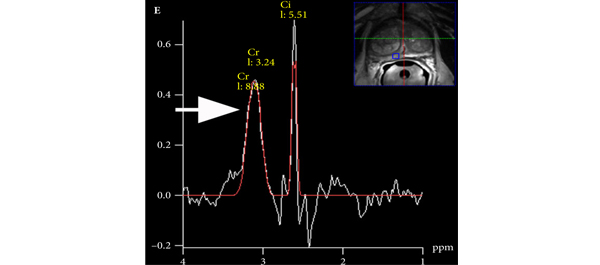
Mullins et al. have attempted to address some of these issues by evaluating the role of multi-parametric MRI (mpMRI) in men followed using active surveillance. The results, albeit preliminary, are very encouraging. The ability of mpMRI to exclude clinically significant prostate cancer found on repeat biopsies reflects those results we have seen from other groups (J Urol 2012, BJU Int 2011). Further, they show that the presence of a lesion on mpMRI more often predicts reclassification on repeat biopsy. This has been supported by others who have demonstrated that the inclusion of mpMRI findings into a nomogram was able to predict clinically insignificant prostate cancer better than models without imaging. Mullins et al. have been appropriately guarded about their own results and point out the weaknesses of their cohort in an open manner so readers can judge the external validity of their findings; however, the significance of these results for the urological community cannot be underestimated, particularly as they point us in the direction of important research questions and clinical trials that need to be formulated to give us the answers we need to improve patient care.

There is an increasing body of evidence pointing to TRUS-guided prostate biopsy as being one of the major problems in the current prostate cancer pathway. As a test, it is both inaccurate, unreliable and has harms. It is inaccurate because about one-third of men with low-risk disease have grade or burden reclassification when a better test (template biopsy) is used. It is unreliable because the status of ‘cancer’ and ‘no cancer’ fluctuates from one biopsy to the next. It is harmful not only because it can cause complications (bleeding, sepsis and pain), but also because it detects clinically insignificant disease the treatment of which the man gains little benefit from. So, the problems with active surveillance do not stem from the fact that surveillance per se is flawed, but rather from its heavy reliance on a deeply flawed diagnostic test.

So, what are the key questions for the field of active surveillance that require a coordinated effort to deliver in a timely fashion? First, could the use of mpMRI before biopsy avoid unnecessary diagnosis of clinically insignificant prostate cancer? Second, if low grade and low-volume lesions were found on an accurate biopsy (template mapping and/or MRI-targeted), could we re-designate these lesions as something other than ‘cancer’? Combined, these two changes could in effect, make active surveillance unnecessary. Third, if mpMRI has a predilection for detecting clinically significant lesions, should the presence of a lesion on imaging lead to a man being excluded from active surveillance? Thus, should all men who are considering active surveillance undergo mpMRI and possibly template mapping biopsies? Fourth, can repeat mpMRI, as opposed to repeat transrectal biopsy, detect disease progression in men on active surveillance, and how is progression defined on imaging? Fifth, is the tissue-preserving strategy of focal therapy an alternative for men suitable for active surveillance or an alternative for those men with intermediate- and high-risk disease who stand to benefit from treatment but wish to minimise the harms of treatment?

It is clear that amongst all of these elements of research we will need to embed health economics to ensure that novel strategies are both clinically and cost-effective. Nonetheless, these are exciting times for those of us who work to innovate in clinical practice and research and improve the care of men with localised prostate cancer.


Hashim U. Ahmed
MRC Clinician Scientist and Clinical Lecturer in Urology, Division of Surgery and Interventional Science, University College London, London, UK

Article of the week: Surgical safety checklist for robotic surgery

Every week the Editor-in-Chief selects the Article of the Week from the current issue of BJUI. The abstract is reproduced below and you can click on the button to read the full article, which is freely available to all readers for at least 30 days from the time of this post.

In addition to the article itself, there is an accompanying editorial written by a prominent member of the urological community. This blog is intended to provoke comment and discussion and we invite you to use the comment tools at the bottom of each post to join the conversation.

If you only have time to read one article this week, it should be this one.

Development and content validation of a surgical safety checklist for operating theatres that use robotic technology

Kamran Ahmed, Nuzhath Khan, Mohammed Shamim Khan and Prokar Dasgupta

MRC Centre for Transplantation, King’s College London, King’s Health Partners, Department of Urology, Guy’s Hospital, London, UK

Read the full article
OBJECTIVES

• To identify and assess potential hazards in robot-assisted urological surgery.

• To develop a comprehensive checklist to be used in operating theatres with robotic technology.

METHODS

• Healthcare Failure Mode and Effects Analysis (HFMEA), a risk assessment tool, was used in a urology operating theatre with innovative robotic technology in a UK teaching hospital between June and December 2011.

• A 15-member multidisciplinary team identified ‘failure modes’ through process mapping and flow diagrams.

• Potential hazards were rated according to severity and frequency and scored using a ‘hazard score matrix’.

• All hazards scoring ≥8 were considered for ‘decision tree’ analysis, which produced a list of hazards to be included in a surgical safety checklist.

RESULTS

• Process mapping highlighted three main phases: the anaesthesia phase, the operating phase and the postoperative handover to recovery phase.

• A total of 51 failure modes were identified, 61% of which had a hazard score ≥8.

• A total of 22 hazards were finalised via decision tree analysis and were included in the checklist.

• The focus was on hazards specific to robotic urological procedures such as patient positioning (hazard score 12), port placement (hazard score 9) and robot docking/de-docking (hazard score 12).

CONCLUSIONS

• HFMEA identified hazards in an operating theatre with innovative robotic technologies which has led to the development of a surgical safety checklist.

• Further work will involve validation and implementation of the checklist.


Read Previous Articles of the Week


Editorial: Sergeant, do you copy?

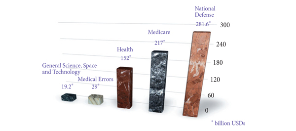
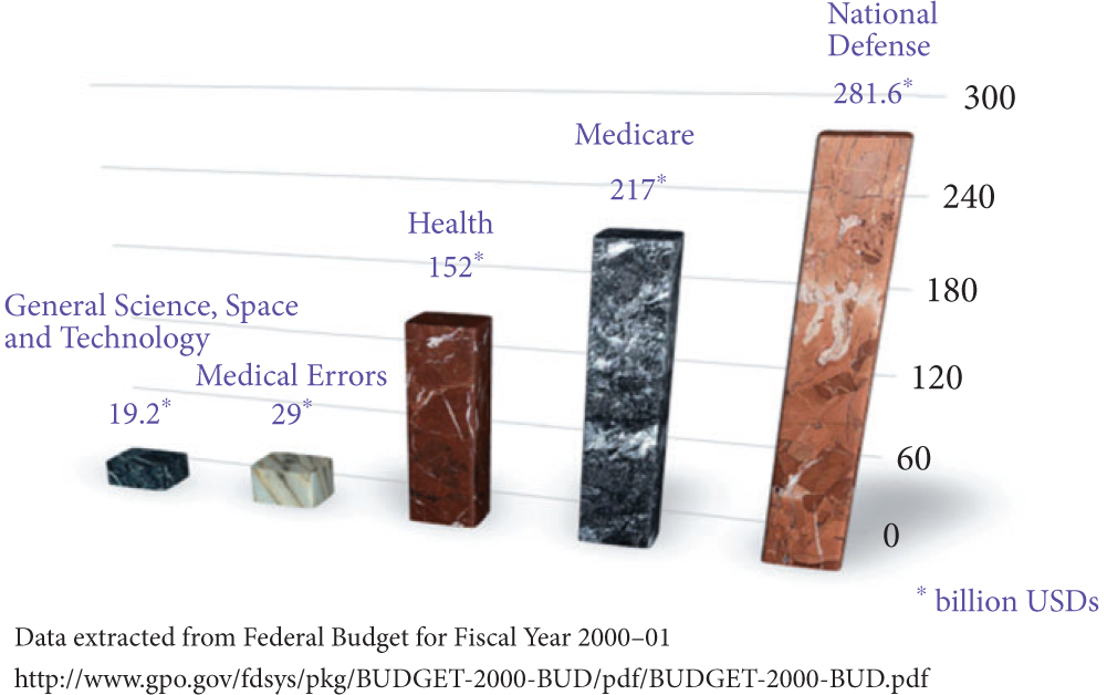
In the Institute of Medicine report published in 1999, it was estimated that 44 000–98 000 patients died annually from preventable medical errors. It was further reported that the annual burden on economy due to preventable medical errors was anywhere between 17–29 billion American dollars. In the USA federal budget 2000–2001, the entire federal resources devoted to general science, space and technology was 19.2 billion American dollars: ≈10 billion less than the cost of medical errors (Fig. 1).

Figure 1. The magnitude of problem caused by medical errors. USDs, American dollars.

On root cause analysis of the errors identified in the Joint Commission on Accreditation and Certification database (2011), it was reported that most of these errors are non-technical, i.e. human factors (72%), leadership (65%), communication breakdown (61%), etc. Furthermore, Greenberg et al. studied the patterns of communication breakdown on the Malpractice Insurers’ Medical Error Prevention Study (MIMEPS) database and concluded that breakdown patterns were similar preoperatively (38%), intraoperatively (30%) and postoperatively (32%). Most errors were due to miscommunication within a single department (78%), as compared with across departments (19%) or institutions (3%). In 49% of the cases, the information was never relayed and in 44% the information relayed was not comprehended appropriately. In all, 29% of these errors involved a surgery attending at transmitting end and 56% at the receiving end of information. In all, 85% of these communications were verbal.

In this issue of BJUI, Ahmed et al. have used the Healthcare Failure Mode and Effect Analysis (HFMEA) model to design a safety checklist specifically for robotic procedures. Checklists have been heavily used in high-risk environments that involve complex technology, e.g. aerospace and nuclear engineering. Robotic surgery is another such high-risk environment, where intraoperative communication is critical. When a surgeon performs a robotic surgery, (s)he is not standing next to the patient (and occasionally not even in the same room!) and relies heavily on his/her assistant. Additionally, the bulky robot takes most of the space around the patient. Small movements of the instruments can cause abrupt and exaggerated movements of the robotic arms, which might injure the bedside assistant, anaesthesiologist, or the patient himself. Last, but not the least, there is a memory clutch on the robotic arms, and its purpose is to ‘remember’ the position of the arms while exchanging the instruments. However, if this clutch is pressed by mistake, all memory is lost and careless insertion of an instrument at this time, making an assumption of memory, can be dangerous and can cause serious injury. The safety checklist described by Ahmed et al. is one of the first checklists specific to robotic surgery. In parallel to this, the Fundamentals of Robotic Surgery (FRS) inter-disciplinary consortium led by Dr Richard Satava has also developed a checklist, specifically for robotic surgery. It will be interesting to study the actual impact of these checklists on prevention of medical errors in robotic surgery. Similar checklists have been validated showing significant clinical correlation using in situ simulation for obstetric emergencies.

Although checklists do help to a certain extent to prevent serious errors, the basics of communications must not be forgotten while communicating to a colleague about patient care. There should be no ambiguity about who is the ‘transmitter’ and who is the ‘receiver’ of information. Both the ‘transmitter’ and ‘receiver’ should have a shared mental model about the purpose of communication (‘transmitter’ is seeking guidance, giving orders, asking for an opinion, referring a case, etc.). Finally, closed-loop communication should be a part of protocol where both the ‘receiver’ and ‘transmitter’ acknowledge the receipt of information, e.g.

Console Surgeon: ‘Please replace the scissors in the right arm with the needle driver’.

Assistant: ‘OK, I am replacing the scissors in your right arm with a needle driver’.

Console Surgeon: ‘Go ahead’.

Assistant: ‘Needle driver coming in’.

Console Surgeon: ‘Perfect. Thank you’.


Sanket Chauhan and Robert M. Sweet
Department of Urology, University of Minnesota Medical School, Minneapolis, MN, USA

Read the full article

Article of the week: Preventing biofilm in wireless capsule bladder endoscopy

Every week the Editor-in-Chief selects the Article of the Week from the current issue of BJUI. The abstract is reproduced below and you can click on the button to read the full article, which is freely available to all readers for at least 30 days from the time of this post.

In addition to the article itself, there is an accompanying editorial written by a prominent member of the urological community. This blog is intended to provoke comment and discussion and we invite you to use the comment tools at the bottom of each post to join the conversation.

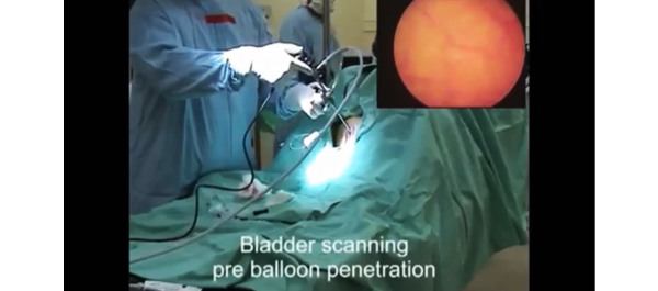
Finally, the third post under the Article of the Week heading on the homepage will consist of additional material or media. This week we feature a video of in-vivo trials in sheep .

If you only have time to read one article this week, it should be this one.

Novel anti-biofilm mechanism for wireless capsule endoscopy in the urinary tract: preliminary study in a sheep model

Amos Neheman*, Claude Schulman and Ofer Yossepowitch†§

*Urology Department, Meir Medical Center, Kfar-Saba, Sackler School of Medicine, Tel-Aviv University, Tel-Aviv, Israel, Department of Urology, University of Brussels, Brussels, Belgium, and §Institute of Urology, Rabin Medical Center, Beilinson, Petah Tikva, Israel

Read the full article
OBJECTIVE

• To develop and test the safety and feasibility of a novel anti-biofilm mechanism configured for wireless capsule endoscopy (WCE) in a sheep bladder model.

MATERIALS AND METHODS

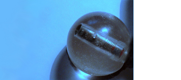
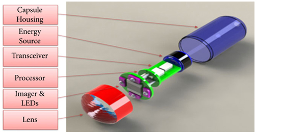
• A WCE mechanism, designed for long-term bladder monitoring, was developed and introduced into a sheep bladder for 5 months.

• The transparency of the surface was assessed by evaluating a resolution target placed inside the capsule at serial intervals using cystoscopy under general anaesthesia.

• Animal behaviour, voiding patterns and urine cultures were monitored throughout the study.

• At study termination, the capsule was extracted and assessed using scanning electron microscopy.

RESULTS

• The resolution target was visualized clearly at all investigation points.

• No notable adverse effects were noted during the entire follow-up period and no urinary tract infection occurred.

• Scanning electron microscopy confirmed the efficacy of the technology to prevent biofilm formation and surface encrustation.

CONCLUSIONS

• We report a novel technology that effectively prevents biofilm formation on the outer surface of foreign objects in the urinary tract.

• Further studies are under way to test the applicability of this technology in bladder WCE to enable high-quality wireless image transmission.


Read Previous Articles of the Week


Editorial: A promising solution for biofilm inhibition in the bladder, but is the application of wireless capsule cystoscopy practical?

The study by Neheman et al. follows up on an idea first proposed in 2009 by Gettman and Swain to adapt wireless capsule endoscopy (WCE) technology for cystoscopy. Unlike the gastrointestinal tract where the small bowel is not endoscopically accessible making WCE appealing and advantageous, the idea of wireless capsule cystoscopy (WCC) competes with a minor procedure, office cystoscopy, that does not require anaesthesia or sedation and takes only a few minutes to perform. Furthermore, although the authors suggest that WCC would shift the labour associated with bladder cancer monitoring from practising urologists to ancillary health team providers, flexible office cystoscopy is a procedure already routinely performed by physician extenders in many offices. Nevertheless, the concept proposed by Neheman et al. is innovative and intriguing. The potential advantage of a wireless capsule cystoscope placed in the bladder safely for up to a 2-year time period, and thereby reducing the inconvenience and cost of repeated cystoscopies, could be a significant advance.

It should be emphasized that despite the title, no WCE was actually performed. The real value to the present study is the novel anti-biofilm mechanism developed that would be needed for any device implanted in the bladder for the long term. The device was housed in a semi-permeable silicone balloon filled with mineral oil that allowed a continuous slow diffusion of oil across the membrane. Based on the evidence provided in only one animal, it seems this continuous permeation of oil can interfere with surface protein adherence and consequently bacteria adhesion and biofilm creation. Certainly, this concept needs to be tested further in additional animals, aggressively exposed to bacteria, and for longer periods.

Although I am unconvinced that the concept of WCC provides significant value, the development of this biofilm inhibition technique could be pioneering. I read this study and wondered if ureteric stents and Foley catheters could be designed and impregnated with mineral oil to be released gradually. Perhaps the balloon of a Foley catheter could be redesigned and filled with mineral oil that is then released along the catheter’s entire length in a similar fashion. The true Holy Grail is the prevention of encrustation and biofilm formation on these relatively mundane devices whose chronic exchange for many patients is more costly than bladder cancer surveillance. I look forward to additional work from the authors exploring the potential of this technology.

Jeffrey A. Cadeddu
Department of Urology, University of Texas Southwestern Medical Center, Dallas, TX, USA

Read the full article

Video: In-Vivo Trials in Sheep


Novel anti-biofilm mechanism for wireless capsule endoscopy in the urinary tract: preliminary study in a sheep model

Amos Neheman*, Claude Schulman and Ofer Yossepowitch†§

*Urology Department, Meir Medical Center, Kfar-Saba, Sackler School of Medicine, Tel-Aviv University, Tel-Aviv, Israel, Department of Urology, University of Brussels, Brussels, Belgium, and §Institute of Urology, Rabin Medical Center, Beilinson, Petah Tikva, Israel

Read the full article
OBJECTIVE

• To develop and test the safety and feasibility of a novel anti-biofilm mechanism configured for wireless capsule endoscopy (WCE) in a sheep bladder model.

MATERIALS AND METHODS

• A WCE mechanism, designed for long-term bladder monitoring, was developed and introduced into a sheep bladder for 5 months.

• The transparency of the surface was assessed by evaluating a resolution target placed inside the capsule at serial intervals using cystoscopy under general anaesthesia.

• Animal behaviour, voiding patterns and urine cultures were monitored throughout the study.

• At study termination, the capsule was extracted and assessed using scanning electron microscopy.

RESULTS

• The resolution target was visualized clearly at all investigation points.

• No notable adverse effects were noted during the entire follow-up period and no urinary tract infection occurred.

• Scanning electron microscopy confirmed the efficacy of the technology to prevent biofilm formation and surface encrustation.

CONCLUSIONS

• We report a novel technology that effectively prevents biofilm formation on the outer surface of foreign objects in the urinary tract.

• Further studies are under way to test the applicability of this technology in bladder WCE to enable high-quality wireless image transmission.


Article of the week: Repeat RAPN: Feasibility and early outcomes

Every week the Editor-in-Chief selects the Article of the Week from the current issue of BJUI. The abstract is reproduced below and you can click on the button to read the full article, which is freely available to all readers for at least 30 days from the time of this post.

In addition to the article itself, there is an accompanying editorial written by a prominent member of the urological community. This blog is intended to provoke comment and discussion and we invite you to use the comment tools at the bottom of each post to join the conversation.

If you only have time to read one article this week, it should be this one.

Repeat robot-assisted partial nephrectomy (RAPN): feasibility and early outcomes

Riccardo Autorino, Ali Khalifeh, Humberto Laydner, Dinesh Samarasekera, Emad Rizkala, Remi Eyraud, Georges-Pascal Haber, Robert J. Stein and Jihad H. Kaouk

Center for Laparoscopic and Robotic Surgery, Glickman Urological and Kidney Institute, Cleveland Clinic, Cleveland, OH, USA

Read the full article
OBJECTIVE

• To demonstrate the feasibility, and to report our single-centre perioperative outcomes of repeat robot-assisted partial nephrectomy (RAPN).

PATIENTS AND METHODS

• From June 2006 to June 2012, 490 patients underwent RAPN for a renal mass at our centre. Of these patients, nine who had undergone previous ipsilateral nephron-sparing surgery (NSS) were included in the analysis.

• Patient charts were reviewed to obtain demographic data, preoperative surgical history, operative details, and postoperative outcomes and follow-up data.

RESULTS

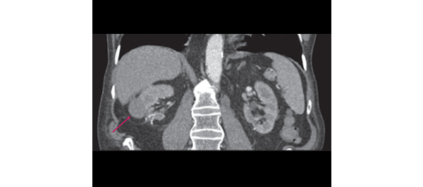
• In all, 12 tumours were removed in nine patients (median age 69 years; six female). A third of the operations were performed on patients with a solitary kidney. The median (range) R.E.N.A.L. nephrometry score for the resected masses was 7 (4–8).

• The warm ischaemia time was 17.5 min and in three of the nine patients an unclamped procedure was performed. No intraoperative complications were registered, whereas only two minor complications occurred postoperatively. There were no renal unit losses. All surgical margins were negative.

• There was no significant difference between mean preoperative and latest postoperative mean estimated glomerular filtration rates (70.5 vs 63.5 mL/min/1.73m2P > 0.05).

• At a mean (sd) follow-up of 8.3 (13) months, eight of the nine patients with a pathology diagnosis of malignant neoplasm were alive and free from disease at the latest follow-up.

CONCLUSION

• Although technically more demanding, repeat RAPN can be safely and effectively performed in patients presenting with local recurrence after primary NSS for kidney cancer.


Read Previous Articles of the Week


Editorial: Robot-assisted partial nephrectomy in patients with recurrent disease: fiction or fact?

In recent decades, the detection of small renal masses (SRMs) has steadily increased with an accompanying shift of treatment towards partial nephrectomy (PN). Indications for PN have successfully expanded to more challenging cases, and robot-assisted PN (RAPN), in particular, has attracted increasing attention (BJUI, Eur Urol); however, despite excellent cure rates for PN, parallel to the increasing number of patients with SRMs undergoing PN, cases of ipsilateral recurrence after PN are also expected to rise. In addition to the incomplete surgical removal of the primary tumour, unknown multifocality or the development of new tumours or metastasis, in a minority of cases recurrence originates at the previous surgical bed and can be considered a proper local recurrence. Retreatment in these patients represents a specific challenge with radical nephrectomy (RN), ablative treatment, repeat PN, and active surveillance in selected cases as therapeutic options. RN should be considered the least attractive option because of the further damage to renal function that it entails, yet it represents one of the most selected options worldwide. Besides ablative techniques, which should be considered under investigational circumstances only, repeat PN is one of the possible options, especially in cases of recurrences attributable to multifocality or bilateral nature.

Repeat open as well as laparoscopic PN (LPN) have been reported sparsely in the literature, but were shown to be associated with good functional and oncological outcomes given adequate laparoscopic experience and patient selection. Such procedures, however, might be challenging and fraught with complications. The role of repeat RAPN (as well as the role of repeat open PN) in this situation is yet to be defined. Nevertheless, given that current data show RAPN to be a more attractive minimally invasive PN technique compared with its standard laparoscopic counterpart, providing equal or better perioperative outcomes, its advantages may even be greater when repeat PN is indicated.

In the current issue of the journal, Autorino et al. report the first study on functional and oncological outcomes and feasibility of repeat RAPN in patients with recurrence after previous PN. Of 490 patients treated with RAPN, nine patients underwent RAPN for recurrent disease at a median time of 39.4 months after previous open or LPN. A total of 12 tumours were removed in these patients, and one third of operations were performed on solitary kidneys. No intra-operative complications were observed, and only two minor complications occurred postoperatively, which were managed conservatively. With regard to functional outcomes, there was a nonsignificant median postoperative decrease in estimated GFR of 7%. More importantly, all patients preserved adequate renal function, which meant that renal replacement therapy was not necessary. With regard to oncological outcomes, all surgical margins were negative and no recurrence was reported.

The results of another study cohort of five patients undergoing repeat RAPN for recurrent disease after open or LPN has recently been published by Jain et al. In their series, surgery was completed in all patients without conversion to RN or an open procedure; furthermore, no complications were reported and the median decrease in GFR was 10%.

Importantly, both series suffer from retrospective evaluation of selective and small sample sizes with a short follow-up, and comparative analysis with other treatment options was not performed; however, the effective comparator for RAPN in this setting has yet to been defined. Bearing in mind current data that demonstrate RAPN to be a preferable minimally invasive PN technique compared with its standard laparoscopic counterparts, the real competitor for RAPN seems to be open surgery. This point was recently also reflected by Mottrie et al. LPN, as a challenging procedure with a long learning curve, limited diffusion and prevalent application in less complex cases, cannot be considered an attractive comparator for RAPN. During the last 8 years, RAPN has become a promising technique which can overcome the technical difficulties of LPN. Three-dimensional vision, ‘endowrist’ technology, and optical magnification allow excellent vision of the operative field and optimum tissue dissection. These technical characteristics help surgeons to minimize ischaemia-time and facilitate accurate tumour excision. Intra-operative ultrasonography, contrast-enhanced sonography, and photodynamic diagnostics can further improve this procedure. It was already shown that the availability of robotic technology is associated with increased use of PN, and, hence, broader diffusion in routine clinical practice may also provide the possibility to outperform results of open PN, even in more complex cases, and will make minimally invasive PN possible and available for more surgeons and patients. Today, the spread of RAPN is only limited by its availability and the associated financial burden. Hence, LPN will currently be considered a cheaper alternative to RAPN in centres with laparoscopic experience and in those which lack the availability of the robot.

Finally, the study from the Cleveland group and the series by Jain et al. provide some valuable support to the feasibility and safety of repeat RAPN and demonstrate that previously performed PN is not a contraindication for RAPN.


Sabine Brookman-May1, Andrea Minervini2, Alessandro Volpe3, Vincenzo Ficarra4, Maciej Salagierski5, Martin Marszalek6,7, Marco Roscigno8, Bülent Akdogan9, Alkuin Vandromme10, Hans Langenhuijsen11, Oscar Rodriguez-Faba12, and Steven Joniau13 for the Renal Cancer Working Group of the Young Academic Urologists (YAU) Working Party of the European Association of Urology (EAU)

1Department of Urology, Ludwig-Maximilians-University, Campus Grosshadern, Munich, Germany, 2Department of Urology, University of Florence, Florence, 3Department of
Urology, University of Eastern Piedmont, Maggiore della Carità Hospital, Novara, 4Department of Oncological and Surgical Sciences, Urologic Unit, University of Padua, Padua,
Italy, 5Department of Urology, Medical University of Łódź, Łódź, Poland, 6Department of Urology and Andrology, Donauspital, Vienna, and 7Department of Urology, Graz Medical University, Graz, Austria, 8Department of Urology, AO Papa Giovanni XXIII, Bergamo, Italy, 9Department of Urology, Hacettepe University, School of Medicine, Ankara, Turkey, 10Klinik für Urologie und Uroonkologie, Klinikum Braunschweig, Germany, 11Laparoscopy, Robotics and Endourology, Radboud University Nijmegen Medical Centre, Nijmegen, The Netherlands, 12Uro-oncology Unit, Fundacio Puigvert, Barcelona, Spain, and 13Department of Urology, University Hospitals Leuven, Leuven, Belgium


Read the full article

Article of the week: ADT increases fracture in high-risk men

Every week the Editor-in-Chief selects the Article of the Week from the current issue of BJUI. The abstract is reproduced below and you can click on the button to read the full article, which is freely available to all readers for at least 30 days from the time of this post.

In addition to the article itself, there is an accompanying editorial written by a prominent member of the urological community. This blog is intended to provoke comment and discussion and we invite you to use the comment tools at the bottom of each post to join the conversation.

Finally, the third post under the Article of the Week heading on the homepage will consist of additional material or media. This week we feature a video of Grace LuYao discussing her paper.

If you only have time to read one article this week, it should be this one.

Fracture after androgen deprivation therapy among men with a high baseline risk of skeletal complications

Yu-Hsuan Shao*, Dirk F. Moore*, Weichung Shih*, Yong Lin*, Thomas L. Jang* and Grace L. Lu-Yao*

*The Cancer Institute of New Jersey, Department of Medicine, The Robert Wood Johnson Medical School, New Brunswick, and ‡Department of Biostatistics, UMDNJ School of Public Health, Piscataway, NJ, USA

Read the full article
OBJECTIVE

• To quantify the impact of androgen deprivation therapy (ADT) in men with a high baseline risk of skeletal complications and evaluate the risk of mortality after a fracture.

PATIENTS AND METHODS

• We studied 75 994 men, aged ≥66 years, with localized prostate cancer from the Surveillance, Epidemiology and End Results–Medicare linked data.

• Cox proportional hazard models were employed to evaluate the risk.

RESULTS

• Men with a high baseline risk of skeletal complications have a higher probability of receiving ADT than those with a low risk (52.1% vs 38.2%, P < 0.001).

• During the 12-year follow-up, more than 58% of men with a high risk and 38% of men with a low risk developed at least one fracture after ADT.

• The dose effect of ADT is stronger among men who received ADT only compared to those who received ADT with other treatments.

• In the high-risk group, the fracture rate increased by 19.9 per 1000 person-years (from 52.9 to 73.0 person-years) for men who did not receive ADT compared to those who received 18 or more doses of gonadotropin-releasing hormone agonist among men who received ADT only, and by 14.2 per 1000 person-years (from 45.2 to 59.4 person-years) among men who received ADT and other treatments.

• Men experiencing a fracture had a 1.38-fold higher overall mortality risk than those who did not (95% CI, 1.34–1.43).

CONCLUSIONS

• Men with a high baseline risk of skeletal complications developed more fractures after ADT.

• The mortality risk is 40% higher after experiencing a fracture.

• Consideration of patient risk before prescribing ADT for long-term use may reduce both fracture risk and fracture-associated mortality.


Read Previous Articles of the Week


Editorial: Androgen deprivation therapy: further confirmation of known harms

Androgen deprivation therapy (ADT) has been an established and effective treatment for men with asymptomatic metastatic prostate cancer for decades. Randomized trials have shown significant survival benefits when ADT is used, coupled with radiotherapy, for patients with locally advanced disease; however it is often used in patients where the benefits are less clear, such as for a rising serum PSA level after radical prostatectomy, and among patients who elect to take a more conservative approach to treatment for low-risk disease. In addition to the absence of data proving benefit, there are a number of adverse consequences attached to androgen deprivation which should be given serious consideration before beginning treatment. Most of the side effects of ADT are linked to its induced hypo-androgenic, and consequently hypo-oestrogenic, state. These include fatigue, vasomotor flushing, loss of muscle mass, weight gain, hyperlipidaemia and insulin resistance.

Osteoporosis and fracture are additional known consequences of ADT with a trend toward greater fracture risk with a higher number of doses of a GnRH agonist and/or longer duration of use. Studies indicate that men with non-metastatic prostate cancer treated with ADT experience an annual loss in bone mineral density of up to nine times that of men in the general population. The use of intermittent ADT, as opposed to continuous use, as a strategy to reduce the negative cardiometabolic and osteoporotic effects is unresolved; however, a report indicating that more recent treatment was associated with a greater risk of fracture, irrespective of cumulative dose, suggests the potential for some reversibility in bone loss post-treatment.

In the present issue of the BJU International, Lu-Yao et al. add to the literature in this area. In a study of nearly 76 000 men with prostate cancer, using data gathered as part of the National Cancer Institute’s Surveillance, Epidemiology and End Results (SEER) cancer registry linked to Medicare claims data, the authors reported that patients at high risk for skeletal complications were, not surprisingly, more likely to experience a fracture associated with ADT use over a 12-year period compared with patients receiving ADT but at low risk for developing such complications. Furthermore, men who experienced a fracture were 40% more likely to die during follow-up than those without fracture. Patients were sorted into risk groups using an index which summed the number of known risk factors for incident fracture identified from Medicare claims in the 12 months before their prostate cancer diagnosis. Unfortunately, owing to the relatively small number of patients with more than one risk factor, the study was limited in its ability to establish a dose – response relationship between the baseline index and fracture risk. SEER-Medicare is an excellent resource to investigate both outcomes and treatment-related expense associated with cancer diagnoses in the USA; however, in this particular study, the reliability of behaviours included in the baseline index (i.e. smoking) is questionable as it would require both patient report of tobacco use as well as physician documentation as a billable claim. Still, one might argue that the heaviest smokers, whose behaviour would most likely be captured as part of a claim, would also be the most important group to capture in an index intended to predict fracture risk.

Interestingly, it was reported that patients at high risk for skeletal complications were significantly more likely to receive ADT than patients at low risk. This was driven in part by the use of primary ADT among elderly men (aged ≥80 years) with prostate cancer and consistent with the notion that when curative treatment is contraindicated (i.e. older patients and those with pre-existing comorbidities) treatment with ADT is more common. Lastly, these findings do not suggest any modification of fracture risk associated with ADT according to baseline risk index, which is consistent with reports of the impact of comorbid conditions and ADT on the risk of incident diabetes and cardiovascular events. This is an important observation in that it says, there is no group that is immune to the adverse effects of ADT – all men are at risk. In absolute terms, however, the men at the greatest risk of an ADT side effect (i.e. a fracture or diabetes) are the men who are at greatest risk of having that side effect even if they were not receiving ADT. The findings of Lu-Yao et al. reinforce the need for careful monitoring of all men receiving ADT. Moreover, when these data are combined with an earlier study that showed that primary ADT was associated with poorer survival than that for men with low-risk prostate cancer who were managed conservatively with observation alone, it should be a wake-up call for us to stop treating non-lethal cancer with lethal and toxic treatments, including ADT.

Jennifer L. Beebe-Dimmer* and Stephen J. Freedland‡§
*Wayne State University School of Medicine, Karmanos Cancer Institute, Detroit, MI, Durham VA Medical Center, and §Duke University School of Medicine, Durham, NC, USA

Read the full article
© 2024 BJU International. All Rights Reserved.