Archive for category: Article of the Week

Article of the week: How useful is FDG-PET/CT in managing carcinoma invading bladder muscle?

Every week the Editor-in-Chief selects the Article of the Week from the current issue of BJUI. The abstract is reproduced below and you can click on the button to read the full article, which is freely available to all readers for at least 30 days from the time of this post.

In addition to the article itself, there is an accompanying editorial written by a prominent member of the urological community. This blog is intended to provoke comment and discussion and we invite you to use the comment tools at the bottom of each post to join the conversation.

Finally, the third post under the Article of the Week heading on the homepage will consist of additional material or media. This week we feature a video of Ms Mertens and Prof Horenblas discussing their findings.

If you only have time to read one article this week, it should be this one.

Impact of 18F-fluorodeoxyglucose (FDG)-positron-emission tomography/computed tomography (PET/CT) on management of patients with carcinoma invading bladder muscle

Laura S. Mertens, Annemarie Fioole-Bruining*, Erik Vegt, Wouter V. Vogel, Bas W. van Rhijn and Simon Horenblas

Departments of Urology, *Radiology and Nuclear Medicine, The Netherlands Cancer Institute, Antoni van Leeuwenhoek Hospital, Amsterdam, The Netherlands

OBJECTIVE

• To evaluate the clinical impact of 18F-fluorodeoxyglucose (FDG)-positron-emission tomography/computed tomography (PET/CT) scanning, compared with conventional staging with contrast-enhanced CT imaging (CECT).

PATIENTS AND METHODS

• The FDG-PET/CT results of 96 consecutive patients with bladder cancer were analysed. Patients included in this study underwent standard CECT imaging of the chest and abdomen/pelvis <4 weeks before FDG-PET/CT.

• Based on the original imaging reports and recorded tumour stage before and after FDG-PET/CT imaging, the preferred treatment strategies before FDG-PET/CT and after FDG-PET/CT were determined for each patient using an institutional multidisciplinary guideline. One of the following treatment strategies was chosen: (i) local curative treatment; (ii) neoadjuvant/induction chemotherapy; or (iii) palliation.

• The changes in management decisions before and after FDG-PET/CT were assessed.

RESULTS

• The median (range) interval between CECT and FDG-PET/CT was 0 (029) days.

• In 21.9% of the patients, stage on FDG-PET/CT and CECT were different. Upstaging by FDG-PET/CT was more frequent than downstaging (19.8 vs 2.1%).

• Clinical management changed for 13.5% of patients as a result of FDG-PET/CT upstaging. In eight patients, FDG-PET/CT detected second primary tumours. This led to changes of bladder cancer treatment in another four of 96 patients (4.2%).

• All the management changes were validated by tissue confirmation of the additional lesions.

CONCLUSIONS

• FDG-PET/CT provides important additional staging information, which influences the treatment of carcinoma invading bladder muscle in almost 20% of cases.

• Patient selection for neoadjuvant/induction chemotherapy was improved and futile attempts at curative treatment in patients found to have metastases were avoided.


Read Previous Articles of the Week


Editorial: Is FDG-PET/CT ready for prime time?

Fluorodeoxyglucose positron-emission tomography (FDG PET)/computed tomography (CT) in bladder cancer

In this month’s issue Mertens et al. [1] present a retrospective analysis of the clinical impact of fluorodeoxyglucose positron-emission tomography (FDG PET)/CT in 96 patients with muscle-invasive bladder cancer. Muscle invasion is present in ≈30% of patients presenting with bladder cancer and is associated with a higher incidence of nodal and metastatic disease than non-muscle-invasive tumours [2]. Accurate staging in this patient group will influence management decisions to proceed to local therapies, to instigate neoadjuvant treatment before local therapy, or to offer palliative chemotherapy where there is imaging evidence and subsequent confirmation of metastatic disease [2].

While there have been a few previous studies investigating FDG PET or FDG PET/CT for staging bladder cancer [3-7], with reported sensitivities and specificities ranging from 60 to 81% and 67 to 94% respectively, to date there are few data describing the impact on clinical management. A recent FDG PET/CT study of 57 patients with bladder cancer [3] reported that management was changed in 68% of cases after PET suggesting that FDG PET/CT has a substantial impact on the management of these patients. However, most patients in that study underwent FDG PET/CT for a suspected recurrence (72%) and the remainder for initial staging (21%) or post-treatment monitoring (chemotherapy or radiotherapy; 7%); 44% of patients had metastatic disease.

In the study reported by Mertens et al. [1], clinical data obtained in 96 patients during the patients’ clinical pathway were reviewed retrospectively. FDG PET/CT staging with standard contrast-enhanced CT was discordant in 22% of cases (21 patients), where PET/CT predominantly upstaged patients, consistent with the previous reports [3, 4]. After PET/CT, the treatment recommendations changed in 13.5% (13 patients) due to disease upstaging. In seven of the 13 patients treatment recommendations altered from local to palliative, due to the presence of metastatic disease, and in the remaining six of the 13 patients, neoadjuvant treatment was recommended in addition to planned local therapy. In another four patients management changed as a consequence of detecting other incidental primary tumours with FDG PET/CT.

However, the final clinical impact of FDG PET/CT may be less. When actual treatment changes were recorded, in only eight of these 13 patients were the recommendations implemented, due to patient co-morbidity or patient wishes in the remainder, e.g. FDG PET/CT changed actual treatment in only 8% in this study (eight of 96 patients). Including the four patients in whom incidental other primary tumours were discovered, the management impact of FDG PET/CT was 12.5%.

There is no doubt that from current published data and supported by this study by Mertens et al. [1] that FDG PET/CT improves staging in bladder cancer due to its higher sensitivity for metastatic disease. However, the actual change in management is relatively low and more prospective data will be required to confirm its clinical and cost effectiveness in terms of outcome, both in a single and multicentre setting.

Vicky Goh* and Gary Cook*
*Division of Imaging Sciences and Biomedical Engineering, King’s College London, Department of Radiology, and Clinical PET Imaging Centre, Guy’s and St Thomas’ Hospitals NHS Foundation Trust, London, UK

Read the full article

References

  1. Mertens L, Fioole-Bruining A, Vegt E, Vogel W, van Rhijn B, Horenblas S. Impact of 18F-fluorodeoxyglucose (FDG)-positron-emission tomography/computed tomography (PET/CT) on management of patients with carcinoma invading bladder muscle. BJU Int 2013; 112: 729–734
  2. Kaufman DS, Shipley WU, Feldman AS. Bladder cancer. Lancet 2009; 374: 239–249
  3. Apolo AB, Riches J, Schoder H et al. Clinical value of fluorine-18 2-fluoro-2-deoxy-d-glucose positron emission tomography/computed tomography in bladder cancer. J Clin Oncol 2010; 28: 3973–3978
  4. Kibel AS, Dehdashti F, Katz MD et al. Prospective study of [18F] Fluorodeoxyglucose positron emission tomography/computed tomography for staging of muscle-invasive bladder carcinoma. J Clin Oncol 2009; 27: 4314–4320
  5. Anjos DA, Etchebehere EC, Ramos CD, Santos AO, Albertotti C, Camargo EE. 18F-FDG PET/CT delayed images after diuretic for restaging invasive bladder cancer. J Nucl Med 2007; 48: 764–770
  6. Drieskens O, Oyen R, Van Poppel H, Vankan Y, Flamen P, Mortelmans L. FDG-PET for preoperative staging of bladder cancer. Eur J Nucl Med Mol Imaging 2005; 32: 1412–1417
  7. Kosuda S, Kison PV, Greenough R, Grossman HB, Wahl RL. Preliminary assessment of fluorine-18 fluorodeoxyglucose positron emission tomography in patients with bladder cancer. Eur J Nucl Med 1997; 24: 615–620

Video: Upstage, downstage: the spotlight on FDG-PET/CT for managing bladder cancer

Impact of 18F-fluorodeoxyglucose (FDG)-positron-emission tomography/computed tomography (PET/CT) on management of patients with carcinoma invading bladder muscle

Laura S. Mertens, Annemarie Fioole-Bruining*, Erik Vegt, Wouter V. Vogel, Bas W. van Rhijn and Simon Horenblas

Departments of Urology, *Radiology and Nuclear Medicine, The Netherlands Cancer Institute, Antoni van Leeuwenhoek Hospital, Amsterdam, The Netherlands

Read the full article
OBJECTIVE

• To evaluate the clinical impact of 18F-fluorodeoxyglucose (FDG)-positron-emission tomography/computed tomography (PET/CT) scanning, compared with conventional staging with contrast-enhanced CT imaging (CECT).

PATIENTS AND METHODS

• The FDG-PET/CT results of 96 consecutive patients with bladder cancer were analysed. Patients included in this study underwent standard CECT imaging of the chest and abdomen/pelvis <4 weeks before FDG-PET/CT.

• Based on the original imaging reports and recorded tumour stage before and after FDG-PET/CT imaging, the preferred treatment strategies before FDG-PET/CT and after FDG-PET/CT were determined for each patient using an institutional multidisciplinary guideline. One of the following treatment strategies was chosen: (i) local curative treatment; (ii) neoadjuvant/induction chemotherapy; or (iii) palliation.

• The changes in management decisions before and after FDG-PET/CT were assessed.

RESULTS

• The median (range) interval between CECT and FDG-PET/CT was 0 (029) days.

• In 21.9% of the patients, stage on FDG-PET/CT and CECT were different. Upstaging by FDG-PET/CT was more frequent than downstaging (19.8 vs 2.1%).

• Clinical management changed for 13.5% of patients as a result of FDG-PET/CT upstaging. In eight patients, FDG-PET/CT detected second primary tumours. This led to changes of bladder cancer treatment in another four of 96 patients (4.2%).

• All the management changes were validated by tissue confirmation of the additional lesions.

CONCLUSIONS

• FDG-PET/CT provides important additional staging information, which influences the treatment of carcinoma invading bladder muscle in almost 20% of cases.

• Patient selection for neoadjuvant/induction chemotherapy was improved and futile attempts at curative treatment in patients found to have metastases were avoided.

Article of the week: Discovery provides new means for regenerative bladder reconstruction

Every week the Editor-in-Chief selects the Article of the Week from the current issue of BJUI. The abstract is reproduced below and you can click on the button to read the full article, which is freely available to all readers for at least 30 days from the time of this post.

In addition to the article itself, there is an accompanying editorial written by a prominent member of the urological community. This blog is intended to provoke comment and discussion and we invite you to use the comment tools at the bottom of each post to join the conversation.

Finally, the third post under the Article of the Week heading on the homepage will consist of additional material or media. This week we feature a video of Dr Verdi discussing his paper.

If you only have time to read one article this week, it should be this one.

Endometrial stem cell differentiation into smooth muscle cell: a novel approach for bladder tissue engineering in women

Alireza Shoae-Hassani*, Shiva Sharif, Alexander M. Seifalian, Seyed Abdolreza Mortazavi-Tabatabaei, Sassan Rezaie§ and Javad Verdi

Departments of Applied Cell Sciences and §Medical Biotechnology, School of Advanced Technologies in Medicine, and *Department of Stem cell and Tissue Engineering, Research Center for Science and Technology in Medicine (RCSTiM), Tehran University of Medical Sciences, Tehran, Islamic Republic of Iran, University College London, UCL Centre for Nanotechnology and Regenerative Medicine, London, UK, and Proteomics Research Center, Faculty of Paramedical Sciences, Shahid Beheshti University of Medical Sciences, Tehran, Islamic Republic of Iran

Read the full article
OBJECTIVE

• To investigate manufacturing smooth muscle cells (SMCs) for regenerative bladder reconstruction from differentiation of endometrial stem cells (EnSCs), as the recent discovery of EnSCs from the lining of women’s uteri, opens up the possibility of using these cells for tissue engineering applications, such as building up natural tissue to repair prolapsed pelvic floors as well as building urinary bladder wall.

MATERIALS AND METHODS

• Human EnSCs that were positive for cluster of differentiation 146 (CD146), CD105 and CD90 were isolated and cultured in Dulbecco’s modified Eagle/F12 medium supplemented with myogenic growth factors.

• The myogenic factors included: transforming growth factor β, platelet-derived growth factor, hepatocyte growth factor and vascular endothelial growth factor.

• Differentiated SMCs on bioabsorbable polyethylene-glycol and collagen hydrogels were checked for SMC markers by real-time reverse-transcriptase polymerase chain reaction (RT-PCR), western blot (WB) and immunocytochemistry (ICC) analyses.

RESULTS

• Histology confirmed the growth of SMCs in the hydrogel matrices.

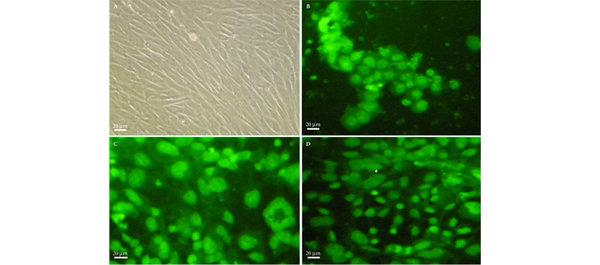
• The myogenic growth factors decreased the proliferation rate of EnSCs, but they differentiated the human EnSCs into SMCs more efficiently on hydrogel matrices and expressed specific SMC markers including α-smooth muscle actin, desmin, vinculin and calponin in RT-PCR, WB and ICC experiments.

• The survival rate of cultures on the hydrogel-coated matrices was significantly higher than uncoated cultures.

CONCLUSIONS

• Human EnSCs were successfully differentiated into SMCs, using hydrogels as scaffold.

• EnSCs may be used for autologous bladder wall regeneration without any immunological complications in women.

• Currently work is in progress using bioabsorbable nanocomposite materials as EnSC scaffolds for developing urinary bladder wall tissue.


Read Previous Articles of the Week

 

Editorial: Reach for the sky – tissue engineering in urology

The work of Verdi et al. [1] published in this issue, shows the continuing quest to find a cellular substrate suitable for producing a tissue engineered replacement for detrusor smooth muscle. This study has identified the regenerative ability of endometrium and with the use of myogenic culture media has sought to differentiate stem cells of endometrial origin to produce the desired smooth muscle cells. The ultimate objective is to produce a functional organ replacement that improves on the current methods of tissue replacement. The current standard for bladder replacement is bowel, both large and small, in various eponymous configurations. All cystoplasties have the potential for long-term consequences including metabolic derangement, UTI, stone formation and mucus secretion [2]. They also suffer the limitation that they will not contract, thus between 10% and 75% will need to self-catheterise. However, many patients do very well after reconstruction with bowel, thus it is important that any substrate designed to replace the current standard matches and improves on that which bowel can offer.

The complex interactions required to achieve a functional bladder replacement are discussed by many authors and include that with urothelium [3], nerve growth and angiogenesis. Despite considerable ingenuity only some of these concerns are solved by previous approaches that have, for example, seeded urothelium onto a vascularised, de-epithelialised flap [4]. The attempt to generate a true composite bladder using cultured urothelium and muscle generated from their native source has been through animal and some clinical exposure but thus far have not gained widespread acceptance and usage – suggesting continued limitations [5, 6].

The pluripotent stem cell approach is attractive, as tissue can be generated from a source distant from the organ needing regeneration, thus bypassing any inherent disease process. The creation of an environment that pushes cellular differentiation along the desired path is the premise by which this works.

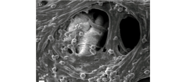
The authors of the current work [1] have analysed the population of generated cells using immunohistochemistry, scanning electron microscopy, gene expression analysis and Western blotting. From this we can learn that the cells are reproducible, viable and appear to exhibit characteristics of the desired smooth muscle cell. That said, all of the current models lack the most desirable of goals – that of controlled, functional similarity to the native bladder. The authors of this paper make the inference that the presence of α-smooth muscle actin suggests that the cells will be contractile. The experiments presented here may imply that but do not confirm it.

The field of tissue engineering remains exciting and authors such as these and others are to be congratulated on continuing to seek innovative approaches to solve a complex problem. The goal is organ replacement and clinical application. Each step along the path to that achievement is valuable but researchers working in the field need to ensure that they remain true to that aim. Cellular markers are only one part of a picture and future work must link them with function in novel cell populations. Once linked with function the means by which function is then controlled becomes important. Before we can safely apply this technology to patients, we must be clear about the functional abilities and limitations of the tissue created, this should be by evidence and not implication. Whilst those undertaking the research convey an optimistic view, the ability to understand the long-term viability and cellular stability remain significant unknowns.

Dan Wood
Adolescent and Paediatric Urology, University College London Hospitals, London, UK

Read the full article

References

  1. Verdi J, Shoae-Hassani A, Sharif S, Seifalian AM, Mortazavi-Tabatabaei SA, Rezaie S. Endometrial stem cell differentiation into smooth muscle cell: a novel approach for bladder tissue engineering in women. BJU Int 2013; 112: 854–863
  2. Biers SM, Venn SN, Greenwell TJ. The past, present and future of augmentation cystoplasty. BJU Int 2012; 109: 1280–1293
  3. Cross WR, Eardley I, Leese HJ, Southgate J. A biomimetic tissue from cultured normal human urothelial cells: analysis of physiological function. Am J Physiol Renal Physiol 2005; 289: F459–468
  4. Fraser M, Thomas DF, Pitt E, Harnden P, Trejdosiewicz LK, Southgate J. A surgical model of composite cystoplasty with cultured urothelial cells: a controlled study of gross outcome and urothelial phenotype. BJU Int 2004; 93: 609–616
  5. Oberpenning F, Meng J, Yoo JJ, Atala A. De novo reconstitution of a functional mammalian urinary bladder by tissue engineering. Nat Biotechnol 1999; 17: 149–155
  6. Atala A, Bauer SB, Soker S, Yoo JJ, Retik AB. Tissue-engineered autologous bladders for patients needing cystoplasty. Lancet 2006; 367: 1241–1246

Video: Endometrial stem cells: new hope for pelvic floor prolapsed?

Endometrial stem cell differentiation into smooth muscle cell: a novel approach for bladder tissue engineering in women

Alireza Shoae-Hassani*, Shiva Sharif, Alexander M. Seifalian, Seyed Abdolreza Mortazavi-Tabatabaei, Sassan Rezaie§ and Javad Verdi

Departments of Applied Cell Sciences and §Medical Biotechnology, School of Advanced Technologies in Medicine, and *Department of Stem cell and Tissue Engineering, Research Center for Science and Technology in Medicine (RCSTiM), Tehran University of Medical Sciences, Tehran, Islamic Republic of Iran, University College London, UCL Centre for Nanotechnology and Regenerative Medicine, London, UK, and Proteomics Research Center, Faculty of Paramedical Sciences, Shahid Beheshti University of Medical Sciences, Tehran, Islamic Republic of Iran

Read the full article
OBJECTIVE

• To investigate manufacturing smooth muscle cells (SMCs) for regenerative bladder reconstruction from differentiation of endometrial stem cells (EnSCs), as the recent discovery of EnSCs from the lining of women’s uteri, opens up the possibility of using these cells for tissue engineering applications, such as building up natural tissue to repair prolapsed pelvic floors as well as building urinary bladder wall.

MATERIALS AND METHODS

• Human EnSCs that were positive for cluster of differentiation 146 (CD146), CD105 and CD90 were isolated and cultured in Dulbecco’s modified Eagle/F12 medium supplemented with myogenic growth factors.

• The myogenic factors included: transforming growth factor β, platelet-derived growth factor, hepatocyte growth factor and vascular endothelial growth factor.

• Differentiated SMCs on bioabsorbable polyethylene-glycol and collagen hydrogels were checked for SMC markers by real-time reverse-transcriptase polymerase chain reaction (RT-PCR), western blot (WB) and immunocytochemistry (ICC) analyses.

RESULTS

• Histology confirmed the growth of SMCs in the hydrogel matrices.

• The myogenic growth factors decreased the proliferation rate of EnSCs, but they differentiated the human EnSCs into SMCs more efficiently on hydrogel matrices and expressed specific SMC markers including α-smooth muscle actin, desmin, vinculin and calponin in RT-PCR, WB and ICC experiments.

• The survival rate of cultures on the hydrogel-coated matrices was significantly higher than uncoated cultures.

CONCLUSIONS

• Human EnSCs were successfully differentiated into SMCs, using hydrogels as scaffold.

• EnSCs may be used for autologous bladder wall regeneration without any immunological complications in women.

• Currently work is in progress using bioabsorbable nanocomposite materials as EnSC scaffolds for developing urinary bladder wall tissue.

Article of the week: Prostate biopsy: shaking up the old standard

Every week the Editor-in-Chief selects the Article of the Week from the current issue of BJUI. The abstract is reproduced below and you can click on the button to read the full article, which is freely available to all readers for at least 30 days from the time of this post.

In addition to the article itself, there is an accompanying editorial written by a prominent member of the urological community. This blog is intended to provoke comment and discussion and we invite you to use the comment tools at the bottom of each post to join the conversation.

Finally, the third post under the Article of the Week heading on the homepage will consist of additional material or media. This week we feature a video of Dr Symons discussing his paper.

If you only have time to read one article this week, it should be this one.

Outcomes of transperineal template-guided prostate biopsy in 409 patients

James L. Symons*, Andrew Huo*, Carlo L. Yuen‡§, Anne-Maree Haynes*, Jayne Matthews, Robert L. Sutherland*, Phillip Brenner‡§ and Phillip D. Stricker†‡§

*Cancer Research Programme, Garvan Institute of Medical Research, St Vincent’s Prostate Cancer Centre, Department of Urology, St. Vincent’s Hospital, and §Department of Urology, St. Vincent’s Clinic, Darlinghurst, NSW, Australia

Read the full article
OBJECTIVE

• To present the template-guided transperineal prostate biopsy (TPB) outcomes for patients of two urologists from a single institution.

PATIENTS AND METHODS

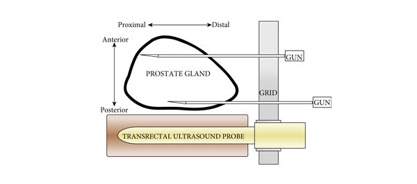
• We conducted a prospective study of 409 consecutive men who underwent TPB between December 2006 and June 2008 in a tertiary referral centre using a standardized 14-region technique.

• The procedure was performed as day surgery under general anaesthesia with fluoroquinolone antibiotic cover.

• Follow-up took place within 2 weeks, during which time men were interviewed using a standardized template.

• Results were compared with those of the Australian national prostate biopsy audits performed by the Urological Society of Australia and New Zealand (USANZ).

RESULTS

• Indications for biopsy included elevated prostate-specific antigen (PSA) level (75%), with a median PSA level of 6.5 ng/mL, abnormal digital rectal examination (8%) and active surveillance (AS) re-staging (18%).

• The mean patient age was 63 years and two-thirds of patients were undergoing their first biopsy.

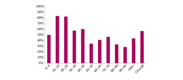
• A positive biopsy was found in 232 men, 74% of whom had a Gleason score of ≥7. The overall cancer detection rate was 56.7% (USANZ 2005 national audit = 56.5%). Stratified between those having their first TPB or a repeat procedure (after a previous negative biopsy), the detection rates were 64.4 and 35.6%, respectively. Significantly higher detection rates were found in prostates <50 mL in volume than in larger prostates (65.2 vs 38.3%, respectively, P < 0.001).

• Haematuria was the most common side effect (51.7%). Others included dysuria (16.4%), acute urinary retention (4.2%) and fever (3.2%). One patient (0.2%) had septicaemia requiring i.v. antibiotics.

• Repeat biopsy was not associated with increased complication rates.

CONCLUSIONS

• TPB is a safe and efficacious technique, with a cancer detection rate of 56.7% in the present series, and a low incidence of major side effects. Stratified by prostate volume, the detection rate of TPB was higher in smaller glands.

• Given the relatively low rate of serious complications, clinicians could consider increasing the number of TPB biopsy cores in larger prostates as a strategy to improve cancer detection within this group. Conversely, in patients on AS programmes, a staging TPB may be a superior approach for patients undergoing repeat biopsy so as to minimize their risk of serious infection.

Editorial: Contemplate the template: a new prostate biopsy approach

Transperineal magnetic resonance imaging – ultrasound fusion targeted biopsies (MRI-US FTB) of the prostate: the future of prostate diagnostics

The prostate cancer diagnostic pathway has remained unchanged for 25 years. At best, laterally directed, peripheral zone (PZ) 12-core transrectal biopsies identify cancer in 44% of cases [1] but transrectal biopsies have an inherent sampling error with a risk of misdiagnosis or mischaracterisation of disease. Of those with negative biopsies who undergo transperineal (TP) biopsies, 30% have cancer, most in the anterior PZ. Active surveillance and the promise of less invasive treatment options are becoming popular because of concerns about ‘over treatment’ for low-risk disease.

Saturation transrectal biopsies have been advocated to improve diagnostic yield but do not address the issue of under sampling of the anterior PZ, particularly in the larger gland [2]. TP biopsies can be used to address the issue of under sampling but prostate template-mapping biopsies are labour intensive and require large numbers of biopsies, often between 60 to 90 cores; however, they have been an essential component of focal therapy trials and the evaluation of novel treatment methods [3].

Primary TP biopsy is the subject of the paper published in this edition of the BJUI titled ‘Outcomes of transperineal template-guided prostate biopsy in 409 patients’ [4]. The authors report a single centre experience of primary TP biopsies. The 14-region protocol described is simpler than prostate template-mapping requiring fewer cores (median of 15 and mean of 19 cores) with a comparable primary diagnostic detection rate of 60% and an encouraging side-effect profile. Unfortunately, the approach still has limitations and the authors admit that their limited biopsy protocol may still mischaracterise disease in the larger gland. In a recent paper from the same group, there was a disappointing correlation between their TP biopsy pathology, MRI abnormalities and radical prostatectomy specimens [5]. Uncertainty prevails, the problem is how best to sample the larger gland. The authors [4] and others, often conclude that more biopsies are necessary for larger glands and resort to mapping protocols and many more biopsies. The solution may not be more biopsies but rather better systematic targeting of the PZ. The impact of hyperplasia within the transition zone (TZ) has a profound effect on PZ anatomy. In the smaller prostate, up to 30 mL, there is little TZ and the PZ is much thicker posteriorly than anteriorly, this difference is even more apparent in glands of 30–50 mL. Above 50 mL TZ expansion causes marked attenuation of the PZ, which becomes much thinner, but the overall volume of the PZ does not change. Less than 4% of cancers originate in the TZ [6], consequently biopsies should be concentrated primarily on the PZ.

The future of prostate cancer diagnosis is likely to be a combination of pre-biopsy multiparametric MRI, followed by targeted biopsies of MRI-identified lesions combined with fewer but better systematic targeted biopsies of the PZ. MRI-ultrasound (MRI-US) fusion techniques have been developed in which axial T2 images of the prostate, diffusion-weighted images and/or dynamic contrast-enhanced MRI images are ‘fused’ with the live US images to allow precise targeting of both regions of interest and the PZ. Commercially available biopsy programs, developed from brachytherapy software systems programs allow individual biopsy sites to be recorded and if combined with inking of the specimen can provide precise pathological localisation of disease within the prostate [7].

There are many potential benefits to this approach. Patients who opt for active surveillance will have an archived record of their disease at a given time to facilitate precise replication of further interval biopsies and assess progression. Improved disease management for an individual should be the aim. The suitability or not for focal or targeted therapies, the planning or boosting of identifed lesions with radiotherapy and/or brachytherapy, and the planning of nerve-sparing surgery or wide excisions should be possible. Feedback to the radiologists of both benign and malignant pathology and grade of disease will improve reporting accuracy and provide imaging sciences with the histopathological characteristics of both MRI ‘visible’ and ‘invisible’ cancer to improve MRI interpretation.

MRI–US fusion targeted biopsies are a significant advance in prostate diagnostics and may resolve some uncertainty within the prostate cancer diagnostic pathway. Benefit vs cost is a recurring issue across health care and questions will continue to be asked about the use of increasingly expensive technology in such an indolent disease. The challenge for investigators will be how to prove the benefit of this approach over standard biopsy protocols and integrate this work in to clinical practice.

Richard Popert
Department of Urology, Guy’s Hospital, London, UK

Read the full article
References
  1. Presti JC, O’Dowd GL, Miller MC et al. Extended peripheral zone biopsy schemes increase cancer detection rates and minimize variance in prostate specific antigen and age related cancer rates: results of a community multi-practice study. J Urol 2003; 169:125–129
  2. Stewart CS, Leibovich BC, Weaver AL, Lieber MM. Prostate cancer diagnosis using a saturation needle biopsy technique after previous negative sextant biopsies. J Urol 2001; 166: 86–92
  3. Onik G, Barzell W. Transperineal 3D mapping biopsy of the prostate: an essential tool in selecting patients for focal prostate cancer therapy. Urol Oncol 2008; 26: 506–510
  4. Symons JL, Huo A, Yuen CL et al. Outcomes of transperineal template-guided prostate biopsy in 409 patients. BJU Int 2013; 112: 585–593
  5. Huo AS, Hossack T, Symons JL et al. Accuracy of primary systematic template guided transperineal biopsy of the prostate for locating prostate cancer: a comparison with radical prostatectomy specimens. J Urol 2012; 187: 2044–2050
  6. Patel V, Merrick GS, Allen ZA et al. The incidence of transition zone prostate cancer diagnosed by transperineal template guided mapping biopsy: implications for treatment planning. Urology 2011; 77: 1148–1152
  7. Hadaschik BA, Kuru TH, Tulea C et al. A novel stereotactic prostate biopsy system integrating pre-interventional magnetic resonance imaging and live ultrasound fusion. J Urol 2011; 186: 2214–2220

Video: Transperineal prostate biopsy: how good is the tumour detection rate?

Outcomes of transperineal template-guided prostate biopsy in 409 patients

James L. Symons*, Andrew Huo*, Carlo L. Yuen‡§, Anne-Maree Haynes*, Jayne Matthews, Robert L. Sutherland*, Phillip Brenner‡§ and Phillip D. Stricker†‡§

*Cancer Research Programme, Garvan Institute of Medical Research, St Vincent’s Prostate Cancer Centre, Department of Urology, St. Vincent’s Hospital, and §Department of Urology, St. Vincent’s Clinic, Darlinghurst, NSW, Australia

Read the full article
OBJECTIVE

• To present the template-guided transperineal prostate biopsy (TPB) outcomes for patients of two urologists from a single institution.

PATIENTS AND METHODS

• We conducted a prospective study of 409 consecutive men who underwent TPB between December 2006 and June 2008 in a tertiary referral centre using a standardized 14-region technique.

• The procedure was performed as day surgery under general anaesthesia with fluoroquinolone antibiotic cover.

• Follow-up took place within 2 weeks, during which time men were interviewed using a standardized template.

• Results were compared with those of the Australian national prostate biopsy audits performed by the Urological Society of Australia and New Zealand (USANZ).

RESULTS

• Indications for biopsy included elevated prostate-specific antigen (PSA) level (75%), with a median PSA level of 6.5 ng/mL, abnormal digital rectal examination (8%) and active surveillance (AS) re-staging (18%).

• The mean patient age was 63 years and two-thirds of patients were undergoing their first biopsy.

• A positive biopsy was found in 232 men, 74% of whom had a Gleason score of ≥7. The overall cancer detection rate was 56.7% (USANZ 2005 national audit = 56.5%). Stratified between those having their first TPB or a repeat procedure (after a previous negative biopsy), the detection rates were 64.4 and 35.6%, respectively. Significantly higher detection rates were found in prostates <50 mL in volume than in larger prostates (65.2 vs 38.3%, respectively, P < 0.001).

• Haematuria was the most common side effect (51.7%). Others included dysuria (16.4%), acute urinary retention (4.2%) and fever (3.2%). One patient (0.2%) had septicaemia requiring i.v. antibiotics.

• Repeat biopsy was not associated with increased complication rates.

CONCLUSIONS

• TPB is a safe and efficacious technique, with a cancer detection rate of 56.7% in the present series, and a low incidence of major side effects. Stratified by prostate volume, the detection rate of TPB was higher in smaller glands.

• Given the relatively low rate of serious complications, clinicians could consider increasing the number of TPB biopsy cores in larger prostates as a strategy to improve cancer detection within this group. Conversely, in patients on AS programmes, a staging TPB may be a superior approach for patients undergoing repeat biopsy so as to minimize their risk of serious infection.

Article of the week: Karakiewicz was right: nomograms are very robust

Every week the Editor-in-Chief selects the Article of the Week from the current issue of BJUI. The abstract is reproduced below and you can click on the button to read the full article, which is freely available to all readers for at least 30 days from the time of this post.

In addition to the article itself, there is an accompanying editorial written by a prominent member of the urological community. This blog is intended to provoke comment and discussion and we invite you to use the comment tools at the bottom of each post to join the conversation.

If you only have time to read one article this week, it should be this one.

Assessing the accuracy and generalizability of the preoperative and postoperative Karakiewicz nomograms for renal cell carcinoma: results from a multicentre European and US study

Luca Cindolo1, Paolo Chiodini2, Sabine Brookman-May8, Ottavio De Cobelli3, Matthias May8, Stefano Squillacciotti4, Cosimo De Nunzio5, Andrea Tubaro5, Ioan Coman7, Bodgan Feciche7, Michael Truss9, Manfred P. Wirth10, Orietta Dalpiaz11, Thomas F. Chromecki11, Shahrock F. Shariat12,13,14, Manuel Sanchez-Chapado15, Maria del Carmen Santiago Martin15, Bernardo Rocco16, Luigi Salzano6, Giuseppe Lotrecchiano6, Francesco Berardinelli1, and Luigi Schips1

1“S. Pio Da Pietrelcina” Hospital, Dept. of Urology, Vasto, Italy, 2Second University of Naples, Dept. of Public Health, Naples, Italy, 3European Institute of Oncology, Dept. of Urology, Milan, Italy, 4“San Carlo” Hospital, Dept. of Urology, Rome, Italy, 5“S. Andrea” Hospital, Dept. of Urology, Rome, Italy, 6“G. Rummo” Hospital, Dept. of Urology, Benevento, Italy, 7Clinical Municipal Hospital, Dept. of Urology, Cluj-Napoca, Romania, 8“Ludwig-Maximilians” University Munich, Dept. of Urology, Campus Grosshadern, Munich, Germany, 9Klinikum Dortmund, Dept. of Urology, Dortmund, Germany, 10Uniklinikum “Carl Gustav Carus”; Dept. of Urology, Dresden, Germany, 11Medical University of Graz, Graz, Austria, 12Weill Cornell Medical Center, New York-Presbyterian Hospital, Dept. of Urology, New York, NY, USA, 13Weill Cornell Medical Center, New York-Presbyterian Hospital, Division of Medical Oncology, New York, NY, USA, 14University of Texas Southwestern, Dept. of Urology, Dallas, TX, USA, 15Department of Urology, Hospital Universitario Principe de Asturias, Alcala de Henares, Spain, and 16Fondazione Ca’ granda, Ospedale Maggiore Policlinico, Dept. of Urology, University of Milan, Milan, Italy

Read the full article
OBJECTIVE

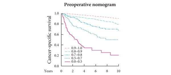
• To assess the accuracy and generalizability of the pre- and postoperative Karakiewicz nomograms for predicting cancer-specific survival (CSS) in patients with renal cell carcinoma (RCC).

PATIENTS AND METHODS

• This retrospective study included 3231 patients from European and US centres, who were treated by radical or partial nephrectomy for RCC between 1992 and 2010.

• Prognostic scores for each patient were calculated and the primary endpoint was CSS.

• Discriminating ability was assessed by Harrell’s c-index for censored data. The ‘validation by calibration’ method proposed by Van Houwelingen was used for checking the calibration of covariate effects. Calibration was graphically explored.

RESULTS

• Local and systemic symptoms were present in 23.2% and 9.1% of the patients, respectively.

• The median follow-up (FU) was 49 months. At the last FU, 408 cancer-related deaths were recorded, Kaplan–Meier estimates of CSS (with 95% confidence intervals [CIs]) at 5 and 10 years were 0.86 (0.84–0.87) and 0.77 (0.75–0.80), respectively.

• Both nomograms discriminated well. Stratified c-indices for CSS were 0.784 (95% CI 0.753–0.814) for the preoperative nomogram, and 0.842 (95% CI 0.816–0.867) for the postoperative one, with a significant difference between the two values (P < 0.001).

• The covariate-based predictions on our data for both nomograms were valid. The calibration plots showed no relevant departures from ideal predictions.

CONCLUSIONS

• The results suggest that the postoperative Karakiewicz nomogram discriminates substantially better than the preoperative one.

• These nomogram-based predictions may be used as benchmark data for pretreatment and postoperative decision-making in patients at various stages of RCC.


Read Previous Articles of the Week
 

© 2024 BJU International. All Rights Reserved.