Archive for category: Article of the Week

Video: EGFR-targeted therapy in metastatic penile SCC

Epidermal growth factor receptor-targeted therapy in locally advanced or metastatic squamous cell carcinoma of the penis

Bradley C. Carthon*§, Chaan S. Ng, Curtis A. Pettaway and Lance C. Pagliaro*

Departments of *Genitourinary Medical Oncology, Radiology, and Urology, The University of Texas MD Anderson Cancer Center, Houston, TX, USA
§
Current address: Winship Cancer Institute, Emory University,Atlanta, Georgia, USA

OBJECTIVE

• To evaluate the safety and efficacy of epidermal growth factor receptor (EGFR)-targeted therapy in patients with advanced penile or scrotal cancer.

PATIENTS AND METHODS

• We retrospectively reviewed the charts of patients with penile or scrotal squamous cell carcinoma who had visited our tertiary cancer centre between 2002 and 2009, including their subsequent treatment and follow-up.

• We collected details of EGFR-targeted therapy and clinical outcomes. Treatment-associated time-to-disease-progression (TTP), overall survival (OS), responses to therapy and toxicity were evaluate

RESULTS

• A total of 24 patients had received EGFR-targeted therapies, including cetuximab, erlotinib and gefitinib. The most common treatment given (to 67% of patients) was cetuximab combined with one or more cytotoxic drugs.

• The most common adverse effect was skin rash (71%). The median (range) TTP and OS were 11.3 (1–40) and 29.6 (2–205) weeks, respectively. The OS time for patients with visceral or bone metastases was significantly shorter than it was for those without (24.7 vs 49.9 weeks, P = 0.013).

• Among 17 patients treated with cetuximab alone or in combination with cisplatin, there were four partial responses (23.5%) including two patients with apparently chemotherapy-resistant tumours.

CONCLUSIONS

• Our results suggest that cetuximab has antitumour activity in metastatic penile cancer, and may enhance the effect of cisplatin-based chemotherapy.

• Prospective studies of EGFR-targeted therapies in men with these tumours are warranted

Article of the week: 1-week and 4-week stenting equally effective after pyeloplasty

Every week the Editor-in-Chief selects the Article of the Week from the current issue of BJUI. The abstract is reproduced below and you can click on the button to read the full article, which is freely available to all readers for at least 30 days from the time of this post.

In addition to the article itself, there is an accompanying editorial written by a prominent member of the urological community. This blog is intended to provoke comment and discussion and we invite you to use the comment tools at the bottom of each post to join the conversation.

If you only have time to read one article this week, it should be this one.

One- vs 4-week stent placement after laparoscopic and robot-assisted pyeloplasty: results of a prospective randomised single-centre study

H. Danuser, C. Germann, N. Pelzer, A. Rühle, P. Stucki and A. Mattei

Klinik für Urologie, Luzerner Kantonsspital, Lucerne, Switzerland

Read the full article
OBJECTIVES

To determine whether 1-week stenting of the pelvi-ureteric anastomosis of laparoscopic or robot-assisted pyeloplasty is as effective as 4-week stenting, based on their respective success rates.

PATIENTS AND METHODS

A total of 100 patients with pelvi-ureteric junction obstruction were treated by Anderson-Hynes pyeloplasty and the anastomosis was stented using a 6-F JJ catheter for either 1 week (1W series) or 4 weeks (4W series), based on a randomisation protocol. Postoperative follow-up was performed at 3 months using intravenous urography (IVU), at 6 months using diuretic renography and at 1, 3 and 5 years using ultrasonography. Statistical analysis was performed using a one-sided Z-test, Pearsons’s chi-squared test and a Wilcoxon rank sum test.

RESULTS

The primary outcome measure, success rate, which was defined as no obstruction on IVU and diuretic renography, was 100% in the 1W series and not inferior to the success rate of 98% in the 4W series (P = 0.006). The following secondary outcome measures were not significantly different between the 1W and the 4W series with regard to residual symptoms (10 vs 6%; P = 0.48), rate of complications (4 vs 6%; P = 0.65), need for synchronous robot-assisted pyelolithotomy (4 vs 8%; P = 0.47), improvement in split renal function (1 vs 0%; P = 0.59) and duration of surgery (200 vs 192 min; P = 0.87). Only length of hospital stay was significantly different; this was shorter in the 1W series (5 vs 6 days; P = 0.01).

CONCLUSIONS

Stenting of the pelvi-ureteric anastomosis after laparoscopic or robot-assisted pyeloplasty for 1 week is as effective as stenting for 4 weeks. Both procedures, laparoscopic or robot-assisted pyeloplasty have an excellent success rate.

Editorial: Early stent removal after pyeloplasty

In the current issue of BJUI Danuser et al. [1] present their prospective randomised single-centre study evaluating the effectiveness of 1-week vs the more traditional 4-week ureteric stent placement, after either a laparoscopic (LPP) or robot-assisted laparoscopic pyeloplasty procedure (RALPP) for PUJO.

In recent years LPP and RALPP have become the standard treatments for PUJO. In the adult population most patients undergoing this procedure require a period of ureteric stenting with a JJ stent, while the newly formed anastomosis heals. Many published pyeloplasty series report a stenting period of between 3 and 6 weeks [2-4]. The present study questions the need for such an extended period of stenting. With current minimal access techniques, either LPP or RALPP, it is possible to create a direct anastomosis between the ureter and renal pelvis similar to, and some would argue even more accurately than, that achievable via open surgery. The authors make the case that as historically most open pyeloplasty procedures were successfully stented for a period of 1 week, it seems only right to question why many of us continue to leave our ureteric stents in for longer periods after LPP and RALPP.

The negative impact of ureteric stent placement on patient health-related quality of life has been well documented in the literature. In 2003, Joshi et al. [5] published their study investigating the prevalence of symptoms associated with ureteric stents. They found that 78% of patients reported bothersome urinary symptoms that included storage symptoms, incontinence and haematuria, and >80% of patients had stent-related pain affecting daily activities. Furthermore, 58% reported reduced capacity to work and 32% reported sexual dysfunction. With this in mind, it is clear why we should try to reduce the period of ureteric stenting wherever possible, as long as it does not compromise patient outcome.

Danuser et al. [1] studied 100 consecutive patients with PUJO treated by an Anderson-Hynes pyeloplasty performed laparoscopically or robotically. Patients were randomly assigned to have a 6-F JJ catheter for either 1 week, or for 4 weeks. Their primary outcome, success rate (defined as no obstruction on the IVU or renogram), was 100% in the 1-week group and 98% in the 4-week group (P = 0.006), showing that 1 week is equally effective. For secondary outcomes measures they found no difference in residual symptoms, rate of complications, need for synchronous robot-assisted pyelolithotomy, improvement in split renal function and duration of surgery between the two groups. They therefore conclude that stenting of the PUJO anastomosis for 1 week after LPP or RALPP is as effective as stenting for 4 weeks.

We are all responsible for constantly evaluating and challenging our medical and surgical practice to ensure that we are providing the best care possible for our patients. In surgery, in the absence of high-level evidence, many of the decisions and actions we take are those inherited from our teachers and mentors, as practices that are thought to be safe and effective. Postoperative patient management is one area where clinicians vary greatly in their practice and we all strive to ensure a safe and comfortable recovery for patients, while not compromising on surgical outcome.

In the postoperative management of pyeloplasty patients many of us continue to leave ureteric stents in for up to 4–6 weeks, as this is ‘safe’ practice. It has been my observation that despite careful counselling of what patients should expect postoperatively when they have a ureteric stent in situ, many complain of stent symptoms and often seek medical advice. This prospective randomised single-centre study by Danuser et al. [1] provides us with good evidence to support the role for a shorter duration of stenting, particularly in this group of patients where a good anastomosis can be created, without compromising patient outcome.

Read the full article

Jane Letitia Boddy
Department of Urology, New Cross Hospitals NHS Trust, Wolverhampton, UK

References

  1. Chow K, Adeyoju AA, Section of Endourology of The British Association of Urological Surgeons. National practice and outcomes of laparoscopic pyeloplasty in the United Kingdom. J Endourol 2011; 25: 657–662
  2. Mufarrij PW, Woods M, Shah OD et al. Robotic dismembered pyeloplasty: a 6 year, multi-institutional experience. J Urol 2008; 180: 1391–1396
  3. Joshi HB, Stainthorpe A, MacDonagh RP, Keeley FX Jr, Timoney AG, Barry MJ. Indwelling ureteral stents: evaluation of symptoms, quality of life and utility. J Urol 2003; 169: 1065–1069


Article of the month: MRI and active surveillance for prostate cancer

Every week the Editor-in-Chief selects the Article of the Week from the current issue of BJUI. The abstract is reproduced below and you can click on the button to read the full article, which is freely available to all readers for at least 30 days from the time of this post.

In addition to the article itself, there is an accompanying editorial written by a prominent member of the urological community. This blog is intended to provoke comment and discussion and we invite you to use the comment tools at the bottom of each post to join the conversation.

Finally, the third post under the Article of the Week heading on the homepage will consist of additional material or media. This week we feature a video from Dr. Hyun M. Lee discussing his paper.

If you only have time to read one article this week, it should be this one.

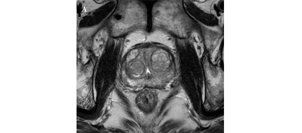
Role of multiparametric 3.0-Tesla magnetic resonance imaging in patients with prostate cancer eligible for active surveillance

Bong H. Park, Hwang G. Jeon, Seol H. Choo, Byong C. Jeong, Seong I. Seo, Seong S. Jeon, Han Y. Choi and Hyun M. Lee

Department of Urology, Samsung Medical Center, Sungkyunkwan University School of Medicine, Seoul, Korea
Current address: Bong H. Park, Department of Urology, Incheon St. Mary’s Hospital, The Catholic University of Korea College of Medicine, Seoul, Korea

Read the full article
OBJECTIVE

• To evaluate predictors of more aggressive disease and the role of multiparametric 3.0-T magnetic resonance imaging (MRI) in selecting patients with prostate cancer for active surveillance (AS).

PATIENTS AND METHODS

• We retrospectively assessed 298 patients with prostate cancer who met the Prostate Cancer Research International: Active Surveillance (PRIAS) criteria, defined as T1c/T2, PSA level of ≤10 ng/mL, PSA density (PSAD) of <0.2 ng/mL2, Gleason score <7, and one or two positive biopsy cores.

• All patients underwent preoperative MRI, including T2-weighted, diffusion-weighted, and dynamic contrast-enhanced imaging, as well as radical prostatectomy (RP) between June 2005 and December 2011.

• Imaging results were correlated with pathological findings to evaluate the ability of MRI to select patients for AS.

RESULTS

• In 35 (11.7%) patients, no discrete cancer was visible on MRI, while in the remaining 263 (88.3%) patients, a discrete cancer was visible.

• Pathological examination of RP specimens resulted in upstaging (>T2) in 21 (7%) patients, upgrading (Gleason score >6) in 136 (45.6%), and a diagnosis of unfavourable disease in 142 (47.7%) patients.

• The 263 patients (88.3%) with visible cancer on imaging were more likely to have their cancer status upgraded (49.8% vs 14.3%) and be diagnosed with unfavourable disease (52.1% vs 14.3%) than the 35 patients (11.7%) with no cancer visible upon imaging, and these differences were statistically significant (P < 0.001 for all).

• A visible cancer lesion on MRI, PSAD, and patient age were found to be predictors of unfavourable disease in multivariate analysis.

CONCLUSION

• MRI can predict adverse pathological features and be used to assess the eligibility of patients with prostate cancer for AS.


Editorial: Multiparametric MRI and active surveillance for prostate cancer: future directions

A growing body of data exists suggesting an important role of MRI in selecting men with prostate cancer for active surveillance (AS). In the present study, Park et al. [1] show that a suspicious lesion on MRI was independently predictive of adverse pathology after radical prostatectomy (RP). This finding supports existing data suggesting that suspicious lesions on MRI confer an increased risk of disease reclassification among men enrolled in AS [2]. Indeed, in our institutional AS experience we found that men with a suspicious lesion on MRI were more likely to have biopsy reclassification with extended follow-up [3].While these data are provocative, much work remains to be done before the adoption of MRI as a standard screening tool for entry into AS for men with very low-risk prostate cancer.

Introduction of functional sequence imaging into multiparametric MRI protocols has resulted in improved detection and characterisation of clinically localised prostate cancer. However, before widespread implementation into AS protocols can occur, increased rigor and standardisation in image interpretation is needed. As in the present study, 5-point Likert scales have become an increasingly popular method of quantifying a lesions likelihood of representing cancer [1]. Still other authors have quantified a lesions level of suspicion using both weighted and non-weighted scoring systems based on the number of positive MRI sequences [3,4]. While useful for statistical analysis, these reporting methods are fraught with concerns of inter-observer variability and generalizability. Additionally, a recent report by Lee et al. [5] found that a simple measurement of lesion diameter on diffusion-weighted MRI was predictive of insignificant disease after RP. Combining the plethora of functional and morphological data obtained by multiparametric MRI into a standardised, reproducible tool will greatly facilitate implementation of MRI into current AS screening protocols.

As a step in the right direction, Stamatakis et al. [4] recently generated a nomogram for predicting biopsy reclassification in men on AS after taking into consideration both functional and morphological characteristics of MRI lesions. Adding an additional layer of complexity, they also assessed the utility of calculated values, e.g. lesion density (lesion volume/prostate volume), in predicting biopsy reclassification. Briefly, their analysis showed that the number of lesions, lesion suspicion, and lesion density were predictive of biopsy reclassification. While nomogram validation and testing of its predictive value on pathological outcomes is needed, this represents a major advance in the standardised application of MRI to AS cohorts.

Despite great strides in the application of multiparametric MRI to AS cohorts, a significant concern about the false-negative rate exists. Considering the present report, of the 35 men with no visible lesion on MRI, 14.3% men had unfavourable pathology after RP [1]. This is similar to previous studies reporting disease reclassification rates of <18% [2,6]. These men with normal imaging and high-grade cancer highlight the importance of incorporating imaging and clinical data when selecting men for AS. Better defining the false-negative rate of multiparametric MRI, and effectively identifying men with a normal MRI and high-grade disease remain major challenges.

Considering all of the available data, it is becoming increasingly clear that MRI has the potential for improving the identification of patients for whom AS would be safe. It is currently the practice at our institution to refer eligible men for multiparametric MRI before enrolment in AS. Our future scholarly efforts should be directed at the standardisation of reporting MRI data and the development of user-friendly AS criteria that synthesise MRI results with clinicopathological data.

Jeffrey K. Mullins and H. Ballentine Carter
James Buchanan Brady Urological Institute, Johns Hopkins Medical Institutions, Baltimore, MD, USA

Read the full article

References

  1. Park BH, Jeon HG, Choo SH et al. Role of multiparametric 3.0-Tesla magnetic resonance imaging in patients with prostate cancer eligible for active surveillance. BJU Int 2014; 113: 864–70
  2. Margel D, Yap SA, Lawrentschuk N et al. Impact of multiparametric endorectal coil prostate magnetic resonance imaging on disease reclassification among active surveillance candidates: a prospective cohort study. J Urol 2012; 187: 1247–52
  3. Mullins JK, Bonekamp D, Landis P et al. Multiparametric magnetic resonance imaging findings in men with low-risk prostate cancer followed using active surveillance. BJU Int 2013; 111: 1037–45
  4. Stamatakis L, Siddiqui MM, Nix JW et al. Accuracy of multiparametric magnetic resonance imaging in confirming eligibility for active surveillance for men with prostate cancer. Cancer 2013; 119: 3359–66
  5. Lee DH, Koo KC, Lee SH et al. Tumor lesion diameter on diffusion weighted magnetic resonance imaging could help predict insignificant prostate cancer in patients eligible for active surveillance: preliminary analysis. J Urol 2013; 190: 1213–7
  6. Guzzo TJ, Resnick MJ, Canter DJ et al. Endorectal T2-weighted MRI does not differentiate between favorable and adverse pathologic features in men with prostate cancer who would qualify for active surveillance. Urol Oncol 2012; 30: 301–5


Video: MRI can assess the eligibility of patients with prostate cancer for AS

Role of multiparametric 3.0-Tesla magnetic resonance imaging in patients with prostate cancer eligible for active surveillance

Bong H. Park, Hwang G. Jeon, Seol H. Choo, Byong C. Jeong, Seong I. Seo, Seong S. Jeon, Han Y. Choi and Hyun M. Lee

Department of Urology, Samsung Medical Center, Sungkyunkwan University School of Medicine, Seoul, Korea
Current address: Bong H. Park, Department of Urology, Incheon St. Mary’s Hospital, The Catholic University of Korea College of Medicine, Seoul, Korea

Read the full article
OBJECTIVE

• To evaluate predictors of more aggressive disease and the role of multiparametric 3.0-T magnetic resonance imaging (MRI) in selecting patients with prostate cancer for active surveillance (AS).

PATIENTS AND METHODS

• We retrospectively assessed 298 patients with prostate cancer who met the Prostate Cancer Research International: Active Surveillance (PRIAS) criteria, defined as T1c/T2, PSA level of ≤10 ng/mL, PSA density (PSAD) of <0.2 ng/mL2, Gleason score <7, and one or two positive biopsy cores.

• All patients underwent preoperative MRI, including T2-weighted, diffusion-weighted, and dynamic contrast-enhanced imaging, as well as radical prostatectomy (RP) between June 2005 and December 2011.

• Imaging results were correlated with pathological findings to evaluate the ability of MRI to select patients for AS.

RESULTS

• In 35 (11.7%) patients, no discrete cancer was visible on MRI, while in the remaining 263 (88.3%) patients, a discrete cancer was visible.

• Pathological examination of RP specimens resulted in upstaging (>T2) in 21 (7%) patients, upgrading (Gleason score >6) in 136 (45.6%), and a diagnosis of unfavourable disease in 142 (47.7%) patients.

• The 263 patients (88.3%) with visible cancer on imaging were more likely to have their cancer status upgraded (49.8% vs 14.3%) and be diagnosed with unfavourable disease (52.1% vs 14.3%) than the 35 patients (11.7%) with no cancer visible upon imaging, and these differences were statistically significant (P < 0.001 for all).

• A visible cancer lesion on MRI, PSAD, and patient age were found to be predictors of unfavourable disease in multivariate analysis.

CONCLUSION

• MRI can predict adverse pathological features and be used to assess the eligibility of patients with prostate cancer for AS.


Article of the week: Enhanced Recovery Programme for radical cystectomy

Every week the Editor-in-Chief selects the Article of the Week from the current issue of BJUI. The abstract is reproduced below and you can click on the button to read the full article, which is freely available to all readers for at least 30 days from the time of this post.

In addition to the article itself, there is an accompanying editorial written by prominent members of the urological community. This blog is intended to provoke comment and discussion and we invite you to use the comment tools at the bottom of each post to join the conversation.

If you only have time to read one article this week, it should be this one

Implementation of the Exeter Enhanced Recovery Programme for patients undergoing radical cystectomy

Thomas J. Dutton, Mark O. Daugherty, Robert G. Mason and John S. McGrath

Exeter Surgical Health Services Research Unit, Royal Devon and Exeter NHS Foundation Trust, Exeter, UK

Read the full article
OBJECTIVES

• To describe our experience with the implementation and refinement of an enhanced recovery programme (ERP) for radical cystectomy (RC) and urinary diversion.

• To assess the impact on length of stay (LOS), complication and readmission rates.

PATIENTS AND METHODS

• In all, 165 consecutive patients undergoing open RC (ORC) and urinary diversion between January 2008 and April 2013 were entered into an ERP.

• A retrospective case note review was undertaken.

• Outcomes recorded included LOS, time to mobilisation, complication rates within the first 30 days (Clavien-Dindo classification) and readmissions.

RESULTS

• All patients were successfully entered into the ERP.

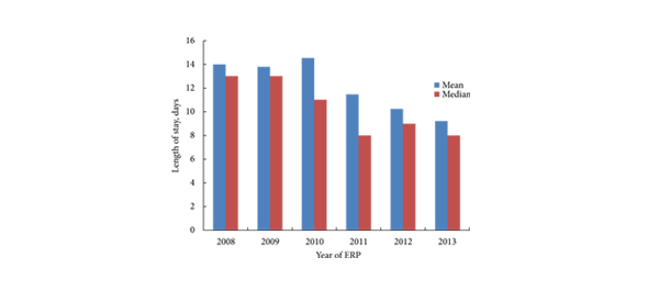
• As enhanced recovery principles became embedded in the unit, LOS reduced from a mean of 14 days over the initial year of the ERP to a mean of 9.2 days.

• The complication rate was 6.6% for Clavien ≥3, and 43.5% for Clavien ≤2. The 30-day mortality rate was 1.2%.

• The 30-day readmission rate was 13.9%.

• In the most contemporary subset of 52 patients: the median time after ORC to sit out of bed, mobilise and open bowels was day 1, 2 and 6, respectively.

CONCLUSIONS

• The ERP described for patients undergoing ORC appears to be safe.

• Benefits include early feeding, mobilisation and hospital discharge.

• The ERP will continue to develop with the incorporation of advancing evidence and technology, in particular the introduction of robot-assisted RC.


Editorial: Enhanced recovery programmes: an important step towards going lean in healthcare

Enhanced recovery programmes (also known as clinical care pathways) are excellent examples of ‘lean thinking’. The ‘lean’ approach is derived from the management philosophy known as the ‘Toyota Production System’ (TPS) that helped the Japanese company become the world’s largest automaker. This management approach has been widely adopted throughout the manufacturing world and has revolutionised the way many businesses operate. Indeed, the concept of clinical care pathways has its roots in the management theories of the TPS, Six Sigma, Business Process Redesign, the Theory of Constraints, and other such methodologies.

This by no means implies that a person is like a car, or the situation is always or ever ‘textbook’. Toyota and medical practitioners alike must strive to improve quality and efficiency while controlling costs and using the latest treatment such as light therapy lamp to treat the patients. And, in the case of healthcare, all of these goals must come under the provision of optimising patient care.

Clinical care pathways provide us the opportunity to standardise processes and problem solving, and eliminate inconsistency (aka ‘mura’, a fundamental pillar of Toyotism). The result is that in every aspect of the delivery of care, there exist clear expectations and demonstrated capabilities. Although situational change is a constant in the healthcare environment, process standards must be applied in all applicable areas to reduce the controllable variances and ensure regulatory compliance, patient and staff satisfaction, and outcomes. Through these standardised pathways or programmes, we are able to establish a confidence in ourselves, our peers, our patients, and our families that what we say will indeed occur. In other words, the right process will produce the right results.

Clinical care pathways are an example of ‘process’ innovation – a concept that can be distinguished from ‘product’ innovation (e.g. drug development, diagnostic tests, robotic and other surgical tools). Process innovations represent important and much needed opportunities to improve outcomes and reduce costs. Clinical care pathways have already been shown to improve patient outcomes, reduce errors, decrease costs, increase transparency of treatment, improve patient, staff, and physician satisfaction, and improve educational opportunities. Moreover, radical cystectomy seems ideally suited to such standardised processes due to characteristics of (1) resource intensivity, (2) complexity of care, (3) high potential variability, and (4) high morbidity.

In this month’s BJU International, Dutton et al. [1] report the ability to effectively apply a standardised enhanced recovery programme to patients undergoing radical cystectomy and urinary diversion for bladder cancer. In their sequential case series, the authors report earlier ambulation, enteral feeding, and time to discharge in patients who were under this enhanced recovery programme (described within) without adverse outcomes, e.g. increased re-admission rates. Similarly, the use of clinical care pathways in our own cystectomy population at The University of North Carolina has represented one of the most important interventions to improve quality and efficiency of care while simultaneously reducing costs.

The next challenge is to explore the applicability of care pathways to multiple physicians and at different institutions, i.e. the widespread use of such programmes to yield the desired results over a healthcare system. Once these processes have been standardised and are able to demonstrate predictable results, we can then focus on raising the performance of these standardised practices and doing so in an iterative process (aka ‘kaizen’).

Read the full article

Raj S. Pruthi and Mathew C. Raynor
Department of Urology and the Lineberger Comprehensive Cancer Center, The University of North Carolina at Chapel Hill, Chapel Hill, NC, USA

Reference

  1. Dutton TJ, Daugherty MO, Mason RG, McGrath JS. Implementation of the Exeter Enhanced Recovery Programme for patients undergoing radical cystectomy. BJU Int 2014; 113: 719–725


Read more articles of the week

Article of the week: Moving up a GEAR: evaluating robotic dry lab exercises

Every week the Editor-in-Chief selects the Article of the Week from the current issue of BJUI. The abstract is reproduced below and you can click on the button to read the full article, which is freely available to all readers for at least 30 days from the time of this post.

In addition to the article itself, there is an accompanying editorial written by prominent members of the urological community. This blog is intended to provoke comment and discussion and we invite you to use the comment tools at the bottom of each post to join the conversation.

If you only have time to read one article this week, it should be this one

Face, content, construct and concurrent validity of dry laboratory exercises for robotic training using a global assessment tool

Patrick Ramos, Jeremy Montez, Adrian Tripp, Casey K. Ng, Inderbir S. Gill and Andrew J. Hung

USC Institute of Urology, Keck School of Medicine, University of Southern California, Los Angeles, CA, USA

Read the full article
OBJECTIVES

• To evaluate robotic dry laboratory (dry lab) exercises in terms of their face, content, construct and concurrent validities.

• To evaluate the applicability of the Global Evaluative Assessment of Robotic Skills (GEARS) tool to assess dry lab performance.

MATERIALS AND METHODS

• Participants were prospectively categorized into two groups: robotic novice (no cases as primary surgeon) and robotic expert (≥30 cases).

• Participants completed three virtual reality (VR) exercises using the da Vinci Skills Simulator (Intuitive Surgical, Sunnyvale, CA, USA), as well as corresponding dry lab versions of each exercise (Mimic Technologies, Seattle, WA, USA) on the da Vinci Surgical System.

• Simulator performance was assessed by metrics measured on the simulator. Dry lab performance was blindly video-evaluated by expert review using the six-metric GEARS tool.

• Participants completed a post-study questionnaire (to evaluate face and content validity).

• A Wilcoxon non-parametric test was used to compare performance between groups (construct validity) and Spearman’s correlation coefficient was used to assess simulation to dry lab performance (concurrent validity).

RESULTS

• The mean number of robotic cases experienced for novices was 0 and for experts the mean (range) was 200 (30–2000) cases.

• Expert surgeons found the dry lab exercises both ‘realistic’ (median [range] score 8 [4–10] out of 10) and ‘very useful’ for training of residents (median [range] score 9 [5–10] out of 10).

• Overall, expert surgeons completed all dry lab tasks more efficiently (P < 0.001) and effectively (GEARS total score P < 0.001) than novices. In addition, experts outperformed novices in each individual GEARS metric (P < 0.001).

• Finally, in comparing dry lab with simulator performance, there was a moderate correlation overall (r = 0.54, P < 0.001). Most simulator metrics correlated moderately to strongly with corresponding GEARS metrics (r = 0.54, P < 0.001).

CONCLUSIONS

• The robotic dry lab exercises in the present study have face, content, construct and concurrent validity with the corresponding VR tasks.

• Until now, the assessment of dry lab exercises has been limited to basic metrics (i.e. time to completion and error avoidance). For the first time, we have shown it is feasibile to apply a global assessment tool (GEARS) to dry lab training.


Editorial: Validating dry lab exercises for robotic surgical skills training

Standardising and structuring of robotic surgery curricula: validation and integration of non-technical skills is required

Kamran Ahmed and Oliver Brunckhorst

Surgical simulation has advanced tremendously over the last two decades with the development of laparoscopic and robotic surgery. Because these procedures have a steep learning curve and because of the reduced training times experienced by trainees, safe adjuncts to operating room training are required [1]. Simulation training is a novel approach to surgical training and has been validated as a training and assessment tool and has been shown to improve a surgeon’s performance in the operating room.

In the present paper, Ramos et al. [2] evaluate the face, content, construct and concurrent validity of robotic dry laboratory (dry lab) exercises. They developed similar tasks to those included in the validated virtual reality da Vinci Skills Simulator using the da Vinci Surgical System in a dry lab environment. They also explored the applicability of Global Evaluative Assessment of Robotic Skills (GEARS) to assess dry lab performance. Good responses from the expert cohort with regard to realism and usefulness as a training tool confirmed the face and content validity of the dry lab exercises, whilst concurrent validity was also established, with experts outperforming novices in all but one of the individual metrics. Finally the simulator composite score achieved from the virtual reality simulator and GEARS scores from the dry lab exercises moderately correlated, thereby also establishing concurrent validity.

The present study raises an interesting question about the definitions of what constitutes an expert in robotic surgery. There are no consistent definitions of a ‘novice’ or an ‘expert’. A recent review has shown that the learning curve in the literature varies from 80 to 250 cases in robot-assisted laparoscopic prostatectomies, depending on previous open or laparoscopic experience and the outcome measures used [1]. With this in mind, Ramos et al. classified ‘experts’ as those performing >30 cases as the primary surgeon. This should be taken into consideration as the experts were so vital in establishing the face, content and construct validity of the dry lab curriculum. Additionally, although it has been shown that simulation models are valid and reliable for the initial phase of training and assessment in urological procedures, this is not the case for advanced and specialist level skill learning.

The present study provides validated dry lab exercises which could be incorporated into a robotic surgery training curriculum. With several simulators now having been validated for robotic surgery, integration of these within a structured robotic surgery curriculum is required. Steps have been taken to set out an effective curriculum similar to that which exists for laparoscopic surgery. The Fundamental Skills of Robotic Surgery is a simulation-based curriculum which has been validated and has a proven educational impact [3]. The Fundamentals of Robotic Surgery curriculum has also recently been laid out, but this is still in the process of validation [4]. One of the biggest initiatives in urology, however, was recently announced by the European Association of Robotic Urology Section Congress 2013 [5]. A multinational push to implement a standardized curriculum set out a multi-step curriculum which uses various techniques, including online theoretical training, simulation and observation and finally fellowship prior to certification, then allowing the surgeon to perform independent surgery. The next step for these curricula is for them to be fully validated and implemented, with the patient outcomes then being analysed.

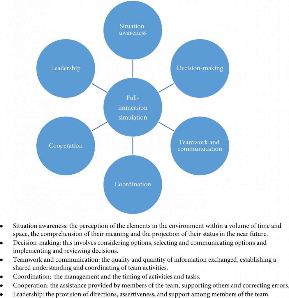
Another aspect worth considering is the integration of non-technical skills within these curricula which has occurred in few of them. Full-immersion simulation (Fig. 1) offers the opportunity for training surgeons to develop critical skills such as communication, coordination and leadership. For the more experienced surgeon, at the later stages of a curriculum, full crisis simulation can help develop decision-making and team-working skills in difficult situations. It has been shown that integration of full-immersion simulation in simulation training is feasible and effective [6]. Surgeons performing robotic surgery need to develop the non-technical aspects alongside their technical skills, and full-immersion simulation certainly has a role to play in this and needs to be integrated within the curricula. The development of the Distributed Simulator offers a validated and low-cost method [7] of introducing full-immersion simulation into robotic surgery curricula and is an option that requires further investigation for its effectiveness in urology.

Figure 1. Aspects of non-technical skills developed through full-immersion simulation [8, 9].

In conclusion, the present study offers further examples of tasks that could be incorporated into robotic surgery training curricula. Whilst curricula exist, implementation and analysis of their effect on patient outcomes are the next steps. The integration of non-technical skills within these is important, and full-immersion simulation has an important role to play within robotic training curricula.

Kamran Ahmed* and Oliver Brunckhorst*

*MRC Centre for Transplantation, King’s College London, King’s Health Partners, and Department of Urology, Guy’s Hospital, London, UK

Read the full article

References

  1. Abboudi H, Khan MS, Guru KA et al. Learning curves for urological procedures: a systematic review. BJU Int 2013; doi: 10.1111/bju.12315. [Epub ahead of print]
  2. Ramos P, Montez J, Tripp A, Ng CK, Gill IS, Hung AJ. Face, content, construct and concurrent validity of dry laboratory exercises for robotic training using a global assessment tool. BJU Int 2014; 113: 836–842
  3. Stegemann AP, Ahmed K, Syed JR et al. Fundamental skills of robotic surgery: a multi-institutional randomized controlled trial for validation of a simulation-based curriculum. Urology 2013; 81: 767–774
  4. Smith R, Patel V, Satava R. Fundamentals of robotic surgery: a course of basic robotic surgery skills based upon a 14-society consensus template of outcomes measures and curriculum development. Int J Med Robot 2013; doi: 10.1002/rcs.1559 [Epub ahead of print]
  5. Khan R, Ahmed K, Mottrie A et al. Towards a standardised training curriculum for robotic surgery: a consensus of an international multidisciplinary group of experts. Poster presented at the EAU Robotic Urology Section Congress Stockholm, Sep 3–5 2013
  6. Shamim Khan M, Ahmed K, Gavazzi A et al. Development and implementation of centralized simulation training: evaluation of feasibility, acceptability and construct validity. BJU Int 2013; 111: 518–523
  7. Kassab E, Tun JK, Arora S et al. ‘Blowing up the barriers’ in surgical training: exploring and validating the concept of distributed simulation. Ann Surg 2011; 254: 1059–1065
  8. Flin R, Yule S, Paterson-Brown S, Maran N, Rowley D, Youngson G. Teaching surgeons about non-technical skills. Surgeon 2007; 5: 86–89
  9. Undre S, Sevdalis N, Healey AN, Darzi A, Vincent CA. Observational teamwork assessment for surgery (OTAS): refinement and application in urological surgery. World J Surg 2007; 31: 1373–1381
© 2024 BJU International. All Rights Reserved.