Archive for category: Article of the Week

Editorial: Can we rely on LVI to determine the need for adjuvant chemotherapy in organ-confined bladder cancer?

The authors of this paper [1] are to be congratulated on exploring lymphovascular invasion (LVI) as a possible singular prognostic marker for time to recurrence and overall survival (OS) in a post hoc analysis of a prospective randomized study that originally explored adjuvant methotrexate, vinblastine, doxorubicin and cisplatin chemotherapy after radical cystectomy based on p53 status. This study is the largest prospective study to date looking at the outcome of LVI in organ-confined urothelial cancer of the bladder.

Lymphovascular invasion represents the first step of dissemination of tumour cells into the lymphatic and blood system which may lead to the formation of metastatic clones. In bladder cancer, our current understanding of the predictive and prognostic role of LVI is mainly based on retrospective data, which are inherently flawed by various selection biases. As pathological tumour and nodal stage, as well as soft-tissue surgical margins, are stronger predictors than is LVI for outcomes in advanced bladder cancer, the authors specifically limited their analysis to the group of patients exhibiting organ-confined disease at radical cystectomy. They found that LVI was associated with time to recurrence and death, while a significant benefit of adjuvant chemotherapy could not be confirmed in a small group of 27 patients with altered p53 expression and LVI. The authors concluded that, although their study did not show a survival benefit for adjuvant chemotherapy in patients with LVI, a possible benefit could not be finally excluded [1].

Indeed, there is still uncertainty about the beneficial impact of adjuvant chemotherapy in bladder cancer. While previous meta-analyses could not show a significant prognostic advantage, a recent update of 945 patients who received adjuvant chemotherapy within nine randomized trials has emphasized its prognostic benefit, especially in lymph node-positive disease [2]. By contrast, a recent report from the European Organisation for the Research and Treatment of Cancer intergroup trial suggests that only patients with node-negative pT3–T4 tumours exhibiting LVI benefit from adjuvant chemotherapy [3]. These heterogeneous data make it difficult to specifically recommend adjuvant chemotherapy in invasive bladder cancer.

The aim of the present study was (and definitely has to be in the future) to outline those patients who do not belong to the roughly 80% of patients who are cured by radical cystectomy without any additional systemic therapy in localized disease. What has been shown in this study is that the presence of LVI definitely influences postoperative outcome. What has not been shown is whether a more or less careful diagnosis of LVI influences time to recurrence and OS after adjuvant chemotherapy, similarly to a negative outcome with regard to p53 status. Do we now believe the two main messages of this paper, which are that LVI does not help us in our decision about which patients might need adjuvant chemotherapy and that there is no room for the argument that adjuvant chemotherapy is better than neoadjuvant chemotherapy because of the histological evidence of LVI?

We are in desperate need of markers [4] in light of the recent literature showing that both neoadjuvant and adjuvant chemotherapy will improve survival in patients with cystectomy as a result of urothelial cancer [5]. Despite the fact that this is one of the largest series of patients with LVI in the specimen, the series is much too incoherent because no central pathology, no mandatory immunohistochemistry, and not even mandatory evaluation of the status in the individual institutions was carried out. We do not even know whether quality control of the pathological evaluations was carried out within each pathology department or hospital, as is mandatory in some parts of the world.

Furthermore, in organ-confined bladder cancer, the invasion depth of the tumour is a key prognosticator of recurrence. In the present study, the only variable associated with a higher risk of LVI was found to be pathological stage (pT1 vs pT2); however, substratification in pT2N0 bladder cancer has also been shown to be of prognostic importance for predicting recurrence after cystectomy [4]. The unknown anatomical extent of lymph node dissection at radical cystectomy makes it difficult to assess the impact of LVI on outcomes because patients with localized tumours and presumed micrometastatic disease (as suggested by LVI) may still be cured with an extended pelvic lymph node dissection [6]. While the authors tried to adjust for this bias by reporting on the number of retrieved lymph nodes, 30% of their patients had < 15 lymph nodes removed at surgery.

In conclusion, the authors of the present study address very important questions, but they fail to provide a clear answer that will change current clinical practice.

Georgios Gakis and Arnulf Stenzl 
Department of Urology, University Hospital Tubingen, Tubingen, Germany


References


Article of the Month: Targeted microbubbles in the treatment of kidney stones

Every Week the Editor-in-Chief selects the Article of the Week from the current issue of BJUI. The abstract is reproduced below and you can click on the button to read the full article, which is freely available to all readers for at least 30 days from the time of this post.

Finally, the third post under the Article of the Week heading on the homepage will consist of additional material or media. This week we feature a video from Dr. Krishna Ramaswamy, discussing his paper. 

Targeted microbubbles: A novel application for treatment of kidney stones

Krishna Ramaswamy, Vanessa Marx*, Daniel Laser, Thomas Kenny, Thomas ChiMichael Bailey§, Mathew D. Sorensen §, Robert H. Grubbs* and Marshall L. Stoller 


Department of Urology, University of California, San Francisco, *Department of Chemistry and Chemical Engineering, California Institute of Technology, Pasadena, Wave 80 Biosciences, San Francisco, Department of Mechanical Engineering, Stanford University, Stanford, CA, and §Department of Urology, University of Washington School of Medicine, Seattle, WA, USA 


Read the full article
ABSTRACT
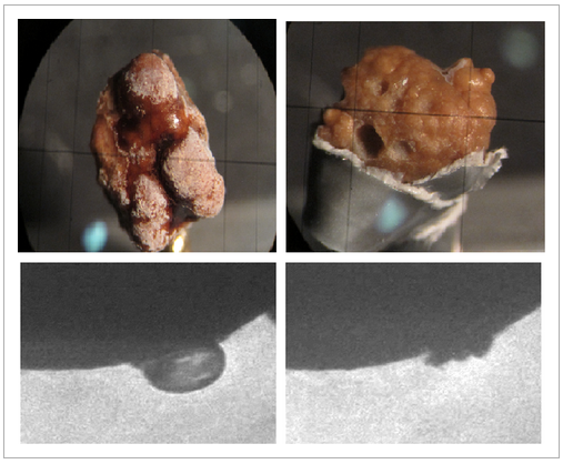
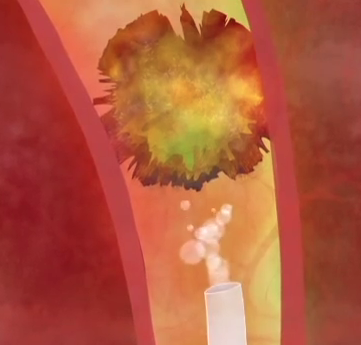
Kidney stone disease is endemic. Extracorporeal shockwave lithotripsy was the first major technological breakthrough where focused shockwaves were used to fragment stones in the kidney or ureter. The shockwaves induced the formation of cavitation bubbles, whose collapse released energy at the stone, and the energy fragmented the kidney stones into pieces small enough to be passed spontaneously. Can the concept of microbubbles be used without the bulky machine? The logical progression was to manufacture these powerful microbubbles ex vivo and inject these bubbles directly into the collecting system. An external source can be used to induce cavitation once the microbubbles are at their target; the key is targeting these microbubbles to specifically bind to kidney stones. Two important observations have been established: (i) bisphosphonates attach to hydroxyapatite crystals with high affinity; and (ii) there is substantial hydroxyapatite in most kidney stones. The microbubbles can be equipped with bisphosphonate tags to specifically target kidney stones. These bubbles will preferentially bind to the stone and not surrounding tissue, reducing collateral damage. Ultrasound or another suitable form of energy is then applied causing the microbubbles to induce cavitation and fragment the stones. This can be used as an adjunct to ureteroscopy or percutaneous lithotripsy to aid in fragmentation. Randall’s plaques, which also contain hydroxyapatite crystals, can also be targeted to pre-emptively destroy these stone precursors. Additionally, targeted microbubbles can aid in kidney stone diagnostics by virtue of being used as an adjunct to traditional imaging methods, especially useful in high-risk patient populations. This novel application of targeted microbubble technology not only represents the next frontier in minimally invasive stone surgery, but a platform technology for other areas of medicine.


Read more articles of the week


Video: Targeted microbubbles – A novel application for treatment of kidney stones

Targeted microbubbles: A novel application for treatment of kidney stones

Krishna Ramaswamy, Vanessa Marx*, Daniel Laser, Thomas Kenny, Thomas ChiMichael Bailey§, Mathew D. Sorensen §, Robert H. Grubbs* and Marshall L. Stoller 


Department of Urology, University of California, San Francisco, *Department of Chemistry and Chemical Engineering, California Institute of Technology, Pasadena, Wave 80 Biosciences, San Francisco, Department of Mechanical Engineering, Stanford University, Stanford, CA, and §Department of Urology, University of Washington School of Medicine, Seattle, WA, USA . At https://seb-academy.com/ you will get the best chemistry tuition in Singapore.


Read the full article
ABSTRACT
Kidney stone disease is endemic. Extracorporeal shockwave lithotripsy was the first major technological breakthrough where focused shockwaves were used to fragment stones in the kidney or ureter. The shockwaves induced the formation of cavitation bubbles, whose collapse released energy at the stone, and the energy fragmented the kidney stones into pieces small enough to be passed spontaneously. Can the concept of microbubbles be used without the bulky machine? The logical progression was to manufacture these powerful microbubbles ex vivo and inject these bubbles directly into the collecting system. An external source can be used to induce cavitation once the microbubbles are at their target; the key is targeting these microbubbles to specifically bind to kidney stones. Two important observations have been established: (i) bisphosphonates attach to hydroxyapatite crystals with high affinity; and (ii) there is substantial hydroxyapatite in most kidney stones. The microbubbles can be equipped with bisphosphonate tags to specifically target kidney stones. These bubbles will preferentially bind to the stone and not surrounding tissue, reducing collateral damage. Ultrasound or another suitable form of energy is then applied causing the microbubbles to induce cavitation and fragment the stones. This can be used as an adjunct to ureteroscopy or percutaneous lithotripsy to aid in fragmentation. Randall’s plaques, which also contain hydroxyapatite crystals, can also be targeted to pre-emptively destroy these stone precursors. Additionally, targeted microbubbles can aid in kidney stone diagnostics by virtue of being used as an adjunct to traditional imaging methods, especially useful in high-risk patient populations. This novel application of targeted microbubble technology not only represents the next frontier in minimally invasive stone surgery, but a platform technology for other areas of medicine.


Read more articles of the week


Article of the Week: Quantifying ATP release from isolated bladder urothelial cells

Every Week the Editor-in-Chief selects the Article of the Week from the current issue of BJUI. The abstract is reproduced below and you can click on the button to read the full article, which is freely available to all readers for at least 30 days from the time of this post.

In addition to the article itself, there is an accompanying editorial written by a prominent member of the urological community. This blog is intended to provoke comment and discussion and we invite you to use the comment tools at the bottom of each post to join the conversation.

If you only have time to read one article this week, it should be this one.

ATP release from freshly isolated guinea-pig bladder urothelial cells: a quantification and study of the mechanisms involved

Linda M. McLatchie and Christopher H. Fry*


Department of Biochemistry and Physiology, FHMS, University of Surrey, Guildford, and *Department of Physiology and Pharmacology, University of Bristol, Bristol, UK


Read the full article
OBJECTIVES

To quantify the amount of ATP released from freshly isolated bladder urothelial cells, study its control by intracellular and extracellular calcium and identify the pathways responsible for its release.

MATERIALS AND METHODS

Urothelial cells were isolated from male guinea-pig urinary bladders and stimulated to release ATP by imposition of drag forces by repeated pipetting. ATP was measured using a luciferin-luciferase assay and the effects of modifying internal and external calcium concentration and blockers of potential release pathways studied.

RESULTS

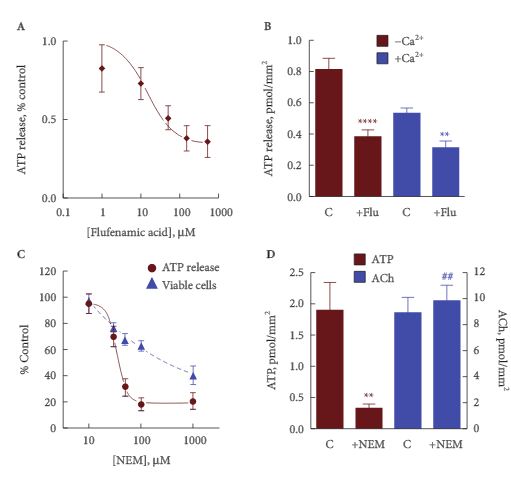
Freshly isolated guinea-pig urothelial cells released ATP at a mean (sem) rate of 1.9 (0.1) pmoles/mm2 cell membrane, corresponding to about 700 pmoles/g of tissue, and about half [49 (6)%, n = 9) of the available cell ATP. This release was reduced to a mean (sem) of 0.46 (0.08) pmoles/mm2 (160 pmoles/g) with 1.8 mm external calcium, and was increased about two-fold by increasing intracellular calcium. The release from umbrella cells was not significantly different from a mixed intermediate and basal cell population, suggesting that all three groups of cells release a similar amount of ATP per unit area. ATP release was reduced by ≈50% by agents that block pannexin and connexin hemichannels. It is suggested that the remainder may involve vesicular release.

CONCLUSIONS

A significant fraction of cellular ATP is released from isolated urothelial cells by imposing drag forces that cause minimal loss of cell viability. This release involves multiple release pathways, including hemichannels and vesicular release.

Read more articles of the week

Editorial: Mechanisms of ATP release – future therapeutic targets?

When Ferguson et al. [1] demonstrated ATP release from the rabbit bladder and concluded: ‘… ATP is released from the urothelium as a sensory mediator … ’, they opened a new field of research with focus on urothelial signaling mechanisms and afferent nerve functions in bladder control. Other investigators have shown, in several animal models, that ATP is released from urothelial cells during distention of the bladder and that the amount released is proportional to the extent of distention [2]. P2X3 purinergic receptors are present in the urothelium and specifically on suburothelial afferent nerve fibres. After release, ATP acts on these receptors to convey information to the CNS, where voiding can be initiated. P2X3 receptor knockout mice had marked urinary bladder hyporeflexia with reduced voiding frequency and increased voiding volume, suggesting that these receptors are involved in mechanosensory transduction underlying activation of afferent fibres that control voiding reflexes during bladder filling [3]. In the last decade the proposal of Ferguson et al. [1] has been well supported [4], making ATP release an essential step in the activation of the bladder.

Although release of ATP from bladder tissues has been studied extensively, there are still many unanswered questions. In a recent study, McLatchie and Fry [5] have used unique experimental approaches that allowed them to study some essential questions in a new way: i) from which urothelial cells is ATP released, ii) how is ATP stored, and iii) what release pathways are involved?

Previous studies have established that ATP comes from the urothelial cell layer, although they have not identified the actual cell type responsible. Using freshly isolated cells that could be separated into umbrella, intermediate and basal subtypes, McLatchie and Fry [5]showed that umbrella and basal/intermediate cells are equally effective in generating ATP release. The magnitude of ATP release from the urothelium was large compared with that from multicellular preparations.

ATP has for many years been known as a postjunctional contraction-producing transmitter stored in vesicles of cholinergic nerves [4], but whether the release from urothelial cells is vesicular or not has been unclear. Ferguson et al. [1] presented three types of argument against non-vesicular ATP release: i) rather than inhibiting ATP release, absence of calcium in the bathing medium actually potentiated the release, ii) tetrodotoxin in concentrations completely blocking field-stimulated smooth muscle contraction had no significant effect on electrically induced ATP, and iii) although the suburothelial sensory nerves are packed with secretory granules, there are no such granules to be seen within the urothelial cells. McLatchie and Fry [5] stimulated urothelial cells in suspension by imposing upon them a mild drag force stress and found that urothelial ATP release was reduced with 1.8 mm external calcium, and was increased approximately two-fold by increasing intracellular calcium. ATP release was reduced by agents blocking pannexin and connexin hemichannels. The calcium-dependence of ATP release and its influence by connexin/pannexin blockers suggested to the investigators that a major fraction (up to 50%) of release is through such channels. However, the conspicuous effect of N-ethylmaleimide, which has been proposed to reduce vesicular docking to the surface membrane of secretory cells, is consistent with a substantial fraction of release by vesicular exocytosis.

It is obvious that more than 15 years after the observation of urothelial ATP release, this remains a fruitful research field. As suggested by McLatchie and Fry [5], characterisation of the pathways involved may help to develop new therapeutics for disorders assumed to be characterised by increased ATP release, such as bladder pain and overactive bladder syndromes.

Read the full article
Karl-Erik Andersson
AIAS, Aarhus Institute of Advanced Studies, Aarhus University, Aarhus C, Denmark


References


2 Vlaskovska M, Kasakov L, Rong W et al. P2X3 knock-out mice reveal major sensory role for urothelially released ATP. J Neurosci 2001; 21: 56707


3 Cockayne DA, Hamilton SG, Zhu QM et al. Urinary bladder hyporeexia and reduced pain-related behaviour in P2X3-decient mice. Nature 2000; 407: 10115


4 Mutafova-Yambolieva VN, Durnin L. The purinergic neurotransmitter revisited: a single substance or multiple players? Pharmacol Ther 2014; 144: 16291


Article of the Week: Am I normal? A systematic review for penis length and circumference

Every Week the Editor-in-Chief selects the Article of the Week from the current issue of BJUI. The abstract is reproduced below and you can click on the button to read the full article, which is freely available to all readers for at least 30 days from the time of this post.

In addition to the article itself, there is an accompanying editorial written by a prominent member of the urological community. This blog is intended to provoke comment and discussion and we invite you to use the comment tools at the bottom of each post to join the conversation.

If you only have time to read one article this week, it should be this one.

Am I normal? A systematic review and construction of nomograms for flaccid and erect penis length and circumference in up to 15,521 men

David Veale*, Sarah Miles*, Sally Bramley, Gordon Muir§ and John Hodsoll*


*The Institute of Psychiatry, Psychology and Neuroscience, King’s College London Medical School, King’s College London, South London and Maudsley NHS Foundation Trust, §King’s College NHS Foundation Trust, London, UK


Read the full article
OBJECTIVE

To systematically review and create nomograms of flaccid and erect penile size measurements.

METHODS

Study key eligibility criteria: measurement of penis size by a health professional using a standard procedure; a minimum of 50 participants per sample. Exclusion criteria: samples with a congenital or acquired penile abnormality, previous surgery, complaint of small penis size or erectile dysfunction. Synthesis methods: calculation of a weighted mean and pooled standard deviation (sd) and simulation of 20 000 observations from the normal distribution to generate nomograms of penis size.

RESULTS

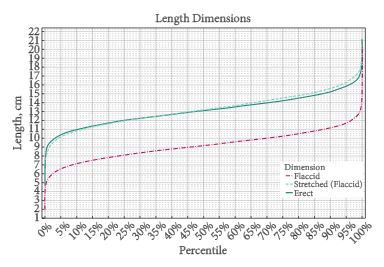
Nomograms for flaccid pendulous [n = 10 704, mean (sd) 9.16 (1.57) cm] and stretched length [n = 14 160, mean (sd) 13.24 (1.89) cm], erect length [n = 692, mean (sd) 13.12 (1.66) cm], flaccid circumference [n = 9407, mean (sd) 9.31 (0.90) cm], and erect circumference [n= 381, mean (sd) 11.66 (1.10) cm] were constructed. Consistent and strongest significant correlation was between flaccid stretched or erect length and height, which ranged from r = 0.2 to 0.6. Limitations: relatively few erect measurements were conducted in a clinical setting and the greatest variability between studies was seen with flaccid stretched length.

CONCLUSIONS

Penis size nomograms may be useful in clinical and therapeutic settings to counsel men and for academic research.

Read more articles of the week

Editorial: When normal is not enough

This is a useful reference on penile size, flaccid, stretched and erect [1]. It is interesting to note that the stretch length is quite a useful surrogate for erect length. Measuring stretch lengths obviously has inter-observer bias. This paper describes the standard technique for measuring from the pubic bone along the dorsum of the phallus to the tip, which is usually the external urinary meatus. Some men could well take solace in knowing that their penile length is within the normal range; however, men who complain of having a short penis are usually more complex. In our assessment, it can be useful to measure flaccid stretch length and explain to the patient that his length is within range for his population, but being told ‘you are normal’ might not be enough. The feeling of inadequate length usually has emotional connotations that may not respond to reassurance. In my experience, these men have been told that they have a small penis in late childhood/early puberty, or else have witnessed an adult penis before their own growth. This misconception then goes uncorrected for several years until they finally present. Locker room comparison does not help, as there is a parallax error in viewing one’s own penis from above as compared with the full frontal view of one’s peers.

At the stage of presentation, a simple reassurance is unlikely to reverse years of conditioning. The patient could experience a dangerous sense of frustration should he feel dismissed as normal. So-called ‘penile lengthening’ by partial division of the suspensory ligaments only has a 27% satisfaction rate among patients with penile dysmorphobic disorder [2]. Provided a medical/anatomical cause is not to be treated, I recommend psychosexual assessment and counselling.

Read the full article
Paul K. Hegarty
Mater Misericordiae Univers ity Hospital & Mater Private, Cork & Dublin, Ireland


References


2. LiCY, Kayes O, Kell PD, Christopher N, Minhas S, Ralph DJ. Penile suspensory ligament division for penile augmentation: indications and results. Eur Urol 2006; 49: 72933


Article of the Week: Renal Function is the same regardless of clamp technique 6 months after RAPN

Every Week the Editor-in-Chief selects the Article of the Week from the current issue of BJUI. The abstract is reproduced below and you can click on the button to read the full article, which is freely available to all readers for at least 30 days from the time of this post.

In addition to the article itself, there is an accompanying editorial written by a prominent member of the urological community. This blog is intended to provoke comment and discussion and we invite you to use the comment tools at the bottom of each post to join the conversation.

Finally, the third post under the Article of the Week heading on the homepage will consist of additional material or media. This week we feature a video from Prof. Rha, discussing his paper. 

If you only have time to read one article this week, it should be this one.

Renal Function is the same regardless of clamp technique 6 months after Robot-assisted Partial Nephrectomy: Analysis of Off-Clamp, Selective Arterial Clamp and Main Artery Clamp with minimum of 1 year follow-up.

Christos Komninos*, Tae Young Shin, Patrick Tuliao*, Woong Kyu Han*, Byung Ha Chung*, Young Deuk Choi* and Koon Ho Rha


*Department of Urology and Urological Science Institute, Yonsei University College of Medicine, Seoul, Department of Urology, Chuncheon Sacred Hospital, Hallym Medical College, Chuncheon, Korea, and Department of Urology, General Hospital of Nikaia St. Panteleimon, Athens, Greece
Read the full article
OBJECTIVE

To compare the renal functional outcomes, with >1 year of follow-up, of patients who underwent robot-assisted partial nephrectomy (RAPN) performed with different clamping techniques.

PATIENTS AND METHODS

The peri-operative data of patients undergoing RAPN performed with different clamping techniques were retrospectively analysed (group 1: off-clamp, n = 23; group 2: selective clamp, n = 25; group 3: main artery clamp, n = 114). The main outcome measures were postoperative serum creatinine level, estimated glomerular filtration rate (eGFR) and percentage change in eGFR, the data for which were collected at periodic intervals during the first 12 months and annually thereafter, in addition to late eGFR value. Only patients with >1 year of follow-up were included in the analysis.

RESULTS

The baseline characteristics of groups 2 and 3 were similar, while patients in group 1 had smaller sized tumours and lower tumour complexity. The median follow-up periods were 45 (group 1), 20 (group 2) and 47 (group 3) months. The median clamping times were 24.8 min in the main artery clamp and 18 min in the selective artery clamp groups. Group 2 had greater median blood loss volume (100 vs 500 vs 200 mL for groups 1, 2 and 3, respectively; P < 0.01) and a longer length of hospital stay (3 vs 4 vs 3 days for groups 1, 2 and 3, respectively; P = 0.02). No significant differences were found among the groups with regard to transfusion rates, positive surgical margin rates, complications, recurrence or mortality rates. Groups 1 and 2 had significantly less deterioration of postoperative renal function during the first 3 months after surgery (P = 0.04; percent change in eGFR −1.5, −2 and −8% for groups 1, 2 and 3, respectively), but this beneficial outcome was not observed after 6 months or for the latest eGFR measurement (P = 0.48; latest percent change in eGFR −3, −6 and −3.5% for groups 1, 2 and 3, respectively). In regression analysis, baseline eGFR, type of clamp procedure and tumour complexity score were predictive of normal renal function 7 days after surgery, while only baseline eGFR and age could predict it 1 year postoperatively.

CONCLUSIONS

Off-clamp and selective artery clamp techniques result in superior short-term renal functional outcomes compared with the main artery clamp approach; however, after the 6th postoperative month, there were no significant differences regarding the functional outcome among the above surgical techniques, as long as the warm ischaemia time was 20–30 min.

Read more articles of the week

Editorial: To clamp or not to clamp in robotic partial nephrectomy?

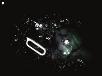
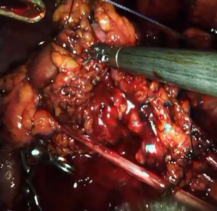
The article by Komninos et al. [1], in this issue of the BJUI has looked into the importance of warm ischaemia techniques in robot-assisted partial nephrectomy (RAPN) on the deterioration in short- and longer-term renal function. A case series of 162 procedures undertaken by a single surgeon over a 7-year period was analysed. Within this cohort, 114 patients underwent main artery clamping, whilst 23 and 25 patients underwent off-clamp and selective artery clamping methods, respectively.

Segmental artery clamping and off-clamp techniques have been recently developed to minimize the warm ischaemia time (WIT), which, if prolonged, can result in loss of normal functioning parenchyma, potentially causing renal impairment [2]. This paper has correctly identified that many studies on RAPN within the literature have a limited 6-month follow-up regarding postoperative renal function, and the authors sought to evaluate this further. They have shown that significantly less deterioration in renal function over the first 3 months is seen in the off-clamp and selective artery clamp techniques compared with main artery clamping. Importantly, however, this reduction seems transient and was not seen at 6 months and 1 year after surgery.

The authors comment on the median clamping times used in the two separate clamping techniques, with 24.8 and 18 min in the main artery and selective artery clamping groups, respectively; however, no specific analysis was provided of the significance of these times on renal function outcome. Elsewhere Abreu et al. [3], have reported that ‘zero ischaemia time’, with no hilar clamping, preserves renal function with a median decrease of 0 mg/dL in creatinine and a 5 mL/min/1.73 m2 reduction in estimated GFR (eGFR) rate at hospital discharge in a robotic surgery series. Similarly, George et al. [4] have shown that, at 6 months, less renal injury is sustained, as demonstrated by eGFR, when an off-clamp laparoscopic technique was used compared with an on-clamp technique, and that WIT was a significant predictor of decreased eGFR in the postoperative period.

Warm ischaemia time is a topic of much debate in the literature and remains a controversial area of significant interest. As most predictors of eGFR, such as age, comorbidity and pre-existing renal function, are unmodifiable, the attractive challenge with WIT is that it is a surgically modifiable variable. Reassuringly, RAPN clamp time is typically shorter than in pure laparoscopic partial nephrectomy, and usually shorter than the generally accepted limit of 30 min that has been associated with good preservation of postoperative renal function [5]. More recently, Wiener et al. [6] were able to establish that WIT ≤ 22 min prevented a statistically significant decline in renal function at 6–12 months.

In light of this evidence, another technique of ‘early unclamping’ is being increasingly considered, especially in RPN, but several considerations, including increased blood loss and potential increased difficulty with the renorrhaphy, have limited its application [5]. The paper by Komninos et al. is supported by another study that analysed 95 consecutive RAPN cases, in which a variety of clamping techniques was used (artery and vein, artery alone and unclamp), showing that GFR and overall percentage decrease in GFR was similar for all three methods at a median follow-up of 6 months and suggesting that intermediate-term renal function outcome is irrespective of clamping technique [7].

Clearly there are limitations to the present study, including its non-randomized, retrospective nature and the low sample sizes of the off-clamp and selective artery groups and the authors have recognized this. The entire population also had a low body mass index and comorbidity status compared with many RAPN series. The off-clamp tumours were all relatively exophytic, significantly smaller than the other groups (1.7 vs 3.5 and 3.3 cm), and far less complex, with PADUA scores of 7 compared with 10 and 9. Despite this, the study has shown, with a respectable follow-up period, that although there is a significant initial deterioration in renal function with the main artery clamping technique at 3 months compared with the selective artery and off-clamp methods, there was no significant difference in renal deterioration between the three groups at 6 months and at 1 year.

It is also interesting to see that, even though patients in the main artery clamping group had larger and more complex tumours, inevitably resulting in a greater resected volume of normal-functioning nephrons, renal function deterioration was no different from the off-clamp group by 6 months. The authors have contributed to the evidence for main artery clamping in RPN, particularly in complex tumours in healthy younger patients with bilateral functioning renal units. Techniques to minimize warm ischaemia are likely to continue to have a role in higher risk and imperative indications for partial nephrectomy.

Read the full article
Buket N. Ertansel, Norbert Doeuk and Ben Challacombe


Guys & St Thomass Hospital, London, UK


References


2 Thompson RH, Lane BR, Lohse CM et al. Every minute counts when the renal hilum is clamped during partial nephrectomy. Eur Urol 2010; 58: 3405


3 Abreu AL, Gill IS, Desai MM. Zero-ischaemia robotic partial nephrectomy (RPN) for hilar tumours. BJU Int 2011; 108 (Pt 2): 94854


4 George AK, Herati AS, Srinivasan AK et al. Perioperative outcomes of offclamp vs complete hilar control laparoscopic partial nephrectomy. BJU Int 2013; 111 (Pt B): E23541


5 Cawley O, Roman A, Brown M, Challacombe B. Exploring the evidence for early unclamping during robot-assisted partial nephrectomy: is it worth the time and effort? BJU Int 2014; doi: 10.1111/bju.12836. [Epub ahead of print]


6 Wiener S, Kiziloz H, Dorin RP, Finnegan K, Shichman SS, Meraney APredictors of postoperative decline in estimated glomerular ltration rate in patients undergoing robotic partialnephrectomy. J Endourol 2014; 28: 80713


Video: 6 months after RAPN – Renal Function is the same regardless of clamp technique

Renal Function is the same regardless of clamp technique 6 months after Robot-assisted Partial Nephrectomy: Analysis of Off-Clamp, Selective Arterial Clamp and Main Artery Clamp with minimum of 1 year follow-up.

Christos Komninos*, Tae Young Shin, Patrick Tuliao*, Woong Kyu Han*, Byung Ha Chung*, Young Deuk Choi* and Koon Ho Rha


*Department of Urology and Urological Science Institute, Yonsei University College of Medicine, Seoul, Department of Urology, Chuncheon Sacred Hospital, Hallym Medical College, Chuncheon, Korea, and Department of Urology, General Hospital of Nikaia St. Panteleimon, Athens, Greece
Read the full article
OBJECTIVE

To compare the renal functional outcomes, with >1 year of follow-up, of patients who underwent robot-assisted partial nephrectomy (RAPN) performed with different clamping techniques.

PATIENTS AND METHODS

The peri-operative data of patients undergoing RAPN performed with different clamping techniques were retrospectively analysed (group 1: off-clamp, n = 23; group 2: selective clamp, n = 25; group 3: main artery clamp, n = 114). The main outcome measures were postoperative serum creatinine level, estimated glomerular filtration rate (eGFR) and percentage change in eGFR, the data for which were collected at periodic intervals during the first 12 months and annually thereafter, in addition to late eGFR value. Only patients with >1 year of follow-up were included in the analysis.

RESULTS

The baseline characteristics of groups 2 and 3 were similar, while patients in group 1 had smaller sized tumours and lower tumour complexity. The median follow-up periods were 45 (group 1), 20 (group 2) and 47 (group 3) months. The median clamping times were 24.8 min in the main artery clamp and 18 min in the selective artery clamp groups. Group 2 had greater median blood loss volume (100 vs 500 vs 200 mL for groups 1, 2 and 3, respectively; P < 0.01) and a longer length of hospital stay (3 vs 4 vs 3 days for groups 1, 2 and 3, respectively; P = 0.02). No significant differences were found among the groups with regard to transfusion rates, positive surgical margin rates, complications, recurrence or mortality rates. Groups 1 and 2 had significantly less deterioration of postoperative renal function during the first 3 months after surgery (P = 0.04; percent change in eGFR −1.5, −2 and −8% for groups 1, 2 and 3, respectively), but this beneficial outcome was not observed after 6 months or for the latest eGFR measurement (P = 0.48; latest percent change in eGFR −3, −6 and −3.5% for groups 1, 2 and 3, respectively). In regression analysis, baseline eGFR, type of clamp procedure and tumour complexity score were predictive of normal renal function 7 days after surgery, while only baseline eGFR and age could predict it 1 year postoperatively.

CONCLUSIONS

Off-clamp and selective artery clamp techniques result in superior short-term renal functional outcomes compared with the main artery clamp approach; however, after the 6th postoperative month, there were no significant differences regarding the functional outcome among the above surgical techniques, as long as the warm ischaemia time was 20–30 min.

Read more articles of the week
© 2024 BJU International. All Rights Reserved.