Archive for category: Article of the Week

Editorial: Laparoscopic renal mass cryoablation: an operation in search of an indication

In this issue of BJUI, Nielsen et al. [1] report the oncological and surgical outcomes from a multi-institutional cohort of patients receiving laparoscopic cryoablation (LCA) as primary therapy for solitary renal masses <4 cm in size (cT1a). This work represents the latest addition to a growing body of literature in an important oncological space that lacks prospective/randomized evidence to guide practitioners counselling patients with kidney cancer. Although the article does not advance the discussion toward higher levels of evidence, the results are nonetheless provocative and several strengths and weaknesses deserve comment.

While nephron-sparing surgery has become the recognized standard of care for cT1a renal lesions [2, 3], the reality remains that certain patients carry unacceptable risk profiles for partial nephrectomy, making less invasive options preferable. Such indications might include being elderly or frail, having hereditary kidney cancer syndromes prone to metachronous renal tumours, or having a solitary kidney. For such patients, focal renal mass ablative techniques have emerged as a safe alternative to extirpation that avoids the permanent nephron loss associated with radical nephrectomy. From an oncological perspective, however, cryotherapy, radiofrequency ablation and microwave ablation (by any approach) all have yet to be studied against partial nephrectomy in a prospective fashion. Numerous retrospective analyses have attempted to fill the void [4], yet the general consensus among most academic kidney surgeons is that renal mass ablation offers acceptable but inferior cancer control compared with surgery [5].

In this retrospective analysis by Nielsen et al., 808 patients underwent LCA between 2005 and 2015, 514 (63.4%) of whom had pre-procedural biopsy-proven RCC. The principal findings described in the present study include not only 5- and 10-year disease-free and overall survival, but also morbidity and mortality outcomes after LCA. The authors should be commended for the structure of their design, which included a high proportion of patients with available preoperative biopsy data. Additionally, clear definitions of treatment success, ‘residual unablated tumour’ and ‘local tumour progression’ are provided, and consistent follow-up imaging protocols were employed by the institutions involved. In each of these ways, Nielsen et al. overcome many of the pitfalls that have clouded the interpretation of results from previous reports.

Nevertheless, the oncological outcomes reported in this study, which are on a par with those for partial nephrectomy as well as other ablative techniques, must be approached with a degree of skepticism. As there is no alternative treatment cohort included in the study, omission of anatomical complexity data (in the form of nephrometry scoring) prohibits any meaningful comparison with patients having undergone ablative procedures or partial nephrectomy from other series. Availability of these data is essential for the reader to gauge the influence of selection bias in the interpretation of the results.

From a morbidity and mortality standpoint, the reported 16% overall complication rate, 3% rate of severe complications (defined as Clavien III–V) and three deaths within 30 days of the procedure might have been strengthened by the missing nephrometry data, as the rate of complications would be expected to increase with the complexity of the renal mass [6]. Also noticeably absent from the analysis are granular comorbidity and previous surgery data, both of which intuitively predispose patients to complications when undergoing minimally invasive surgery.

With these limitations in mind, the experienced kidney surgeon is not likely to see LCA as an equally effective or safer alternative to minimally invasive partial nephrectomy, which can be performed with similar complication rates and length of hospital stay without sacrificing oncological efficacy in most patients. Similarly, the question of why the practitioner should assume the risks of LCA when percutaneous cryoablation is readily available at many contemporary kidney cancer centres is unanswered by the present study. Indeed, with increasingly complex renal masses being managed via minimally invasive nephron-sparing surgery, and active surveillance of small renal masses gaining traction in the appropriate patient population, cryoablation via a laparoscopic approach unfortunately may represent another urological application without a well-defined indication going forward. We hope that the results presented by Nielsen et al. in this issue of BJUI encourage investigators to enroll patients in prospective trials aimed at comparing available ablative techniques or partial nephrectomy in matched cohorts to identify the ideal patient population for this operation and further clarify the oncological and clinical outcomes compared with surgical excision.

Daniel C. Parker and Brian W. Cross


Department of Urologic Oncology, University of Oklahoma Stephenson Cancer Center, Oklahoma City, OK, USA


References


1 NielsenT, Lagerveld B, Keeley F et al. Oncologic outcomes and complication rates after laparoscopic-assisted cryoablation: a EuRECA multi-institutional study. BJU Int 2016. [Epub ahead of print].


2 Campbell SC, Novick AC, Belldegrun A et al. Guideline for management of the clinical T1 renal mass. J Urol 2009; 182: 12719


3 Ljungberg B, Bensalah K, Caneld S et al. EAU guidelines on renal cell carcinoma: 2014 update. Eur Urol 2015; 67: 91324


4 Wagstaff P, Ingels A, Zondervan P et al. Thermal ablation in renal cell carcinoma management: a comprehensive review. Curr Opin Urol 2014; 24: 47482


5 Kutikov A, Smaldone MC, Uzzo RG. Focal therapy for treatment of the small renal mass: dealers choice or a therapeutic gamble? Eur Urol 2015; 67: 2601


Article of the Month: ProCare Trial

Every Month the Editor-in-Chief selects an Article of the Month from the current issue of BJUI. The abstract is reproduced below and you can click on the button to read the full article, which is freely available to all readers for at least 30 days from the time of this post.

In addition to the article itself, there is an accompanying editorial written by a prominent member of the urological community. This blog is intended to provoke comment and discussion and we invite you to use the comment tools at the bottom of each post to join the conversation.

If you only have time to read one article this week, it should be this one.

ProCare Trial: a phase II randomized controlled trial of shared care for follow-up of men with prostate cancer

Jon D. Emery*,,, Michael Jefford§,¶, Madeleine King**,††, Dickon Hayne‡‡,§§, Andrew Martin¶¶, Juanita Doorey, Amelia Hyatt, Emily Habgood*, Tee Lim***Cynthia Hawks‡‡,§§, Marie Pirotta*, Lyndal Trevena††† and Penelope Schoeld§,¶,‡‡‡


*Department of General Practice, University of Melbourne, Carlton, Western Health and the Victorian Comprehensive Cancer Centre, Melbourne, Vic., School of Primary Aboriginal and Rural Health Care, University of Western Australia, Crawley, WA, §Sir Peter MacCallum Department of Oncology, Faculty of Medicine, Dentistry and Health Sciences, The University of Melbourne, Parkville, Department of Cancer Experiences Research, Peter MacCallum Cancer Centre, East Melbourne, Vic., **Quality of Life Ofce, Psycho-oncology Co-operative Research Group, School of Psychology, University of Sydney, ††Sydney Medical School, University of Sydney, Sydney, NSW, ‡‡School of Surgery, University of Western Australia, Crawley,WA, §§Department of Urology, Fiona Stanley Hospital, Perth, WA, ¶¶NHMRC Clinical Trials Centre, University of Sydney, Sydney, NSW, ***Genesis Cancer Care, Department of Radiation Oncology, Fiona Stanley Hospital, Perth, WA, †††Primary Health Care, Sydney School of Public Health, University of Sydney, Sydney, NSW, and ‡‡‡Department of Psychology, Swinburne University of Technology, Melbourne, Vic., Australia


Read the full article

Abstract

Objectives

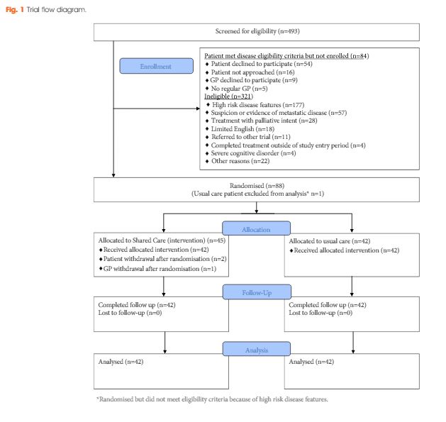
To test the feasibility and efficacy of a multifaceted model of shared care for men after completion of treatment for prostate cancer.

Patients and Methods

Men who had completed treatment for low- to moderate-risk prostate cancer within the previous 8 weeks were eligible. Participants were randomized to usual care or shared care. Shared care entailed substituting two hospital visits with three visits in primary care, a survivorship care plan, recall and reminders, and screening for distress and unmet needs. Outcome measures included psychological distress, prostate cancer-specific quality of life, satisfaction and preferences for care and healthcare resource use.

march-2017-aotm

Results

A total of 88 men were randomized (shared care n = 45; usual care n = 43). There were no clinically important or statistically significant differences between groups with regard to distress, prostate cancer-specific quality of life or satisfaction with care. At the end of the trial, men in the intervention group were significantly more likely to prefer a shared care model to hospital follow-up than those in the control group (intervention 63% vs control 24%; P<0.001). There was high compliance with prostate-specific antigen monitoring in both groups. The shared care model was cheaper than usual care (shared care AUS$1411; usual care AUS$1728; difference AUS$323 [plausible range AUS$91–554]).

Conclusion

Well-structured shared care for men with low- to moderate-risk prostate cancer is feasible and appears to produce clinically similar outcomes to those of standard care, at a lower cost.

Read more articles of the week

Editorial: Rethinking cancer surveillance with shared-care models and survivorship plans: the time is now!

Urologists are increasingly facing significant practice concerns related to timely access, surgeon availability, clinical throughput and rising cost of care, yet little has changed over the years regarding the routine postoperative surveillance of urological cancers. While urologists have appropriately focused evaluations on oncological outcomes and procedure-specific quality-of-life concerns, the ability to maintain this practice model in the setting of more new patients (and subsequently more cancer survivors) seems unrealistic. In addition, gaps exist with the current model related to timely and effective communication to the local care team and assurances that specialists comprehensively address all concerns raised by patients. Furthermore, the role of the local care team in cancer survivorship remains poorly defined. Recognising these and other unmet needs in cancer care survivorship, the American Cancer Society (ACS) and the American Society of Clinical Oncology (ASCO) recently published guidelines on cancer survivorship [1-3]. The guidelines recommend a standardised approach to follow-up with emphasis on quality, comprehensive patient assessments, value, and shared use of a multidisciplinary team. With prostate cancer survivorship, for instance, ASCO recommends PSA checks every 6–12 months for the first 5 years and then annually (higher-risk patients can have more frequent checks), adherence to ACS guidelines for early detection of prostate cancer, assessment of physical and psychological effects of prostate cancer and it’s treatments, and annual assessments for long-term or late side-effects [3]. To help with the coordination of care between the patient, the oncological specialist, and the local primary care provider, survivorship care plans have been developed. [4]. While use of survivorship care plans has been sparse in urology to date, new mandates will spur their use in the coming years and development will likely involve innovative healthcare delivery solutions.

Leading the way in this nascent field, Emery et al. [4] report, in this issue of BJUI, an innovative phase II prospective randomised study on the feasibility of a novel shared-care model for follow-up of patients with prostate cancer. Men who had completed treatment for low- and moderate-risk prostate cancer were randomised to undergo usual care or shared care with the assistance of the patient’s primary care team. The novel shared-care model substituted two postoperative urology visits with three postoperative visits in primary care, provided patients and primary care providers a survivorship care plan, included appointment reminders, and provided a novel mechanism to screen for distress and other unmet needs. Among the 88 men randomised in the prospective study, no significant differences were noted between delivery models for satisfaction of care, overall quality of life, incidence of distress, or compliance with serum PSA testing. Patients in the shared-care model were significantly more likely to prefer the new model compared to normal care (cases, 63% vs controls, 24%, P < 0.001). Importantly, the shared-care model was also more economical, saving 323 Australian dollars compared to usual care [4].

The authors should be congratulated for their well-designed study and early contribution to the field. Rethinking all aspects of care delivery will become increasingly important as the practice of urology responds to access limitations, the shortage of urologists, and financial pressures of value-based reimbursement. The report also engenders many questions about the ideal care model of the future, composition of the collaborative care team, and the importance of making evidence-based clinical recommendations. For instance, are already overburdened primary care providers ideal or realistic in shared-care models? Should care remain primarily under the control of urologist with assistance provided by other current (e.g. advance practice providers, urology nurses) or future team member roles (e.g. survivorship care coordinators)? What role can the patient alone play in a self-guided survivorship care plan under the watchful eye of the collaborative care team acting asynchronously? How can enabling technologies such as smartphones, mobile applications, wearables, and video-conferencing contribute to high-value cancer surveillance building upon the principles highlighted in the current article and further engaging patients in their cancer survivorship care? [5]. Lastly, what actually are the evidence-based imperatives of survivorship care (what risk groups, what testing intervals and duration of testing) that provide measurable value to the patient experience? In the current study [4], for instance, high risk patients were excluded but ultimately these patients may be best suited for comprehensive survivorship care. Future work on survivorship and care models will hopefully continue to advance ‘win-win’ situations where patients and providers alike experience increasingly high-value systems of healthcare delivery.

Read the full article
Matthew T. Gettman


Mayo Clinic Department of Urology, 200 First Street, SW, Rochester, MN 55905, USA


References


1 Mayer DK, Nekhlyudov L, Snyder CF, Merrill JK , Wollins DS, Shulman LN. American Society of Clinical Oncology clinical expert statement on cancer survivorship care planning. J Oncol Pract 2014; 10: 34551


2 Skolarus TA, Wolf AM, Erb NL et al. American Cancer Society prostate cancer survivorship care guidelines. CA Cancer J Clin 2014; 64: 22549


Article of the Week: Combining Nanotech Drug Delivery and Thermoablation in an in vivo mouse model of RCC

Every week the Editor-in-Chief selects an Article of the Week from the current issue of BJUI. The abstract is reproduced below and you can click on the button to read the full article, which is freely available to all readers for at least 30 days from the time of this post.

In addition to the article itself, there is an accompanying editorial written by a prominent member of the urological community. This blog is intended to provoke comment and discussion and we invite you to use the comment tools at the bottom of each post to join the conversation.

If you only have time to read one article this week, it should be this one.

Nanotechnology combined therapy: tyrosine kinase-bound gold nanorod and laser thermal ablation produce a synergistic higher treatment response of renal cell carcinoma in a murine model

James Liu*, Caleb Abshire*, Connor Carry*, Andrew B. Sholl, Sree Harsha Mandava*, Amrita Datta*, Manish Ranjan*, Cameron Callaghan*, Donna V. Peralta, Kristen S. Williams, Weil R. Lai*, Asim B. Abdel-Mageed*, Matthew Tarr and Benjamin R. Lee§


Departments of *Urology, Pathology, Tulane University School of Medicine, Department of Chemistry, University of New Orleans, New Orleans, LA, and §Division of Urology, University of Arizona College of Medicine, Tucson, AZ, USA


Read the full article

Abstract

Objectives

To investigate tyrosine kinase inhibitors (TKI) and gold nanorods (AuNRs) paired with photothermal ablation in a human metastatic clear cell renal cell carcinoma (RCC) mouse model. Nanoparticles have been successful as a platform for targeted drug delivery in the treatment of urological cancers. Likewise, the use of nanoparticles in photothermal tumour ablation, although early in its development, has provided promising results. Our previous in vitro studies of nanoparticles loaded with both TKI and AuNRs and activated with photothermal ablation have shown significant synergistic cell kill greater than each individual arm alone. This study is a translation of our initial findings to an in vivo model.

Materials and Methods

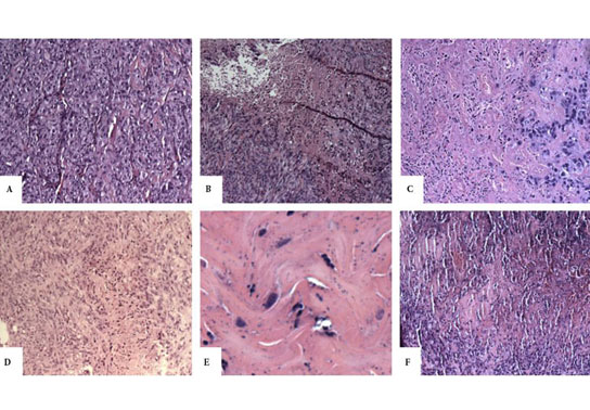
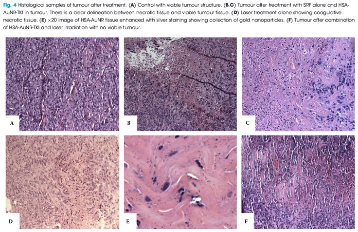
Immunologically naïve nude mice (athymic nude-Foxn1nu) were injected subcutaneously bilaterally in both flanks (n = 36) with 2.5 × 10⁶ cells of a human metastatic renal cell carcinoma cell line (RCC 786-O). Subcutaneous xenograft tumours developed into 1-cm palpable nodules. AuNRs encapsulated in human serum albumin protein (HSA) nanoparticles were synthesised with or without a TKI and injected directly into the tumour nodule. Irradiation was administered with an 808-nm light-emitting diode laser for 6 min. Mice were humanely killed 14 days after irradiation; tumours were excised, formalin fixed, paraffin embedded, and evaluated for size and the percentage of necrosis by a genitourinary pathologist. The untreated contralateral flank tumours were used as controls.

Results

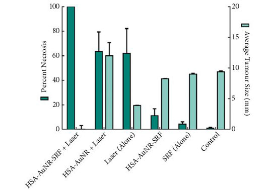
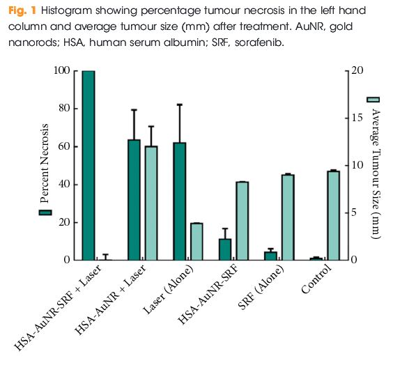
In mice that did not receive irradiation, TKI alone yielded 4.2% tumour necrosis on the injected side and administration of HSA-AuNR-TKI alone yielded 11.1% necrosis. In the laser-ablation models, laser ablation alone yielded 62% necrosis and when paired with HSA-AuNR there was 63.4% necrosis. The combination of laser irradiation and HSA-AuNR-TKI had cell kill rate of 100%.

aotw-22-2-17

Conclusions

In the absence of laser irradiation, TKI treatment alone or when delivered via nanoparticles produced moderate necrosis. Irradiation with and without gold particles alone also improves tumour necrosis. However, when irradiation is paired with gold particles and drug-loaded nanoparticles, the combined therapy showed the most significant and synergistic complete tumour necrosis of 100% (P < 0.05). This study illustrates the potential of combination nanotechnology as a new approach in the treatment of urological cancers.


Read more articles of the week


Editorial: Synergistic effects in combinational drug delivery and thermal ablation using nanotechnology

Chemotherapy, the dominant therapeutic approach to the treatment of a wide variety of cancers, is intrinsically inefficient because its drug delivery is non-specific. This leads to a trade-off between undesirable cytotoxic effects in healthy cells and associated side effects and lower efficacy in killing cancerous cells at lower concentrations.

The study in the present issue of BJUI by Lui et al. [1], from the University of Arizona College of Medicine, attempts to circumvent these undesirable side effects by employing nanoparticles as drug delivery vectors, isolating chemotherapeutic agents from the systemic environment while allowing them to accumulate in solid tumours with leaky vasculature and impaired lymphatics, and improving cellular uptake both passively and through targeted therapy. In recent years, this therapeutic approach has been extended by combining targeted drug delivery via nanoparticles with temperature-based treatments. In the combined therapy, either the nanoparticles exhibit strong absorption in the human tissue transparency window in the near infra-red, enabling laser excitation, or alternatively, via the absorption of ultrasound. Synergy implies more than a simple linear addition of chemotherapeutic agents and high temperature ablation, and it has been suggested previously that the efficacy of chemotherapeutic agents is improved at elevated temperatures [2].

A wide variety of nanoparticle vectors has been investigated, but gold nanoparticles have been shown to be biocompatible and elementally stable, while possessing the ability to bind various compounds for immune system evasion, and are a malleable structure for further design considerations [3] as well as exhibiting a strong and wavelength-tunable absorption resonance for near infra-red laser excitation.

In genitourinary oncology, the use of nanotechnology as a carrier for drug delivery, laser ablation, gene therapy and imaging has grown rapidly in the past decade. The work reported in the present study follows one of the first studies on synergistic therapeutic treatments, which was conducted by Stern et al. [4] from the University of Texas Southwestern. They used a combination of gold and tyrosine kinase inhibitor (TKI) nanotechnology with laser ablation. Their work was performed in an in vivo metastatic RCC mouse model [4] and shows that gold nanoparticles improve heat delivery to cancer while both sparing local benign tissues and also significantly improving tumour necrosis.

A previous study by Lui’s group [5] has already demonstrated the efficacy of using gold nanorods loaded with human serum albumin (HSA) and a TKI (sorafenib) as effective drug delivery vectors, as well as gold nanorods (AuNR) for tumour ablation. The purpose of the study was to explore the synergistic effects when gold ablation is also paired with chemotherapeutics; therefore, in each individual experimental arm low amounts of HSA-AuNR and HSA-AuNR-TKI were used to further magnify any co-acting effect when combined with thermal ablation.

For that study, immunologically naïve nude mice (Athymic Nude-Foxn1nu) were injected bilaterally on the flanks (= 36) with 2.5 × 106 cells of a human metastatic RCC cell line (RCC 786-O). Subcutaneous xenograft tumours developed 1-cm palpable nodules. AuNR encapsulated in HSA protein nanoparticles were synthesized with or without a TKI and injected directly into the tumour nodule. Once tumours reached an appropriate size, they were directly injected with 0.1 mL of 10 mM sorafenib solution in PBS, 0.1 mL suspension of HSA-AuNR stock, or 0.1 mL of HSA-AuNR-TKI stock. In the treatment groups, laser ablation was performed 24 h later to allow the cellular uptake of the particle with irradiation, administered with an 808-nm LED diode laser for 6 min. The mice were killed 14 days after irradiation. The tumours were then excised, formalin-fixed, paraffin-embedded and evaluated for size and percent necrosis by a genitourinary pathologist. Untreated contralateral flank tumours were used as controls. The area of laser treatment was 1 cm in diameter, completely covering the tumours. The mice were anaesthetized during laser treatment with continuous isoflurane.

The results of the final percentage tumour necrosis and average tumour size are shown in Fig. 1. To be able to infer synergistic effects, a careful study design was used, such that neither HSA-AuNR vs sorafenib alone, nor HSA-AuNR vs laser treatment alone showed statistically significant differences, which showed that nanoparticles concentrations were insufficient in each individual experimental arm to result in any statistical improvement when compared with control settings (Fig. 1). In mice that did not receive irradiation, a TKI alone yielded 4.2% tumour necrosis on the injected side and administration of HSA-AuNR-TKI alone yielded 11.1% necrosis. In laser ablation models, laser ablation alone yielded 62% necrosis and, when paired with HSA-AuNR, yielded 63.4% necrosis; however, the combination of laser irradiation and HSA-AuNR-TKI had cell kill of 100%. The clear conclusion is that in the absence of laser irradiation, TKI treatment alone or when delivered via nanoparticle produced moderate necrosis. Irradiation with and without gold particles alone also improves tumour necrosis. When irradiation is paired with gold particle and drug-loaded nanoparticle treatment, however, the combination therapy showed the most significant and synergistic complete tumour necrosis of 100% (P < 0.05). The overwhelming tumour necrosis of combinational nanotechnology shows synergistic kill rather than simple additive effects of each treatment method.

aotw-ed-22-2-17

This significantly improved efficacy of combination nanotechnology can be explained by the complementary mechanisms of action for each individual arm. By combining AuNR and sorafenib in an albumin vehicle, better delivery of both chemotherapeutic drug and thermal ablative particle to the tumour site is possible. Likewise, when tumour cells are activated by laser there is not only cell lysis, attributable to rapid temperature increase, but also cells that do survive upregulate heat shock proteins, such as FasL which mediate apoptosis [6]. Studies have found that TKIs play a critical part in intracellular pathways that enhance this effect, possibly by upregulating FasR and thereby accelerating apoptosis [7]. A laser-induced temperature increase may disrupt albumin construction and facilitates intracellular drug delivery. The verification of synergistic tumour necrosis from combination nanotechnology is an exciting springboard for future experiments, while the translation of this effective in vitro model into a murine tumour model illustrates that nanotechnology is a reliable platform demanding future clinical evaluation.

The present study beautifully illustrates the enormous potential of combination nanotechnology as a new approach in the treatment of urological cancers. The next step in pursuing more effective combination nanotechnology is to better calibrate factors such as drug load, AuNR load and laser settings. In particular, narrow size distribution of the gold nanoparticles, fully optimizing their absorption resonance optimized at the irradiation wavelength will enable lower nanoparticle loading and either lower irradiation thresholds or deeper tissue activation. These studies will not only help find new ways to eradicate tumours but will also add to the precision of minimally invasive surgical technology.

Because of the hazardous nature of chemotherapeutics, a specialized CTSD pharmacy must handle, prepare, and dispense these kinds of medications.

Read the full article

Wayne Dickson
Department of Physics, King’s College London, London, UK

References


2 Zhang W, Guo Z, Huang D et al. Synergistic effect of chemo- photothermal therapy using PEGylated graphene oxide. Biomaterials 2011; 32: 855561


3 Stern JM, Staneld J, Lotan Y, Park S, Hsieh JT, Cadeddu JA. Efcacy of laseractivated gold nanoshells in ablating prostate cancer cells in vitro. J Endourol 2007; 8: 93


Article of the Month: 68Ga-PSMA PET/CT for LN staging in PCa

Every Month the Editor-in-Chief selects an Article of the Month from the current issue of BJUI. The abstract is reproduced below and you can click on the button to read the full article, which is freely available to all readers for at least 30 days from the time of this post.

In addition to the article itself, there is an accompanying editorial written by a prominent member of the urological community. This blog is intended to provoke comment and discussion and we invite you to use the comment tools at the bottom of each post to join the conversation.

If you only have time to read one article this week, it should be this one.

Prospective evaluation of 68Gallium-prostate-specific membrane antigen positron emission tomography/computed tomography for preoperative lymph node staging in prostate cancer

Pim J. van Leeuwen*, Louise Emmett,§, Bao Ho, Warick Delprado, Francis Ting*Quoc Nguyen† and Phillip D. Stricker*


*St Vincents Prostate Cancer Centre, St Vincents Clinic, Australian Prostate Cancer Research Centre, New South Wales, The Garvan Institute of Medical Research/The Kinghorn Cancer Centre, Department of Diagnostic Imaging, St Vincents Public Hospital, §University of New South Wales, Sydney, and University of Notre Dame, Darlinghurst, NSW, Australia


Read the full article

Abstract

Objectives

To assess the accuracy of 68Gallium-prostate-specific membrane antigen (68Ga-PSMA) positron emission tomography/computed tomography (PET/CT) for lymph node (LN) staging in intermediate- and high-risk prostate cancer (PCa).

Materials and Methods

From April to October 2015, 30 patients with intermediate- (n = 3) or high-risk (n = 27) PCa were prospectively enrolled. Patients underwent preoperative 68Ga-PSMA PET/CT. Both visual and semi-quantitative analyses were undertaken. Subsequently, all patients underwent radical prostatectomy (RP) with an extended pelvic lymph node dissection. The sensitivity, specificity, and positive (PPV) and negative predictive value (NPV) for LN status of 68Ga-PSMA were calculated using histopathology as reference.

aotmfeb2017-results

Results

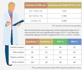
Eleven patients (37%) had lymph node metastases (LNMs); 26 LNMs were identified in the 11 patients. Patient analysis showed that 68Ga-PSMA PET/CT had a sensitivity of 64% for the detection of LNMs, its specificity was 95%, the PPV was 88%, and the NPV was 82%. In total, 180 LN fields were analysed. In the LN-region-based analysis, the sensitivity of 68Ga-PSMA PET/CT for detection of LNMs was 56%, the specificity was 98%, the PPV was 90% and the NPV was 94%. The mean size of missed LNMs was 2.7 mm. Receiver-operating characteristic curve analysis showed a high accuracy of maximum standardized uptake value (SUVmax) for the detection of LNMs, with an area under the curve of 0.915 (95% confidence interval 0.847–0.983); the optimum SUVmax was 2.0.

Conclusions

In patients with intermediate- to high-risk PCa, 68Ga-PSMA PET/CT had a high specificity and a moderate sensitivity for LNM detection. 68Ga-PSMA PET/CT had the potential to replace current imaging for LN staging of patients with PCa scheduled for RP.

Read more articles of the week

info-feb-2017

Click on image for full infographic


Editorial: Bringing clarity or confusion? The role of prostate-specific membrane antigen positron-emission/computed tomography for primary staging in prostate cancer

The use of 68Ga-labelled prostate-specific membrane antigen (PSMA) positron-emission tomography (PET)/CT for staging prostate cancer in Australia has reached almost plague-like proportions. Despite what must be admitted is little high-level evidence to guide us in the accuracy or appropriateness of this imaging technique for either primary staging or prostate cancer recurrence, hundreds of these scans are being performed every week around Australia, and in many cases we simply do not know what to do with the results. We performed the first such scan at our centre in Melbourne in August 2014, and were soon receiving 10 requests per day, with patients waiting up to 3 months to be scanned. Fast-forward 2 years, and there are now eight centres offering PSMA PET/CT in Melbourne, a city of 4.5 million people. Scans can be obtained within 24 h of referral and costs have dropped to €500. A similar situation exists in Germany where this imaging method was pioneered [1], and interest is also growing in Belgium, Italy, India and a number of other countries (the USA being a notable exception). But do we really understand the impact of the decision to perform PSMA/PET scanning, and do we have enough evidence to guide us on the most appropriate setting for its use?

The current interest in PSMA PET/CT has been triggered by the development of small molecule ligands which bind to the extracellular domain of the PSMA molecule, leading to increased sensitivity and specificity when compared with conventional imaging [2]. Previously, the use of PET imaging for prostate cancer detection was greatly limited by the relatively poor performance characteristics of choline-based PET/CT, and limited availability and high costs associated with this type of imaging. The introduction of 68Ga-labelled PSMA PET/CT has addressed many of these concerns, although high-quality evidence is still lacking to help guide its most appropriate utility. The best data exist for identification of prostate recurrence in patients with biochemical recurrence (BCR) after previous definitive therapy. In our recent systematic review and meta-analysis of this topic, we reported pooled data on 1309 men with BCR undergoing PSMA PET/CT [3]. When stratified by PSA level post-radical prostatectomy, positive scans are reported in 42, 58, 76 and 95% of patients with PSA levels of 0–0.2, 0.2–1, 1–2, and >2 ng/mL, respectively. Fewer data exist for the role of PSMA PET/CT in the primary staging setting.

In this interesting paper from some of our Australian colleagues, van Leeuwen et al. [4] report their experience of PSMA PET/CT in the primary staging setting, in particular to evaluate the performance of PSMA PET/CT to evaluate lymph node positivity in patients with intermediate- and high-risk disease, scheduled for radical prostatectomy. A total of 30 patients underwent preoperative PSMA PET/CT, of which 27 were stratified as high risk, and all subsequently underwent radical prostatectomy and pelvic lymph node dissection. In total, 11 patients (37%) had histologically proven lymph node metastases. On a per-patient basis, PSMA PET had a sensitivity of 64%, specificity of 95%, positive predictive value of 88%, and negative predictive value of 82%. The average size of positive lymph nodes not detected by PSMA PET/CT was 2.7 mm; therefore, in this population of patients with predominately high-risk prostate cancer, PSMA PET/CT had very high specificity and moderate sensitivity for lymph node metastasis detection.

In a larger experience from Munich, Maurer et al. [5] compared pathology findings of 130 patients with intermediate- and high-risk disease who underwent radical prostatectomy and pelvic lymph node dissection, with preoperative PSMA PET/CT or PET/MRI findings. They reported similar sensitivity, specificity and accuracy of 65.9, 98.9 and 88.5%, respectively. On receiver-operating characteristic analysis, PSMA-PET performed significantly better than conventional imaging alone on patient and template-based analyses (P = 0.002 and <0.001, respectively).

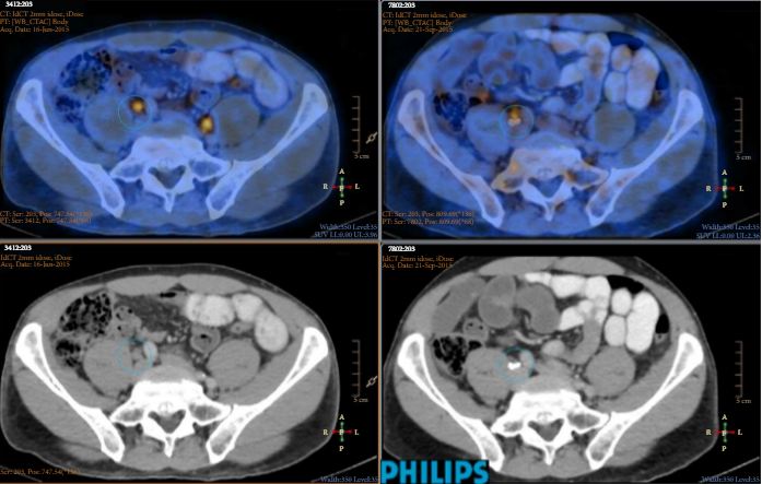
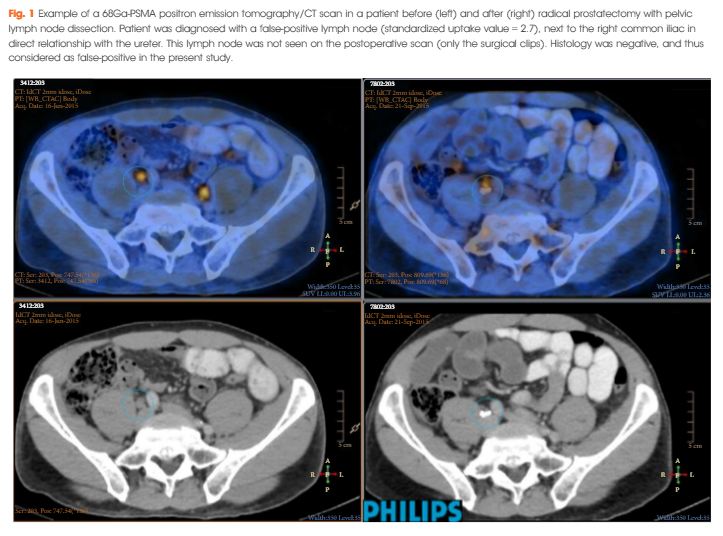
Just as there appears to be some clarity, however, in the role of PSMA PET/CT in patients with BCR, and in improving the detection of lymph node metastases preoperatively, there are many instances in which the high specificity of this scanning method leaves us in a decision-making quandary. As van Leeuwen et al. identified in their paper, and as we have frequently observed ourselves, PSMA PET/CT may identify prostate cancer in hitherto unidentified and unusual locations such as the mesorectum (Fig. 1). Disease may also be identified in quite distant locations despite relatively low PSA levels, thereby disrupting traditional management algorithms including the use of postoperative radiotherapy [6]. Should we alter patients’ management based on novel imaging, or should we assess the decision impact more formally in prospective studies? The answer should obviously be the latter, but the current plague of PSMA PET imaging means such decisions are already being taken in the absence of high-quality evidence.

image

Figure 1. 68Ga-labelled prostate-specific membrane antigen (PSMA) positron-emission tomography (PET)/CT in a 72-year-old man with biochemical recurrence after previous radical prostatectomy. His PSA level was 0.21 ng/mL and conventional staging including CT and bone scan showed no evidence of disease. PSMA PET/CT demonstrates intense avidity in an 11-mm mesorectal node near the recto-sigmoid junction on the left side. (a) CT demonstrates non-specific findings in area of subsequent avidity; (b) PSMA PET raw data demonstrating avidity in mesorectal node; (c) fused PSMA PET/CT image provides anatomical correlation; (d) coronal fused PET/CT image.

Nonetheless, PSMA PET imaging is here to stay, and will doubtless have a positive impact in improving decision-making in prostate cancer management as a result of the more accurate staging which it heralds. We must await more formal evaluation of the decision impact before defining the patient population who will benefit the most from this exciting imaging method.

Read the full article
Declan G. Murphy, Urologist*,, Michael Hofman, Nuclear Medicine Physician, Nathan Lawrentschuk, Urologist*,§ and Tobias Maurer, Urologist


*Division of Cance r Surgery, Peter MacCallum Cancer Centre, University of Melbourne, Epworth Prostate Centre, Epworth Hospital, Department of Cancer Imaging, Peter MacCallum Cancer Centre, Melbourne, §Department of Surgery, The Austin Hospital, University of Melbourne, Heidelberg, Vic.Australia and Department of Urology, Technische Universitat Munchen, Klinikum rechts der Isar, Munich, Germany


References


Infographic: 68Ga-PSMA PET/CT for LN staging in PCa

Infographic to accompany the February 2017 Article of the Month

68ga-psma-pet-ct-infographic

Read the full article

See more infographics

Article of the Week: TRT and rates of PCa or LUTS

Every Week the Editor-in-Chief selects an Article of the Week from the current issue of BJUI. The abstract is reproduced below and you can click on the button to read the full article, which is freely available to all readers for at least 30 days from the time of this post.

In addition to the article itself, there is an accompanying editorial written by a prominent member of the urological community. This blog is intended to provoke comment and discussion and we invite you to use the comment tools at the bottom of each post to join the conversation.

If you only have time to read one article this week, it should be this one.

Testosterone treatment is not associated with increased risk of prostate cancer or worsening of lower urinary tract symptoms: prostate health outcomes in the Registry of Hypogonadism in Men

Frans M.J. Debruyne*, Hermann M. Behre, Claus G. Roehrborn, Mario Maggi§Frederick C.W. Wu, Fritz H. Schroder**, Thomas Hugh Jones††, Hartmut Porst‡‡Geoffrey Hackett§§, Olivia A. Wheaton¶¶, Antonio Martin-Morales***, Eric J. Meuleman†††, Glenn R. Cunningham‡‡‡, Hozefa A. Divan¶¶ and Raymond C. Rosen ¶¶ for the RHYME Investigators


*Andros Mens Health Institutes, Arnhem, The Netherlands, Center for Reproductive Medicine and Andrology, Martin Luther University Halle-Wittenberg, Halle, Germany, Southwestern Medical Center, University of Texas, Dallas, TX, USA, §Sexual Medicine and Andrology Unit, Department of Experimental and Clinical Biomedical Sciences, University of Florence, Florence, Italy, ¶ University of Manchester, Manchester, UK, **Erasmus Medical Center, Rotterdam, The Netherlands, ††Barnsley Hospital NHS Foundation Trust, Barnsley, UK, ‡‡Private Practice of Urology/Andrology, Hamburg, Germany, §§Holly Cottage Clinic, Licheld, Staffordshire, UK, ¶¶New England Research Institutes, Inc., Watertown, MA, USA, ***Carlos Haya University Hospital, Malaga, Spain, †††VU Medical Center, Amsterdam, The Netherlands, and ‡‡‡Baylor College of Medicine, St. Lukes Episcopal Hospital, Houston, TX, USA


Read the full article

Abstract

Objectives

To evaluate the effects of testosterone-replacement therapy (TRT) on prostate health indicators in hypogonadal men, including rates of prostate cancer diagnoses, changes in prostate-specific antigen (PSA) levels and lower urinary tract symptoms (LUTS) over time.

Patients and Methods

The Registry of Hypogonadism in Men (RHYME) is a multi-national patient registry of treated and untreated, newly-diagnosed hypogonadal men (n = 999). Follow-up assessments were performed at 3–6, 12, 24, and 36 months. Baseline and follow-up data collection included medical history, physical examination, blood sampling, and patient questionnaires. Prostate biopsies underwent blinded independent adjudication for the presence and severity of prostate cancer; PSA and testosterone levels were measured via local and central laboratory assays; and LUTS severity was assessed via the International Prostate Symptom Score (IPSS). Incidence rates per 100 000 person-years were calculated. Longitudinal mixed models were used to assess effects of testosterone on PSA levels and IPSS.

feb-aotw-2-results

Results

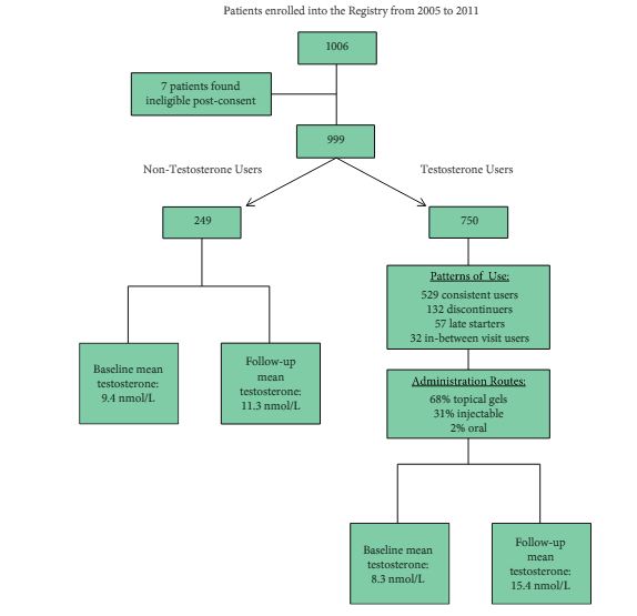
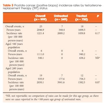
Of the 999 men with clinically diagnosed hypogonadism (HG), 750 (75%) initiated TRT, contributing 23 900 person-months of exposure. The mean testosterone levels increased from 8.3 to 15.4 nmol/L in treated men, compared to only a slight increase from 9.4 to 11.3 nmol/L in untreated men. In all, 55 biopsies were performed for suspected prostate cancer, and 12 non-cancer related biopsies were performed for other reasons. Overall, the proportion of positive biopsies was nearly identical in men on TRT (37.5%) compared to those not on TRT (37.0%) over the course of the study. There were no differences in PSA levels, total IPSS, or the IPSS obstructive sub-scale score by TRT status. Lower IPSS irritative sub-scale scores were reported in treated compared to untreated men.

Conclusions

Results support prostate safety of TRT in newly diagnosed men with HG.


Read more articles of the week

Editorial: Mythology and Reality Should Never Be Confused (But Often Are)

The debating halls of learned urological societies often attempt to define theories based on little substance. Unfortunately, although this type of scientific discourse should be encouraged, it can create both content for unrepeatable ‘late breaking’ abstracts and headlines for mass circulation newspapers. Once started, the momentum can make a perception become a ‘reality’ that is invariably difficult to dislodge. An area of particular current interest to both the scientific and lay press, and indeed the regulatory authorities in the USA and the European Union, is testosterone replacement. Within this context, an important potential myth-buster is described in the article by DeBruyne et al. [1]. The hypothesis (aka myth) that was being examined was that application of exogenous testosterone, irrespective of formulation, could exacerbate both benign and malignant prostatic disease. To understand the importance of this type of definitive study we must start at the beginning.

The foundation of the mythology, or in reality a Canadian-US urological tragedy, was ironically in the Nobel Prize winning work of Charles B Huggins. The ‘good news’ was that prostatic tumour regression could be affected by androgen deprivation, a finding certainly worthy of global recognition and acceptance; however, this became embellished to a certain extent, incorporating other aliquots of somewhat circumstantial evidence, leading to a suggestion that testosterone replacement could increase the probability and/or rate of prostate cancer progression. As a result, various societies perhaps understandably adopted their normal conservative approach of inserting a warning (with varying degrees of emphasis) in their guidelines [2]. Over the last decade, with the increasing use of testosterone replacement in the treatment of men with hypogonadism, the issue of benefit–risk has become more relevant, indeed is often transposed to risk–benefit, and is increasingly likely to feature in the popular press. Within the last couple of years, we have seen the spectre of testosterone and cardiovascular risk played out in full public view, but the issue of testosterone and exacerbation of prostatic disease has continued to smoulder in the background.

The RHYME study [1], although it is largely confirmatory as other studies have described similar conclusions, is of considerable ‘real-life’ value. The power of the study is that it should be considered to be representative of the potential hypogonadal population likely to present to the physician. Although not used for regulatory approval purposes, a registry study is considered to be the ‘gold standard’ for this type of analysis. So what are the conclusions of this and the majority of other studies? In essence, there is no evidence that restoration of testosterone to the normal range does increase the incidence or progression of either benign (BPH) or malignant prostatic disease. Any slight changes in PSA level were considered not to be clinically relevant and likely to be as a consequence of the direct pharmacology of increased testosterone levels [3]. This does not imply that exacerbations will never be seen or that appropriate monitoring should not be carried out; more that the conventional wisdom on testosterone and prostatic disease has gone (or should go) the way of antimuscarinic agents, invariably causing urinary retention in patients with BPH. It is to be hoped that studies such as the RHYME study will continue to be undertaken and help provide perspective for specialist and primary care physicians alike in areas of emergent clinical interest.

Read the full article
Michael Wyllie
Global Pharma Consulting Ltd, Stratton House, Shenington, Banbury, Oxfordshire


References


© 2024 BJU International. All Rights Reserved.