Archive for category: Article of the Week

Article of the Week: Decision-Making by PCa Physicians During AS

Every Week the Editor-in-Chief selects an Article of the Week from the current issue of BJUI. The abstract is reproduced below and you can click on the button to read the full article, which is freely available to all readers for at least 30 days from the time of this post.

In addition to the article itself, there is an accompanying editorial written by a prominent member of the urological community. This blog is intended to provoke comment and discussion and we invite you to use the comment tools at the bottom of each post to join the conversation.

If you only have time to read one article this week, it should be this one.

Qualitative study on decision-making by prostate cancer physicians during active surveillance

Stacy Loeb*,,, Caitlin Curnyn, Angela Fagerlin¶,**, Ronald Scott Braithwaite
Mark D. Schwartz, Herbert Lepor*, Herbert Ballentine Carter†† and Erica Sedlander


Departments of *Urology, Population Health, Laura and Isaac Perlmutter Cancer Center, New York University, §Manhattan Veterans Affairs Medical Center, New York, NY, Department of Population Health Sciences, University of Utah, Salt Lake City, UT, **Informatics, Decision Enhancement, and Surveillance (IDEAS) Center, Salt Lake City VA, UT, and ††Brady Urological Institute, Johns Hopkins Hospital, Baltimore, MD, USA


How to Cite

Loeb, S., Curnyn, C., Fagerlin, A., Braithwaite, R. S., Schwartz, M. D., Lepor, H., Carter, H. B. and Sedlander, E. (2017), Qualitative study on decision-making by prostate cancer physicians during active surveillance. BJU International, 120: 32–39. doi: 10.1111/bju.13651

Abstract

Objective

To explore and identify factors that influence physicians’ decisions while monitoring patients with prostate cancer on active surveillance (AS).

Subjects and Methods

A purposive sampling strategy was used to identify physicians treating prostate cancer from diverse clinical backgrounds and geographic areas across the USA. We conducted 24 in-depth interviews from July to December 2015, until thematic saturation was reached. The Applied Thematic Analysis framework was used to guide data collection and analysis. Interview transcripts were reviewed and coded independently by two researchers. Matrix analysis and NVivo software were used for organization and further analysis.

Results

Eight key themes emerged to explain variation in AS monitoring: (i) physician comfort with AS; (ii) protocol selection; (iii) beliefs about the utility and quality of testing; (iv) years of experience and exposure to AS during training; (v) concerns about inflicting ‘harm’; (vi) patient characteristics; (vii) patient preferences; and (viii) financial incentives.

Conclusion

These qualitative data reveal which factors influence physicians who manage patients on AS. There is tension between providing standardized care while also considering individual patients’ needs and health status. Additional education on AS is needed during urology training and continuing medical education. Future research is needed to empirically understand whether any specific protocol is superior to tailored, individualized care.

Editorial: AS in PCa- New Efforts, New Voices, New Hope

In January 2016, in his final State of the Union address, US President Barack Obama tasked Vice President Joseph Biden with heading up a new national mission, the Cancer Moonshot, to expedite advances in cancer prevention, diagnosis and treatment. One of the blue-ribbon panel recommendations was to minimize the side effects of cancer treatment.

There is no better target for that goal than prostate cancer, the cancer that leads all others in the toll of Americans annually diagnosed with cancer, and the fourth most common worldwide. Many men with low-risk prostate cancer undergo unnecessary treatments, including prostatectomy and radiation therapy, which are unlikely to affect their survival, even if their disease were left untreated. A case in point is the ProtecT study [1], which showed at a median of 10 years that there was no difference in prostate cancer-specific mortality between treatment with surgery or radiation therapy and no treatment [1]. Although there has been a paradigm shift in the management of low-risk prostate cancer with an increased uptake of active surveillance (AS) [2], the fact is that only ~40% of men with low-risk prostate cancer choose AS.

Because of equivalency in effectiveness of treatment options in low-risk prostate cancer, an explication of the steps involved in the clinical decision-making process were long overdue. In an innovative study in the present issue of BJUI, Loeb et al. [3] report a qualitative analysis using a purposive sampling strategy to explore the decision-making process of physicians caring for patients with prostate cancer undergoing AS. This study used qualitative interviews and investigators then analysed responses to identify factors influencing therapeutic decision-making. It is noteworthy that despite the fact that AS acceptance rates have increased and it is an established therapeutic approach, significant differences still remain with regard to when physicians enroll and how they monitor patients on AS. These findings align with those from a Surveillance Epidemiology and End Results (SEER) registry study of 12 068 men with low-risk prostate cancer whose urologists and radiation oncologists reported a spectrum of observation practices [4]. Neither study accounted for patients’ preference or perspectives.

Although there are many national guidelines for AS, no consensus on optimum AS management exists, but Movember–GAP3 (https://au.movember.com/report-cards/view/id/3372/gap3-prostate-cancer-active-surveillance), an international effort comprising 25 institutions with AS programmes, may change that. It seeks to establish standard guidelines for patient selection and monitoring and to find agreement on a trigger for treatment. The tumour heterogeneity and possible lack of linearity in early disease progression that we can glean from the next-generation sequencing studies in advanced prostate cancer [5] will not make that easy. Given the promise of precision medicine, we anticipate a decision-making process that by integrating clinical and pathological data, imaging, and biomarkers prognostic of risk of disease progression as well as patient comorbidity effectively removes guesswork from the calculation.

As this international effort and the vice president’s work proceed, we urge all to listen to the voices of patients and ensure they are heard as clearly as those of the experts. We know the paternalistic model of medicine, in which physicians are the exclusive decision-makers, has long been outmoded [6]. With so much at stake, let us now act like it.

Spyridon P. Basourakos* Karen Hoffman† and Jeri Kim*


*Department of Genitourinary Medical Oncology, and Department of Radiation Oncology, University of Texas MD Anderson Cancer Center, Houston, TX, US


References


1 Hamdy F, Donovan J, Lane J et al. 10-year outcomes after monitoring, surgery, or radiotherapy for localized prostate cancer. N Engl J Med 2016; 375: 141524


3 Loeb S, Curnyn C, Fagerlin A et al. Qualitative study on decision- making by prostate cancer physicians during active surveillance. BJU Int 2017; 120: 329


4 Hoffman K, Niu J, Shen Y et al. Physician variation in management of low-risk prostate cancer: a population-based cohort study. JAMA Intern Med 2014; 174: 14509


5 Robinson D, Van Allen E, Wu Y et al. Integrative clinical genomics of advanced prostate cancer. Cell 2015; 161: 121528


Video: Decision-Making by PCa Physicians During AS

Qualitative study on decision-making by prostate cancer physicians during active surveillance


Abstract

Objective

To explore and identify factors that influence physicians’ decisions while monitoring patients with prostate cancer on active surveillance (AS).

Subjects and Methods

A purposive sampling strategy was used to identify physicians treating prostate cancer from diverse clinical backgrounds and geographic areas across the USA. We conducted 24 in-depth interviews from July to December 2015, until thematic saturation was reached. The Applied Thematic Analysis framework was used to guide data collection and analysis. Interview transcripts were reviewed and coded independently by two researchers. Matrix analysis and NVivo software were used for organization and further analysis.

Results

Eight key themes emerged to explain variation in AS monitoring: (i) physician comfort with AS; (ii) protocol selection; (iii) beliefs about the utility and quality of testing; (iv) years of experience and exposure to AS during training; (v) concerns about inflicting ‘harm’; (vi) patient characteristics; (vii) patient preferences; and (viii) financial incentives.

Conclusion

These qualitative data reveal which factors influence physicians who manage patients on AS. There is tension between providing standardized care while also considering individual patients’ needs and health status. Additional education on AS is needed during urology training and continuing medical education. Future research is needed to empirically understand whether any specific protocol is superior to tailored, individualized care.

View more videos

Article of the Week: Value of 111In-PSMA-RGS for salvage lymphadenectomy in recurrent PCa

Every Week the Editor-in-Chief selects an Article of the Week from the current issue of BJUI. The abstract is reproduced below and you can click on the button to read the full article, which is freely available to all readers for at least 30 days from the time of this post.

In addition to the article itself, there is an accompanying editorial written by a prominent member of the urological community. This blog is intended to provoke comment and discussion and we invite you to use the comment tools at the bottom of each post to join the conversation.

Finally, the third post under the Article of the Week heading on the homepage will consist of additional material or media. This week we feature a video discussing the paper.

If you only have time to read one article this week, it should be this one.

Value of 111In-prostate-specific membrane antigen (PSMA)-radioguided surgery for salvage lymphadenectomy in recurrent prostate cancer: correlation with histopathology and clinical follow-up

Isabel Rauscher*, Charlotte Duwel, Martina Wirtz, Margret SchotteliusHans-Jurgen Wester, Kristina Schwamborn§, Bernhard Haller, Markus Schwaiger*, Jurgen E. Gschwend, Matthias Eiber* and Tobias Maurer


*Departments of Nuclear Medicine, Urology, Technical University of Munich, Klinikum rechts der Isar, Munich, Institute of Pharmaceutical Radiochemistry, Technical University of Munich, Garching, §Department of Pathology, and Institute of Medical Statistics and Epidemiology, Technical University of Munich, Klinikum rechts der Isar, Munich, Germany


Read the full article

How to Cite

Rauscher, I., Düwel, C., Wirtz, M., Schottelius, M., Wester, H.-J., Schwamborn, K., Haller, B., Schwaiger, M., Gschwend, J. E., Eiber, M. and Maurer, T. (2017), Value of 111In-prostate-specific membrane antigen (PSMA)-radioguided surgery for salvage lymphadenectomy in recurrent prostate cancer: correlation with histopathology and clinical follow-up. BJU International, 120: 40–47. doi: 10.1111/bju.13713

Abstract

Objectives

To evaluate the use of 111In-labelled prostate-specific membrane antigen (PSMA)-I&T-based radioguided surgery (111In-PSMA-RGS) for salvage surgery in recurrent prostate cancer (PCa) using comparison of intra-operative gamma probe measurements with histopathological results of dissected specimens. In addition, to determine the success of 111In-PSMA-RGS with regard to postoperative prostate-specific antigen (PSA) responses, PCa-specific treatment-free survival rates and postoperative complication rates.

Patients and Methods

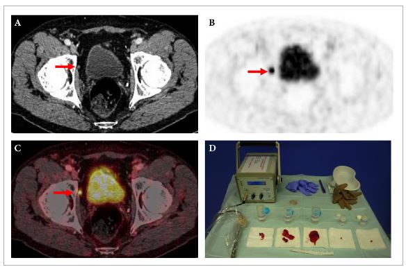
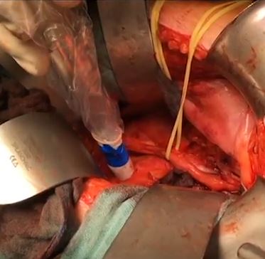
A total of 31 consecutive patients with localized recurrent PCa undergoing salvage surgery with PSMA-targeted radioguided surgery using a 111In-labelled PSMA ligand between April 2014 and July 2015 were retrospectively included in this study. The preoperative (interquartile range; range) median PSA level was 1.3 (0.57–2.53 ng/mL; 0.2–13.9 ng/mL). Results of ex vivo radioactivity rating (positive vs negative) of resected tissue specimens were compared with findings of postoperative histological analysis. Best PSA response without additional treatment was determined after 111In-PSMA-RGS, and salvage-surgery-related postoperative complications and PCa-specific additional treatments were recorded.

aotw-jul-2017-3-results

Results

In 30/31 patients, 111In-PSMA-RGS allowed intra-operative identification of metastatic lesions. In total, 145 surgical specimens were removed and 51 showed metastatic involvement at histological analysis. According to 111In-PSMA-RGS ex vivo measurements, 48 specimens were correctly classified as metastatic and 87 as cancer-free, four were false-negative and six were false-positive compared with histological evaluation. Follow-up information was available for 30/31 patients. PSA declines of >50% and >90% were observed in 23/30 patients and in 16/30 patients, respectively. In 18/30 patients, a PSA decline to <0.2 ng/mL was observed. In 10/30 patients further PCa-specific treatment was given after a median (range) of 125 (48–454) days post-111In-PSMA-RGS. The remaining 20 patients remained treatment-free at a median (range) follow-up of 337 (81–591) days. Of 30 patients, 10 presented with surgery-related complications (Clavien–Dindo grade 1, n = 6, Clavien–Dindo grade 3b, n = 4).

Conclusion

111In-PSMA-RGS proved to be of high value for intra-operative detection of even small metastatic lesions in patients with PCa scheduled for salvage lymphadenectomy. It allows the exact localization and resection of metastatic tissue during 111In-PSMA-RGS and is therefore anticipated to have a beneficial influence on further disease progression; however, identification of suitable patients on the basis of PSMA-positron-emission tomography imaging as well as clinical variables is essential for satisfactory results to be obtained.

Editorial: PSMA-RS – a promising utility

There is no doubt that, in the field of prostate cancer, few recent topics have been the subject of as much captivation and discussion as prostate-specific membrane antigen (PSMA) positron-emission tomography (PET) imaging. The body of literature on this imaging technique, the majority on the use of 68Ga-PSMA-HBED-CC as a radiotracer, is growing unceasingly [1], and includes data to support the superior accuracy of PSMA PET/CT for lymph node staging in prostate cancer [2] and in identifying patients unlikely to benefit from radiotherapy after radical prostatectomy [3].

In the present paper, Rauscher et al. [4] present their data on the use of an 111In-PSMA-I&T tracer during salvage lymphadenectomy for recurrent prostate cancer. In a previous study by the same research group, 111In-PSMA-I&T has already proven to be a high-affinity radiotracer, with enhanced internalization efficiency compared with other molecules and significant accumulation in prostate cancer tissue [5].

In the present pilot study, salvage lymphadenectomy was performed in 31 patients with recurrent prostate cancer after primary treatment. Using intra-operative γ-probe measurements and comparing these with the histopathological results of the specimens, the authors found that these correlated well, resulting in a sensitivity of 92.3%, a specificity of 93.5% and an accuracy of 93.1%, with a positive predictive value of 88.9% and a negative predictive value of 95.6%.

These findings also translated well into PSA response after surgery. Postoperative PSA reductions of >50% were observed in 76.7% of patients and of >90% in 53.3% of patients. Further cancer-specific treatment was given to 33% of patients at a median of 125 days after surgery. The remaining patients remained free of treatment at a median follow-up of 337 days.

The study sample was relatively small and the analysis was conducted retrospectively. These are obvious drawbacks; nonetheless the intra- and postoperative results presented are promising. However, as the authors of the present paper point out, careful patient selection is important, and the follow-up period for these patients is still quite short. We will need to wait several years to compare the outcomes of this series with those reported in the literature with regard to salvage lymphadenectomy without prior PSMA PET CT. These data were recently published in a review by Heidenreich et al. [6]. They reported 5-year biochemical recurrence-free survival of 19–25% after salvage lymphadenectomy without the use of PSMA PET, and a median time to systemic treatment of 20–30 months [6].

As physicians, of course, our primary goal is to do the best for our patients. Certainly, we would like to believe that aggressively treating every single lesion made visible with this new imaging technique would be to the patient’s benefit. It is certainly tempting to chase these colourful lesions now demonstrated so nicely by PSMA PET/CT, but we owe it to ourselves as scientists to gather the facts and the evidence to determine whether or not our current course of action makes sense. PSMA PET radio-guided surgery is no exception to the rule, and only the evidence will tell what exact role this new technology is to have in the treatment of prostate cancer. A number of questions need to be addressed. Can we justify putting patients through surgical procedures with the morbidity associated with them? Do we not need to define oligometastatic disease in the molecular imaging era? Should we then start using PSMA PET in primary staging of prostate cancer patients? What of those tumours that do not express PSMA? Can this approach be offered laparoscopically or robotically?

It seems the introduction of PSMA PET has, instead of giving us all the answers, given rise to even more questions.

Nicolas Geurts,*Alastair D. Lamb,*† Nathan Lawrentschuk*†‡ and Declan G. Murphy*§¶


*Division of Cancer Surgery, University of Melbourne, Peter MacCallum Cancer Centre, Melbourne, Department of Surgery, Austin Hospital, University of Melbourne, Heidelberg§ Australian Prostate Cancer Research Centre, Epworth Healthcare, and
Sir Peter MacCallum Department of Oncology, University of Melbourne, Melbourne, Vic., Australia


Read the full article

How to Cite

Geurts, N., Lamb, A. D., Lawrentschuk, N. and Murphy, D. G. (2017), Prostate-specific membrane antigen radioguided surgery: a promising utility. BJU International, 120: 5–6. doi: 10.1111/bju.13838

References


Video: Value of 111In-PSMA-RGS for salvage lymphadenectomy in recurrent PCa

Value of 111In-prostate-specific membrane antigen (PSMA)-radioguided surgery for salvage lymphadenectomy in recurrent prostate cancer: correlation with histopathology and clinical follow-up


Read the full article


Abstract

Objectives

To evaluate the use of 111In-labelled prostate-specific membrane antigen (PSMA)-I&T-based radioguided surgery (111In-PSMA-RGS) for salvage surgery in recurrent prostate cancer (PCa) using comparison of intra-operative gamma probe measurements with histopathological results of dissected specimens. In addition, to determine the success of 111In-PSMA-RGS with regard to postoperative prostate-specific antigen (PSA) responses, PCa-specific treatment-free survival rates and postoperative complication rates.

Patients and Methods

A total of 31 consecutive patients with localized recurrent PCa undergoing salvage surgery with PSMA-targeted radioguided surgery using a 111In-labelled PSMA ligand between April 2014 and July 2015 were retrospectively included in this study. The preoperative (interquartile range; range) median PSA level was 1.3 (0.57–2.53 ng/mL; 0.2–13.9 ng/mL). Results of ex vivo radioactivity rating (positive vs negative) of resected tissue specimens were compared with findings of postoperative histological analysis. Best PSA response without additional treatment was determined after 111In-PSMA-RGS, and salvage-surgery-related postoperative complications and PCa-specific additional treatments were recorded.

Results

In 30/31 patients, 111In-PSMA-RGS allowed intra-operative identification of metastatic lesions. In total, 145 surgical specimens were removed and 51 showed metastatic involvement at histological analysis. According to 111In-PSMA-RGS ex vivo measurements, 48 specimens were correctly classified as metastatic and 87 as cancer-free, four were false-negative and six were false-positive compared with histological evaluation. Follow-up information was available for 30/31 patients. PSA declines of >50% and >90% were observed in 23/30 patients and in 16/30 patients, respectively. In 18/30 patients, a PSA decline to <0.2 ng/mL was observed. In 10/30 patients further PCa-specific treatment was given after a median (range) of 125 (48–454) days post-111In-PSMA-RGS. The remaining 20 patients remained treatment-free at a median (range) follow-up of 337 (81–591) days. Of 30 patients, 10 presented with surgery-related complications (Clavien–Dindo grade 1, n = 6, Clavien–Dindo grade 3b, n = 4).

Conclusion

111In-PSMA-RGS proved to be of high value for intra-operative detection of even small metastatic lesions in patients with PCa scheduled for salvage lymphadenectomy. It allows the exact localization and resection of metastatic tissue during 111In-PSMA-RGS and is therefore anticipated to have a beneficial influence on further disease progression; however, identification of suitable patients on the basis of PSMA-positron-emission tomography imaging as well as clinical variables is essential for satisfactory results to be obtained.

View more videos

Article of the Week: Ureteric stent dwelling time: a risk factor for post-ureteroscopy sepsis

Every Week the Editor-in-Chief selects an Article of the Week from the current issue of BJUI. The abstract is reproduced below and you can click on the button to read the full article, which is freely available to all readers for at least 30 days from the time of this post.

In addition to the article itself, there is an accompanying editorial written by a prominent member of the urological community. This blog is intended to provoke comment and discussion and we invite you to use the comment tools at the bottom of each post to join the conversation.

If you only have time to read one article this week, it should be this one.

Ureteric stent dwelling time: a risk factor for post-ureteroscopy sepsis

Amihay Nevo*, Roy Mano*, Jack Baniel*† and David A. Lifshitz*


*Department of Urology, Rabin Medical Centre, Petach Tikva, Israel, and Sackler Faculty of Medicine, Tel Aviv University, Tel Aviv, Israel


Read the full article

Abstract

Objectives

To evaluate the association between stent dwelling time and sepsis after ureteroscopy, and identify risk factors for sepsis in this setting.

Patients and Methods

The prospectively collected database of a single institution was queried for all patients who underwent ureteroscopy for stone extraction between 2010 and 2016. Demographic, clinical, preoperative and operative data were collected. The primary study endpoint was sepsis within 48 h of ureteroscopy. Logistic regressions were performed to identify predictors of post-ureteroscopy sepsis in the ureteroscopy cohort and specifically in patients with prior stent insertion.

aotw-jul-2

Results

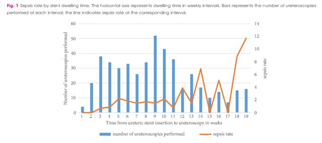
Between October 2010 and April 2016, 1 256 patients underwent ureteroscopy for stone extraction. Risk factors for sepsis included prior stent placement, female gender and Charlson comorbidity index. A total of 601 patients had a ureteric stent inserted before the operation and were included in the study cohort, in which the median age was 56 years, 90 patients were women (30%), and 97 patients were treated for positive preoperative urine cultures (16.1%). Postoperative sepsis, <48 h after surgery, occurred in eight (1.2%) non-stented patients and in 28 patients (4.7%) with prior stent insertion. Sepsis rates after stent dwelling times of 1, 2, 3 and >3 months were 1, 4.9, 5.5 and 9.2%, respectively. On multivariate analysis, stent dwelling time, stent insertion because of sepsis, and female gender were significantly associated with post-ureteroscopy sepsis in patients with prior stent placement.

Conclusions

Patients who undergo ureteroscopy after ureteric stent insertion have a higher risk of postoperative sepsis. Prolonged stent dwelling time, sepsis as an indication for stent insertion, and female gender are independent risk factors. Stent placement should be considered cautiously, and if inserted, ureteroscopy should be performed within 1 month.

Editorial: Pre-stenting and the risk of postoperative sepsis: a shorter dwell time is better

In this edition of the BJUI, Nevo et al. [1] report their retrospective review of 1256 patients who underwent ureteroscopy (URS)/flexible ureterorenoscopy (FURS) for stone disease and identified an overall sepsis rate of 2.8% within 48 h of surgery. About half of the cohort had a previously placed JJ stent, and the key finding of the study was the association between this and postoperative sepsis. In particular, the risk of sepsis in unstented patients was 1.2%, compared with 4.7% in those with a stent, such that overall, 80% of the patients who developed sepsis had a prior JJ stent in situ. Furthermore, this risk increased cumulatively with longer stent dwell-time before definitive surgery, increasing the risk of sepsis to 9.2% in patients who had a stent in situ for >3 months before the treatment of their stone.

Pre-stenting before ureteroscopic stone treatment can be for ‘absolute’ reasons (e.g. achieving ureteric drainage following presentation with an obstructed/infected kidney, or for an inaccessible ureter during the initial attempt at ureteroscopic stone treatment) or ‘relative’ reasons (e.g. emergency pain relief from ureteric colic if stone clearance cannot be offered immediately, or strategically inserted to allow passive ureteric dilatation to facilitate a subsequent definitive procedure). Data from the Clinical Research Office of the Endourological Society (CROES) URS Global Study showed that pre-stenting occurred in 36.4% of patients with rena l stones, and was associated with an increased stone-free rate (SFR), with a small but significant decrease in intraoperative complications. Pre-stenting was less common in ureteric stone management (11.9% of 8189 patients) and showed no difference in the SFR or complications, but was associated with a shorter length of stay [2]. Similarly, Jessen et al. [3] have reported that pre-stenting conferred a significant improvement in SFR for renal stones (83% vs 60%) but not for ureteric stones (94% vs 90%), although complications were reduced for pre-stented vs unstented patients in both renal stones (8.7% vs 19.4%) and ureteric stones (3.1% vs 10.7%) in that study. The advantage of pre-stenting appears to be greatest for larger stones: as a consequence of ureteric dilatation, and therefore improved renal access, in patients with stones >1 cm in whom a multi-phased approach was anticipated, Chu et al. [4] found that pre-stenting significantly reduced the operative time of the first URS, as well as the total operative time to stone clearance, including the need to re-operate at all in some patients.

The study in this edition of the BJUI [1] has shown that these advantages must be balanced against the increased risk of postoperative sepsis in patients with a pre-placed JJ stent, particularly in cases where the stent has been in situ for >1 month. The ability to identify patients who are at greater risk of post-URS/FURS infections is clearly useful: female gender, diabetes mellitus, ischaemic heart disease, an American Society of Anesthesiologists (ASA) score of ≥II, a large-volume stone burden, and same-session bilateral URS, have already been established as significant risk factors for postoperative infection [5, 6]. In addition, preoperative infections, either as a positive midstream specimen of urine (MSU) or previous sepsis also increase the risk of postoperative infectious complications. Specifically, Blackmur et al[6]reported that patients with a positive preoperative MSU were about five-times more likely to have postoperative urosepsis, even if they had been treated with an appropriate course of antibiotics before their stone surgery. Consistent with this, Youssef et al. [7] reported that the complication rate in patients undergoing URS after previous sepsis increased to 20% compared to 7% in matched non-septic controls, with an associated increased length of stay and duration of postoperative antibiotics.

In the present article [1], the overall sepsis rate was low at 2.8%, (and was comparable to recent CROES data that reported an overall prevalence of postoperative fever/UTI after URS or FURS of ≤2.2% [5]), but patients who had stents inserted for sepsis in the presence of an obstructing stone had a four-times higher postoperative infection rate when their stones were eventually treated than pre-stented patients without prior sepsis [1].

Taken together, these studies suggest that pre-stenting may have value in improving SFR, total operation time and reducing complication rates for large renal stones (i.e. >1 cm), but offers less advantage to stone clearance rates or complications in ureteric stones. Given the findings of increased risk of sepsis with stent-dwell time (increasing from 2.2% at 30 days, to 4.9% at 60 days, 5.5% at 90 days and 9.2% for >90 days) the authors recommendation ‘to keep stent dwelling time as short as possible’, is both practically beneficial to the patient and evidence-based. Furthermore, in addition to an awareness of the recognised risk factors for postoperative sepsis mentioned above, patients who have had stents inserted for prior sepsis, patients with positive preoperative MSU (even if treated), and patients with prolonged stent duration before definitive treatment should be counselled of the greater risk of postoperative sepsis, and watched cautiously in the early postoperative period.

In this study [1], patients with a stent in situ for <1 month had a similar risk of UTI to unstented patients. It would therefore seem reasonable to conclude that patients who have a stent inserted, especially for more ‘relative’ reasons to facilitate future surgery, should be scheduled for their definitive procedure within a month to achieve the benefits of pre-stenting, whilst minimising the potential for postoperative septic complications that Nevo et al. [1] have highlighted.

Daron Smith


Institute of Urology, University College Hospital, London, UK


Read the full article


References


Residents’ Podcast: Long term follow up of erectile dysfunction after RP using nerve grafts

 Jesse Ory, Kyle Lehmann and Jeff Himmelman

Department of Urology, Dalhousie University
Halifax, NS, Canada

Read the full article

Abstract

Objective

To study a novel penile reinnervation technique using four sural nerve grafts and end-to-side neurorraphies connecting bilaterally the femoral nerve and the cavernous corpus and the femoral nerve and the dorsal penile nerves.

Patients and Methods

Ten patients (mean [± sd; range] age 60.3 [± 4.8; 54–68] years), who had undergone radical prostatectomy (RP) at least 2 years previously, underwent penile reinnervation in the present study. Four patients had undergone radiotherapy after RP. All patients reported satisfactory sexual activity prior to RP. The surgery involved bridging of the femoral nerve to the dorsal nerve of the penis and the inner part of the corpus cavernosum with sural nerve grafts and end-to-side neurorraphies. Patients were evaluated using the International Index of Erectile Function (IIEF) questionnaire and pharmaco-penile Doppler ultrasonography (PPDU) preoperatively and at 6, 12 and 18 months postoperatively, and using a Clinical Evolution of Erectile Function (CEEF) questionnaire, administered after 36 months.

Results

The IIEF scores showed improvements with regard to erectile dysfunction (ED), satisfaction with intercourse and general satisfaction. Evaluation of PPDU velocities did not reveal any difference between the right and left sides or among the different time points. The introduction of nerve grafts neither caused fibrosis of the corpus cavernosum, nor reduced penile vascular flow. CEEF results showed that sexual intercourse began after a mean of 13.7 months with frequency of sexual intercourse varying from once daily to once monthly. Acute complications were minimal. The study was limited by the small number of cases.

Conclusions

A total of 60% of patients were able to achieve full penetration, on average, 13 months after reinnervation surgery. Patients previously submitted to radiotherapy had slower return of erectile function. We conclude that penile reinnervation surgery is a viable technique, with effective results, and could offer a new treatment method for ED after RP.

Article of the Month: Identification of non-invasive biomarkers of UCPPS: findings from the MAPP Research Network

Every Month the Editor-in-Chief selects an Article of the Month from the current issue of BJUI. The abstract is reproduced below and you can click on the button to read the full article, which is freely available to all readers for at least 30 days from the time of this post.

In addition to the article itself, there is an accompanying editorial written by a prominent member of the urological community. This blog is intended to provoke comment and discussion and we invite you to use the comment tools at the bottom of each post to join the conversation.

If you only have time to read one article this week, it should be this one.

Identification of novel non-invasive biomarkers of urinary chronic pelvic pain syndrome: findings from the Multidisciplinary Approach to the Study of Chronic Pelvic Pain (MAPP) Research Network

Adelle Dagher*, Adam Curatolo*, Monisha Sachdev*, Alisa J. Stephens, Chris Mullins§, J. Richard Landis, Adrie van Bokhoven, Andrew El-Hayek*, John W. Froehlich**, Andrew C. Briscoe**, Roopali Roy*,††, Jiang Yang*,††, Michel A. Pontari‡‡David Zurakowski††§§, Richard S. Lee**††, Marsha A. Moses*,†† for the MAPP Research Network 
*Vascular Biology Program, Department of Surgery, Boston Childrens Hospital, Boston, MA,


Department of Biostatistics and Epidemiology, Perelman School of Medicine at the University of Pennsylvania, Philadelphia, PA, §National Institute of Diabetes and Digestive and Kidney Diseases, National Institutes of Health, Bethesda, MD, Department of Pathology, University of Colorado Anschutz Medical Campus, Aurora, CO, **Department of Urology, Boston Childrens Hospital, Boston, MA, ††Department of Surgery, Harvard Medical School, Boston, MA, ‡‡Lewis Katz School of Medicine at Temple University, Philadelphia, PA, and §§Department of Anesthesia, Boston Childrens Hospital, Boston, MA, USA


Read the full article

How to Cite

Dagher, A., Curatolo, A., Sachdev, M., Stephens, A. J., Mullins, C., Landis, J. R., van Bokhoven, A., El-Hayek, A., Froehlich, J. W., Briscoe, A. C., Roy, R., Yang, J., Pontari, M. A., Zurakowski, D., Lee, R. S., Moses, M. A. and the MAPP Research Network (2017), Identification of novel non-invasive biomarkers of urinary chronic pelvic pain syndrome: findings from the Multidisciplinary Approach to the Study of Chronic Pelvic Pain (MAPP) Research Network. BJU International, 120: 130–142. doi: 10.1111/bju.13832

Abstract

Objective

To examine a series of candidate markers for urological chronic pelvic pain syndrome (UCPPS), selected based on their proposed involvement in underlying biological processes so as to provide new insights into pathophysiology and suggest targets for expanded clinical and mechanistic studies.

Methods

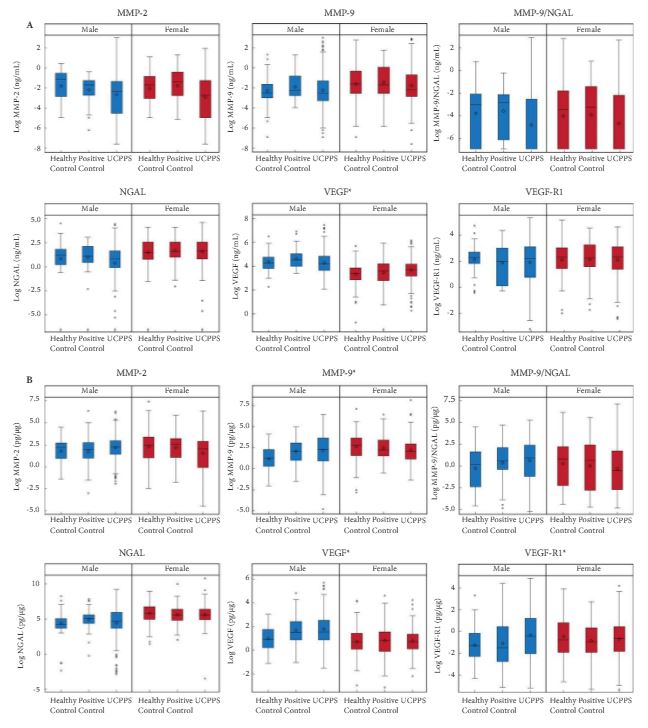
Baseline urine samples from Multidisciplinary Approach to the Study of Chronic Pelvic Pain (MAPP) Research Network study participants with UCPPS (n = 259), positive controls (PCs; chronic pain without pelvic pain, n = 107) and healthy controls (HCs, n = 125) were analysed for the presence of proteins that are suggested in the literature to be associated with UCPPS. Matrix metalloproteinase (MMP)-2, MMP-9, MMP-9/neutrophil gelatinase-associated lipocalin (NGAL) complex (also known as Lipocalin 2), vascular endothelial growth factor (VEGF), VEGF receptor 1 (VEGF-R1) and NGAL were assayed and quantitated using mono-specific enzyme-linked immunosorbent assays for each protein. Log-transformed concentration (pg/mL or ng/mL) and concentration normalized to total protein (pg/μg) values were compared among the UCPPS, PC and HC groups within sex using the Student’s t-test, with P values adjusted for multiple comparisons. Multivariable logistic regression and receiver-operating characteristic curves assessed the utility of the biomarkers in distinguishing participants with UCPPS and control participants. Associations of protein with symptom severity were assessed by linear regression.
jul-aotw-1

Results

Significantly higher normalized concentrations (pg/μg) of VEGF, VEGF-R1 and MMP-9 in men and VEGF concentration (pg/mL) in women were associated with UCPPS vs HC. These proteins provided only marginal discrimination between UCPPS participants and HCs. In men with UCCPS, pain severity was significantly positively associated with concentrations of MMP-9 and MMP-9/NGAL complex, and urinary severity was significantly positively associated with MMP-9, MMP-9/NGAL complex and VEGF-R1. In women with UCPPS, pain and urinary symptom severity were associated with increased normalized concentrations of MMP-9/NGAL complex, while pain severity alone was associated with increased normalized concentrations of VEGF, and urinary severity alone was associated with increased normalized concentrations of MMP-2. Pain severity in women with UCPPS was significantly positively associated with concentrations of all biomarkers except NGAL, and urinary severity with all concentrations except VEGF-R1.

Conclusion

Altered levels of MMP-9, MMP-9/NGAL complex and VEGF-R1 in men, and all biomarkers in women, were associated with clinical symptoms of UCPPS. None of the evaluated candidate markers usefully discriminated UCPPS patients from controls. Elevated VEGF, MMP-9 and VEGF-R1 levels in men and VEGF levels in women may provide potential new insights into the pathophysiology of UCPPS.

© 2024 BJU International. All Rights Reserved.