Archive for category: Article of the Week

Editorial: RARP and a Parachute

Radical prostatectomy is probably the most scrutinized, debated and re‐invented procedure in the field of urology. This is with good reason. Dr Hugh Hampton Young gave the first formal account of a radical treatment for prostate cancer in 1905. It was a procedure performed perineally with the prime intention of total cancer extirpation. The oncological results were tremendous given the nascency of the procedure. Functional outcomes, however, were less so, with only ~25% of the patients achieving urinary continence and almost none achieving potency 1. Seminal studies undertaken by Drs Patrick C. Walsh and Pieter J. Donker in the 1980s led to the next major advance in technique: nerve‐sparing. It lead to dramatic improvements in sexual and urinary function preservation, with urinary continence achieved in upwards of 90% and potency in upwards of 60% of patients 23. Nerve‐sparing retropubic radical prostatectomy was rapidly adopted by urologists across the world. No randomized trial was conducted as the operation ‘made sense’, and it would have been unethical to offer patients an alternative, inferior operation. Retropubic radical prostatectomy, however, remained a morbid operation that was difficult to master; 1 200 mL blood loss and 20% incontinence, 15% stricture and 60–90% erectile dysfunction rates were the norm. Robot‐assisted surgery changed much of this. This surgery was perfected and popularized by Dr Mani Menon and his team at the Henry Ford Hospital. Blood loss dropped to ~100 mL, and incontinence and stricture rates today are ~1–5% with potency rates between 70% and 80% 4.

In the systematic review by Ilic et al. 5 trials that compared open with minimally invasive radical prostatectomy were evaluated. The authors are commended for combing through vast quantities of data to arrive at the final sample of two clinical trials (one laparoscopic, one robot‐assisted). The authors found no data on oncological endpoints. With respect to functional outcomes, the data reported in their systematic review are essentially from the recent Yaxley trial. It is true that randomized controlled trials form the backbone of medical progress, but they must be interpreted with care. The Yaxley trial has been criticized for comparing surgeons of vastly varying surgical expertise (1 500‐case experience for open prostatectomies, 200‐case experience for robot‐assisted prostatectomies), having a small sample size (~150 patients in each arm) and having a limited follow‐up (3 months). In line with this, Dr Gordon Smith has eloquently argued about the judicious use of randomized trials in evidence‐based medicine in his classic paper on parachutes and gravitational challenge 6. It is our sincere belief that robot‐assisted radical prostatectomy is a procedure in the service of the patients, and we believe that anyone who has performed both an open radical prostatectomy and a robot‐assisted radical prostatectomy would agree. Operating deep in the pelvis and being able to visualize the anatomy up‐close with robotic assistance ‘makes sense’, much like the anatomical radical prostatectomy did in the 1980s 23.

‘If it ain’t broke, don’t fix it’, the old saying goes. In case of radical prostatectomy, the converse has been true, for the most part. The procedure is still not perfect, and nuances need to be worked out. Nonetheless, over the last century, radicalprogress has been made in the surgery for prostate; the two issues that now remain to be solved are: improving potency and improving cost‐effectiveness. With the potential arrival of competing robotic systems and improvements in technology, the costs associated with robotic surgery may become less of an issue in the future. With regard to potency, focal therapies, whether surgical or otherwise, hold promise.

Akshay SoodFiras Abdollah, and Mani Menon
Vattikuti Urology Institute, Henry Ford Hospital, Detroit, MI, USA


References
  • Young HH. The cure of cancer of the prostate by radical perineal prostatectomy (prostato‐seminal Vesiculectomy): History, Literature and Statistics of Young’s Operation1J Urol 194553: 188–252


  • Menon M, Dalela D, Jamil M et al. Functional recovery, oncologic outcomes and postoperative complications after robot‐assisted radical prostatectomy: an evidence‐based analysis comparing the Retzius sparing and standard approachesJ Urol 2018199: 1210–7


  • Ilic D, Evans SM, Allan CA, Jung JH, Murphy D, Frydenberg M. Laparoscopic and robot‐assisted vs open radical prostatectomy for the treatment of localized prostate cancer: a Cochrane systematic reviewBJU Int 2018121: 845–53


  • Smith GC, Pell JP. Parachute use to prevent death and major trauma related to gravitational challenge: systematic review of randomised controlled trialsBMJ 200320: 1459–61


Article of the Month: BAUS consensus on priapism

Every Month, the Editor-in-Chief selects an Article of the Month from the current issue of BJUI. The abstract is reproduced below and you can click on the button to read the full article, which is freely available to all readers for at least 30 days from the time of this post.

In addition to the article itself, there is an accompanying editorial written by a prominent member of the urological community. This blog is intended to provoke comment and discussion and we invite you to use the comment tools at the bottom of each post to join the conversation.

If you only have time to read one article this week, it should be this one.

BAUS consensus document for the management of male genital emergencies: priapism


Asif Muneer, Gareth Brown, Trevor Dorkin, Marc Lucky, Richard Pearcy, Majid Shabbir, Chitranjan J. Shukl,a Rowland W. Rees & Duncan J. Summerton

BAUS Section of Andrology Genitourethral Surgery


Read the full article


Abstract

Male genital emergencies relating to the penis and scrotum are rare and require prompt investigation and surgical intervention. Clinicians are often unfamiliar with the management of these conditions and may not work in a specialist centre with on‐site expertise in genitourethral surgery. A series of consensus statements have been developed by an expert consensus committee comprising members of the BAUS Section of Andrology and Genitourethral Surgery together with experts from urology units throughout the UK. Priapism requires prompt assessment and treatment and these consensus statements provide guidance for UK practice.

Read more articles of the week

Editorial: The BAUS consensus documents on andrology

In 2018, the BAUS returns to Liverpool and we have taken this opportunity to renew the lasting friendship between the BAUS and the BJUI. We also celebrate a monumental achievement for the city of Liverpool itself – the Knighthood of Sir Ringo Starr. This has finally happened, 50 years after his MBE and is richly deserved. We therefore decided to feature Liverpool and The Beatles on the front cover of your journal.

This year, Duncan Summerton, a well‐respected Urologist and Andrologist, starts his 2‐year term as the President of the BAUS. In our ‘Guidelines’ section, we have featured two BAUS consensus documents from the Andrology Section on priapism [1] and testicular trauma [2]. The former has an excellent flow chart on management of priapism with timelines of presentation, which every urologist will find clinically useful.

We have also included two excellent UK articles on renal trauma [34], which BAUS members and beyond can learn from.

Finally, renal oncocytoma and its management may pose its own challenges as recorded by Neves et al. [5]. We also present the BAUS radical prostatectomy audit, which is publicly accessible and reassures readers (and the public) that the majority of these operations are being performed in high‐volume centres (164/centre) by high‐volume surgeons with good outcomes [6]. Nearly three in four operations are now performed robotically, which was certainly not the case when I started 15 years ago.

We look forward to meeting you at lunchtime on the Monday and Tuesday of the BAUS conference at the BJUI stand. I am particularly excited about the BJUI lecture and the National Clinical Entrepreneurship Programme, led by my friend Tony Young, on the second day of the meeting (https://www.baus.org.uk/agm/programme.aspx).


Prokar Dasgupta

MRC Centre for Transplantation, King’s College London, London, UK


Read the full article


References


  • Lucky M, Brown G, Dorkin T et al. British Association of Urological Surgeons (BAUS) consensus document for the management of male genital emergencies – testicular traumaBJU Int 2018121: 840–4


  • Wong KY, Jeeneea R, Healey A et al. Management of paediatric high‐grade blunt renal trauma: a 10‐year single‐centre UK experienceBJU Int 2018121: 923–7


  • Hadjipavlou M, Grouse E, Gray R et al. Managing penetrating renal trauma: experience from two major trauma centres in the UKBJU Int 2018121: 928–34


  • Neves JB, Withington J, Fowler S et al. Contemporary surgical management of renal oncocytoma: a nation’s outcomeBJU Int 2018121: 893–9


  • Khadhouri S, Miller C, Fowler S et al. The British Association of Urological Surgeons (BAUS) radical prostatectomy audit 2014/2015 – an update on current practice and outcomes by centre and surgeon case‐volumeBJU Int 2018121: 886–92


Article of the Week: Focal irreversible electroporation as primary treatment for localized prostate cancer

Every Week, the Editor-in-Chief selects an Article of the Week from the current issue of BJUI. The abstract is reproduced below and you can click on the button to read the full article, which is freely available to all readers for at least 30 days from the time of this post.

In addition to the article itself, there is an accompanying editorial written by a prominent member of the urological community. This blog is intended to provoke comment and discussion and we invite you to use the comment tools at the bottom of each post to join the conversation.

If you only have time to read one article this week, it should be this one.

Focal irreversible electroporation as primary treatment for localized prostate cancer


Willemien van den Bos*†‡, Matthijs J. Scheltema*†‡, Amila R. Siriwardana*Anton M.F. Kalsbeek*, James E. Thompson, Francis Ting*, Maret Bohm*,
Anne-Maree Haynes*, Ron Shnier§, Warick Delprado¶ and Phillip D. Stricker


*Garvan Institute of Medical Research and Kinghorn Cancer Centre, St Vincents Prostate Cancer Centre, Darlinghurst, NSW, Australia, Academic Medical Centre, University of Amsterdam, Amsterdam, The Netherlands, §Southern Radiology, Randwick, and Douglass Hanly Moir Pathology, Macquarie Park, NSW, Australia


Read the full article

Abstract

Objectives

To determine the safety, quality of life (QoL) and short‐term oncological outcomes of primary focal irreversible electroporation (IRE) for the treatment of localized prostate cancer (PCa), and to identify potential risk factors for oncological failure.

Patients and Methods

Patients who met the consensus guidelines on patient criteria and selection methods for primary focal therapy were eligible for analysis. Focal IRE was performed for organ‐confined clinically significant PCa, defined as high‐volume disease with Gleason sum score 6 (International Society of Urological Pathology [ISUP] grade 1) or any Gleason sum score of 7 (ISUP grades 2–3). Oncological, adverse event (AE) and QoL outcome data, with a minimum of 6 months’ follow‐up, were analysed. Patient characteristics and peri‐operative treatment variables were compared between patients with and without oncological failure on follow‐up biopsy. Wilcoxon’s signed rank test, Wilcoxon’s rank sum test and the chi‐squared test were used to assess statistically significant differences in paired continuous, unpaired continuous and categorical variables respectively.

Results

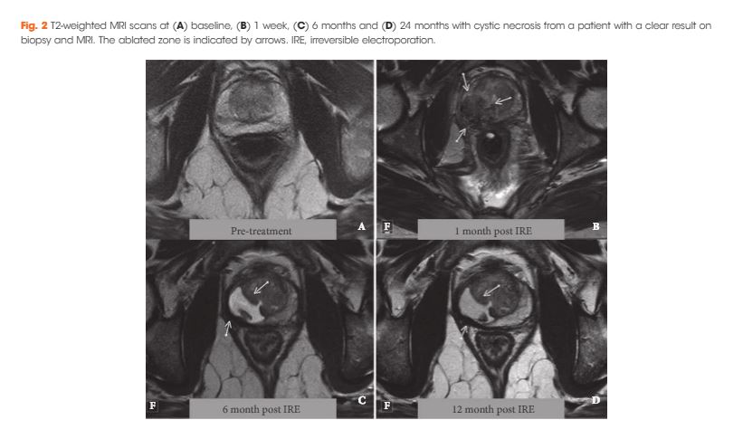
A total of 63 patients met all eligibility criteria and were included in the final analysis. No high‐grade AEs occurred. QoL questionnaire analysis demonstrated no significant change from baseline in physical (P = 0.81), mental (P = 0.48), bowel (P = 0.25) or urinary QoL domains (P = 0.41 and P = 0.25), but there was a mild decrease in the sexual QoL domain (median score 66 at baseline vs 54 at 6 months; P < 0.001). Compared with baseline, a decline of 70% in prostate‐specific antigen level (1.8 ng/mL, interquartile range 0.96–4.8 ng/mL) was seen at 6–12 months. A narrow safety margin (P = 0.047) and system errors (P = 0.010) were identified as potential early risk factors for in‐field oncological failure. In‐field and whole‐gland oncological control on follow‐up biopsies was 84% (38/45 patients) and 76% (34/45 patients); this increased to 97% (38/39 patients) and 87% (34/39 patients) when patients treated with a narrow safety margin and system errors were excluded.

Conclusion

Our data support the safety and feasibility of focal IRE as a primary treatment for localized PCa with effective short‐term oncological control in carefully selected men.

Read more articles of the week

Editorial: Has tailored, tissue‐selective tumour ablation in men with prostate cancer come of age?

There are two principal challenges that face the growing number of clinical investigators that are evaluating tissue‐preserving therapies in men with prostate cancer.

The first is that every man’s prostate is different. So different and so unique are the personal attributes of a man’s prostate that it would, just like the iris or fingerprint, qualify as a unique identifier. It is just a few practical considerations that prevent it from doing so. This is a challenge that the clinician treating the liver, kidney or brain does not face – as these organs do not exhibit the between patient variability that we see in the prostate.

The second relates to within‐patient (or within‐prostate) differences. The nature of the tissue being treated will depend on which part of the prostate is being treated – peripheral, transition or central zone. Each of these zones will, in turn, be dependent on the age of the prostate, the extent of BPH, exhibit calcification and or cysts, and may or may not be infiltrated by acute and/or chronic inflammation.

These two sets of variability present considerable challenges to investigators that seek to selectively ablate a given zone of tissue, given that the nature of the target volume will be different in every man treated and exist in a context that is specific to the that man.

Add to this challenge the variability in prostate cancer tumour attributes – volume, location, heterogeneity (genetic, radiological, and histological), degree of immune infiltrate, and the extent of microscopic extension, we begin to get the picture 1.

The paper in this issue of the BJUI by van den Bos et al. 2 describes a modern attempt to overcome these challenges and attempt and achieve personalised care to individuals, their prostate glands, and their cancer.

The team used irreversible electroporation (IRE) to create a selective ablation zone around a given tumour volume, embracing a margin of 5–10 mm. This method of ablation has certain attributes that lend itself to the task. It can be applied to any zone of the prostate. It is not limited by the size of the prostate. It can create lesions of variable volume. The treatment is quick and therefore not overly affected by prostate gland swelling. Because the treatment uses an interstitial approach (needle based) the effectors of the treatment move with the prostate during respiration and changes in rectal fullness. These attributes mitigate most of the challenges generated by between‐patient variability.

The authors describe the methods by which they manage tumour‐specific differences. These important but rather technical constraints (to the non‐expert) comprise: tumour‐volume dependent variable needle load; individualised tissue impedance‐based energy adjustment; minimising variability in needle–needle distance; application of a 10‐mm margin; and near term verification of tissue change with post‐treatment MRI.

These conditions seem to have paid off. Although every patient underwent a treatment that was bespoke to both their prostate and their prostate cancer, the results were most promising for this truly personalised sub‐specialty of uro‐oncology.

The treatment was safe. There were no high‐grade adverse events reported in the 63 men included in the analysis. The disease‐specific and generic quality of life was not compromised by the range of interventions administered except in relation to the sexual quality of life domain that was marginally affected – a median score of 66 prior to therapy diminished to 54 when measured again 6‐months after treatment.

The authors managed to get a high proportion of men to undergo verification biopsy after treatment. From this they derived two oncological outcomes. These comprised freedom from clinically significant prostate cancer (high‐volume exclusive Gleason pattern 3 and/or any Gleason pattern 4 or 5) within and on the edge of field on the one hand and out of field on the other.

In patients who were free of any technical failure in relation to the administration of IRE and had a 10‐mm margin incorporated, the results were very promising with most of the patients evaluated free of disease both within (97% [38/39 men]) and out of field (87% [34/39 men]).

Although the numbers of patients reported upon are relatively small, the overall results represent a welcome improvement on previously published phase I clinical trial data using IRE, probably as a result of better patient selection and optimisation of energy delivery 3. The results, however, are reassuringly similar to previous case‐series that used alternative energy sources but were predicated on an anatomical‐based approach to tissue preservation 4. The tumour‐based approach reported upon by van den Bos et al. 2 is much more challenging as it exposes any subtle deficiencies in the base‐line risk‐stratification and imposes exacting constraints on the reliability of the energy source in creating irreversible cell kill where cell kill is intended.

Mark Emberton
Division of Surgery and Interventional Science, UCL, London, UK


Read the full article
References
  • Linch M, Goh G, Hiley C et al. Intratumoural evolutionary landscape of high‐risk prostate cancer: the PROGENY study of genomic and immune parametersAnn Oncol201728: 2472–80


  • van den Bos W, Scheltema MJ, Siriwardana AR et al. Focal irreversible electroporation as primary treatment for localized prostate cancerBJU Int 2018121: 716–24


  • Valerio M, Dickinson L, Ali A et al. Nanoknife electroporation ablation trial: a prospective development study investigating focal irreversible electroporation for localized prostate cancerJ Urol 2017197: 647–54


  • Ahmed HU, Hindley RG, Dickinson L et al. Focal therapy for localised unifocal and multifocal prostate cancer: a prospective development studyLancet Oncol 201213: 622–32


Article of the Week: Multiple Growth Periods of SRMs Predict Unfavourable Pathology

Every Week, the Editor-in-Chief selects an Article of the Week from the current issue of BJUI. The abstract is reproduced below and you can click on the button to read the full article, which is freely available to all readers for at least 30 days from the time of this post.

In addition to the article itself, there is an accompanying editorial written by a prominent member of the urological community. This blog is intended to provoke comment and discussion and we invite you to use the comment tools at the bottom of each post to join the conversation.

Finally, the third post under the Article of the Week heading on the homepage will consist of additional material or media. This week we feature a video discussing the paper.

If you only have time to read one article this week, it should be this one.

Multiple growth periods predict unfavourable pathology in patients with small renal masses

Alex Jang , Hiten D. Patel, Mark Riffon, Michael A. Gorin , Alice SemerjianMichael H. Johnson, Mohamad E. Allaf and Phillip M. Pierorazio


Department of Urology, James Buchanan Brady Urological Institute, Johns Hopkins University School of Medicine, Baltimore, MD, USA


Read the full article

Abstract

Objective

To use the number of positive growth periods as a characterization of the growth of small renal masses in order to determine potential predictors of malignancy.

Patients and Methods

Patients who underwent axial imaging at multiple time points prior to surgical resection for a small renal mass were queried. Patients were categorized based on their pathological tumour grade and stage: favourable (benign, chromophobe and low‐grade pT1–2 renal cell carcinoma [RCC]) vs unfavourable (high‐grade of any stage and low‐grade pT3–4 RCC). A positive growth period was counted each time the difference in greatest tumour diameters between two images was positive. The Cochran–Armitage trend test and Somers’ D association were used to determine if the number of positive growth periods was correlated with unfavourable pathology.

Results

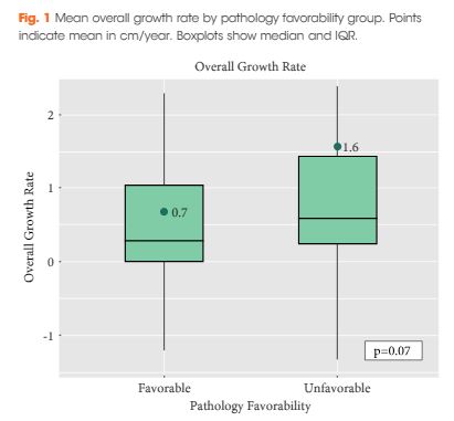
Of the 124 patients, 86 (69.4%) had favourable pathology and 38 (30.6%) had unfavourable pathology. Those who had favourable pathology were younger than those who had unfavourable pathology: median (interquartile range [IQR]) 61.0 (52.2–66.0) vs 68.5 (61.5–77.0); P < 0.001. The overall growth rate was higher in the unfavourable group, but was not statistically significant: mean (sd) 0.7 (1.7) vs 1.6 (2.8) cm/year; P = 0.07. There was a significant trend difference in the number of positive growth periods between favourability groups (P = 0.02). An association between increased number of positive growth periods and unfavourable pathology was observed: 0.15 (95% confidence interval 0.02, 0.29). The ratios of favourable to unfavourable pathology were 1.8, 1.0, 0.66, 0.59 and 0 as the number of positive growth periods increased from 0 to 4, respectively.

Conclusion

While overall growth rate was not predictive of pathology favourability, there was a positive association between the number of positive growth periods and unfavourable pathology. The number of positive growth periods may be a potential parameter for malignant potential in patients undergoing active surveillance for small renal masses.

Read more articles of the week

Editorial: Growth spurts of small renal masses correlate with pathology

A rapid growth rate has long been known to be a harbinger of aggressive tumour pathology and clinical behaviour of small renal masses (SRMs) 1. However, this association has not been confirmed in prospective studies of patients with SRMs on active surveillance (AS) 2. Jewett et al. 2, in a multicentre prospective phase 2 clinical trial of 209 patients with a SRM, found that despite an average growth rate of 0.13 cm/year, pathology did not impact growth. Clinically, the growth rates of SRMs are most commonly variable, rather than the assumed steady slow growing rate that is often reported 3. Due to the discrepancies in the literature, the use of average growth rate for a SRM can be misleading. Finding a clinically effective tool to identify potentially aggressive cancers remains an important (and unmet) need for patients undergoing AS.

Jang et al. 4 provide insight into this need by evaluating the number of growth periods over time as a risk factor for renal masses under surveillance. They retrospectively reviewed renal masses that were initially <4 cm at diagnosis and were followed for a variable time period before undergoing surgical therapy. Two cohorts were grouped into ‘favourable‐’ (benign tumours, chromophobe RCC and low grade, pT1–2 RCC) and ‘unfavourable’‐risk tumours (high‐grade RCC of any stage and low‐grade, pT3–4 RCC), finding no difference in the amount of interval imaging between the two groups. There was a significant difference in the number of positive growth periods between the ‘favourable’ and ‘unfavourable’ pathology groups (P = 0.02) for the entire cohort (all pT stages) and a similar finding when examining only pT1a tumours (P < 0.05). Additionally, there was a positive association between increased number of positive growth periods and unfavourable pathology (odds ratio 0.15, 95% CI 0.02–0.29).

Active surveillance is considered an acceptable initial option for the management of all patients with SRMs, not just those with limited life expectancy or poor performance, as highlighted in the 2017 AUA guidelines 5. Optimal management of patients on AS would appear to require a good understanding of growth kinetics, pathological details, and risk–benefit analysis. The previously held dogma of using linear growth rate as a guide for the aggressiveness of a tumour has been challenged recently 6 and again put into question by this study 4. The Delayed Intervention and Surveillance for Small Renal Masses (DISSRM) study showed that there was a higher growth rate in patients who had delayed intervention compared with those on AS, but there was no significant association (P = 0.15) 6. However, in the same study, the proportion of patients who had a positive growth rate (>0 cm/year) or a recorded ‘growth spurt’ were significantly more likely to have delayed intervention than AS patients (P < 0.01) 6.

Growth spurts, rather than growth rate over time, as a marker for malignancy, correlates best with clinical experience. Average growth rate can be artificially high with one large growth and multiple stable imaging intervals. One of the important elements of AS is regular interval imaging. The development of protocols that can be evaluated to determine the optimal type, interval, and duration of follow‐up imaging is a clinical need in this space. Whilst the cohort from Jang et al. 4 did not have regular intervals for imaging, there were an equal number of studies performed on patients with ‘favourable’ and ‘unfavourable’ pathology. Without clear protocols, using growth spurts rather than growth rate, also provides a simplified message for patients and will aid in counselling.

The identification of growth spurts as a risk factor for potentially aggressive features of SRMs under surveillance provides a useful tool when determining the need for intervention of a SRM. The uniform reporting of pathological data for patients under study is a strength, but several weaknesses limit the generalisability of the findings including the retrospective study design and inconsistent manner in which AS was performed. The clinical impact of growth spurts in the AS population warrants further investigation before definitive conclusions can be drawn.

Conrad M. Tobert* and Brian R. Lane
*Department of Urology, University of Iowa, Iowa City, IAUSA, Division of Urology, Spectrum Health Medi cal GroupGrand Rapids, MI, USA and Michigan State University College of Human Medicine, Grand Rapids, MI, USA


Read the full article
References
  • Lee SW, Sung HH, Jeon HG et al. Size and volumetric growth kinetics of renal masses in patients with renal cell carcinomaUrology 201690: 119–24


  • Chawla SN, Crispen PL, Hanlon AL, Greenberg RE, Chen DY, Uzzo RG. The natural history of observed enhancing renal masses: meta‐analysis and review of the world literatureJ Urol 2006175: 425–31


  • Jang A, Patel HD, Riffon M et al. Multiple growth periods predict unfavourable pathology in patients with small renal massesBJU Int 2018121: 732–6


Video: Multiple Growth Periods of SRMs Predict Unfavourable Pathology


Multiple growth periods predict unfavourable pathology in patients with small renal masses


Read the full article

Abstract

Objective

To use the number of positive growth periods as a characterization of the growth of small renal masses in order to determine potential predictors of malignancy.

Patients and Methods

Patients who underwent axial imaging at multiple time points prior to surgical resection for a small renal mass were queried. Patients were categorized based on their pathological tumour grade and stage: favourable (benign, chromophobe and low‐grade pT1–2 renal cell carcinoma [RCC]) vs unfavourable (high‐grade of any stage and low‐grade pT3–4 RCC). A positive growth period was counted each time the difference in greatest tumour diameters between two images was positive. The Cochran–Armitage trend test and Somers’ D association were used to determine if the number of positive growth periods was correlated with unfavourable pathology.

Results

Of the 124 patients, 86 (69.4%) had favourable pathology and 38 (30.6%) had unfavourable pathology. Those who had favourable pathology were younger than those who had unfavourable pathology: median (interquartile range [IQR]) 61.0 (52.2–66.0) vs 68.5 (61.5–77.0); P < 0.001. The overall growth rate was higher in the unfavourable group, but was not statistically significant: mean (sd) 0.7 (1.7) vs 1.6 (2.8) cm/year; P = 0.07. There was a significant trend difference in the number of positive growth periods between favourability groups (P = 0.02). An association between increased number of positive growth periods and unfavourable pathology was observed: 0.15 (95% confidence interval 0.02, 0.29). The ratios of favourable to unfavourable pathology were 1.8, 1.0, 0.66, 0.59 and 0 as the number of positive growth periods increased from 0 to 4, respectively.

Conclusion

While overall growth rate was not predictive of pathology favourability, there was a positive association between the number of positive growth periods and unfavourable pathology. The number of positive growth periods may be a potential parameter for malignant potential in patients undergoing active surveillance for small renal masses.

Read more articles of the week

Article of the Week: More PLND template at RP detects metastases in the common iliac region and in the fossa of Marcille

Every Month, the Editor-in-Chief selects an Article of the Month from the current issue of BJUI. The abstract is reproduced below and you can click on the button to read the full article, which is freely available to all readers for at least 30 days from the time of this post.

In addition to the article itself, there is an accompanying editorial written by a prominent member of the urological community. This blog is intended to provoke comment and discussion and we invite you to use the comment tools at the bottom of each post to join the conversation.

If you only have time to read one article this week, it should be this one.

More extended lymph node dissection template at radical prostatectomy detects metastases in the common iliac region and in the fossa of Marcille


Lydia Maderthaner, Marc A. Furrer, Urs E. Studer, Fiona C. BurkhardGeorge N. Thalmann and Daniel P. Nguyen


Department of Urology, Inselspital, Bern University Hospital, University of Bern, Bern, Switzerland
Read the full article


Abstract

Objectives

To assess the effect of adding lymph nodes (LNs) located along the common iliac vessels and in the fossa of Marcille to the extended pelvic LN dissection (PLND) template at radical prostatectomy (RP).

Patients and Methods

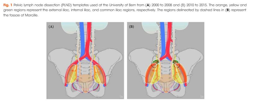
A total of 485 patients underwent RP and PLND at a referral centre between 2000 and 2008 (historical cohort: classic extended PLND template) and a total of 268 patients between 2010 and 2015 (contemporary cohort: extended PLND template including LNs located along the common iliac vessels and in the fossa of Marcille). Descriptive analyses were used to compare baseline, pathological, complication and functional data between the two cohorts. A logistic regression model was used to assess the template’s effect on the probability of detecting LN metastases.

Results

Of 80 patients in the historical cohort with pN+ disease, the sole location of metastasis was the external iliac/obturator fossa in 23 (29%), and the internal iliac in 18 (23%), while 39 patients (49%) had metastases in both locations. Of 72 patients in the contemporary cohort with pN+ disease, the sole location of metastasis was the external iliac/obturator fossa in 17 patients (24%), the internal iliac in 24 patients (33%), and the common iliac in one patient (1%), while 30 patients (42%) had metastases in >1 location (including fossa of Marcille in five patients). Among all 46 patients in the contemporary cohort with ≤2 metastases, three had one or both metastases in the common iliac region or the fossa of Marcille. The adjusted probability of detecting LN metastases was higher, but not significantly so, in the contemporary cohort. There were no differences between the two cohorts in complication rates and functional outcomes.

Conclusion

A more extended template detects LN metastases in the common iliac region and the fossa of Marcille and is not associated with a higher risk of complications; however, the overall probability of detecting LN metastases was not significantly higher.

Read more articles of the week


Editorial: PLND during RP for PCa: extending the template in the right patients without increasing complications

It took long time and consistent evidence to endorse the staging role of extended pelvic lymph node dissection (PLND) in prostate cancer (PCa). The poor performance of both conventional and functional imaging in identifying preoperative nodal status has contributed to making extended PLND the most accurate nodal staging procedure in PCa 1.

Current available guidelines recommend a standard extended PLND template that includes external, internal iliac and obturator lymph nodes 24; however, where does the need for a more extended template originate? Observational data suggest that a standard extended PLND template intercepts ~75% of all anatomical landing sites 4. Extending the anatomical template by adding nearby nodal stations would further minimize the risk of missing positive lymph nodes; however, it has previously been shown that a more accurate staging (i.e. a more extended template) might come at the price of longer operating time and a higher risk of procedure‐related complications 1.

According to the study by Maderthaner et al5, in the current issue of BJUI, an experienced academic surgical team is able to further extend the PLND template (including common iliac and the fossa of Marcille lymph nodes) without significantly increasing the risk of complications. In their study, 17% and 7% of the included men with pN+ disease had positive common iliac and fossa of Marcille lymph nodes, respectively.

Before celebrating this super‐extended template as safe and effective, however, at least three points need to be considered. First, these results were obtained by a group of skilled surgeons with longstanding experience in anatomical pelvic nodal dissection. It should not be taken for granted that this template in the hands of other surgeons would result in no additional complications, especially during the learning curve.

Second, >80% of men submitted to the super‐extended template did not have positive nodes outside the standard extended template boundaries, indicating possible overtreatment in a substantial proportion of men. Notably, extended PLND in this study was offered apparently without upfront preoperative lymph node invasion risk stratification.

Third, as a consequence, patient selection has a role to play. In other words, is super‐extended PLND appropriate for every patient? The use of available risk stratification tools in everyday clinical practice allows a more accurate decision process; this is the case for the Briganti nomogram concerning the need to perform an extended PLND 6. Could a similar approach be used in the setting of a super‐extended template to identify the best candidates? Recently, Gandaglia et al. 7 analysed data from 471 men with high‐risk PCa treated with radical prostatectomy and a super‐extended PLND including common iliac and pre‐sacral nodes in order to identify those men who really require such a super‐extended PLND. Interestingly, although not specifically designed for this task, the Briganti nomogram was able to provide a patient selection strategy: only 5% of patients with a nomogram‐derived N+ risk of <30% had positive common iliac and pre‐sacral nodes, indicating that the super‐extended PLND template should perhaps be considered exclusively in men with an N+ risk ≥30%.

In conclusion, a critical assessment of super‐extended staging PLND template would be welcome, allowing selection of the proper candidates, and a proper balance between accurate staging and the risk of treatment‐related complications.


Eugenio Vent imiglia, *Alberto Briganti,*† and Francesco Montorsi,*
*University Vita-Salute San Raffaele, Milan, Italy and Division of Experimental Oncology/Unit of Urology, URI, IRCCS Ospedale San Raffaele, Milan, Italy


Read the full article


References


  • Fossati N, Willemse P‐PM, Van den Broeck T et al. The benefits and harms of different extents of lymph node dissection during radical prostatectomy for prostate cancer: a systematic reviewEur Urol 201772: 84–109


  • Santis D, Henry A, Joniau S et al. Prostate Cancer EAU ESTRO SIOG Guidelines on 2017.


  • Mattei A, Fuechsel FG, Bhatta Dhar N et al. The template of the primary lymphatic landing sites of the prostate should be revisited: results of a multimodality mapping studyEur Urol 200853: 118–25


  • Maderthaner L, Furrer MA, Studer UE, Burkhard FC, Thalmann GN, Nguyen DP. More extended lymph node dissection template at radical prostatectomy detects metastases in the common iliac region and in the fossa of MarcilleBJU Int 2018121: 725–31


  • Briganti A, Larcher A, Abdollah F et al. Updated nomogram predicting lymph node invasion in patients with prostate cancer undergoing extended pelvic lymph node dissection: the essential importance of percentage of positive coresEur Urol 201261: 480–7


  • Gandaglia G, Zaffuto E, Fossati N et al. Identifying the candidate for super extended staging pelvic lymph node dissection among patients with high‐risk prostate cancerBJU Int 2018121: 421–7


© 2024 BJU International. All Rights Reserved.