Archive for category: Article of the Week

Article of the Week: TIND implantation in benign prostatic obstruction

Every Week, the Editor-in-Chief selects an Article of the Week from the current issue of BJUI. The abstract is reproduced below and you can click on the button to read the full article, which is freely available to all readers for at least 30 days from the time of this post.

In addition to the article itself, there is an accompanying editorial written by a prominent member of the urological community. This blog is intended to provoke comment and discussion and we invite you to use the comment tools at the bottom of each post to join the conversation.

Finally, the third post under the Article of the Week heading on the homepage will consist of additional material or media. This week we feature a podcast discussing the paper.

If you only have time to read one article this week, it should be this one.

3‐Year follow‐up of temporary implantable nitinol device implantation for the treatment of benign prostatic obstruction


Francesco Porpiglia , Cristian Fiori, Riccardo Bertolo, Andrea GiordanoEnrico Checcucci, Diletta Garrou, Giovanni Cattaneo, Stefano De Luca and Daniele Amparore
Division of Urology, Department of Oncology School of Medicine, San Luigi Hospital, University of Turin, Orbassano (Turin), Italy


Abstract

Objectives

To report 3‐year follow‐up results of the first implantations with a temporary implantable nitinol device (TIND®; Medi‐Tate Ltd., Or Akiva, Israel) for the treatment of lower urinary tract symptoms (LUTS) secondary to benign prostatic hyperplasia (BPH).

Patients and Methods

In all, 32 patients with LUTS were enrolled in this prospective study. The study was approved by the local Ethics Committee. Inclusion criteria were: age >50 years, International Prostate Symptom Score (IPSS) ≥10, peak urinary flow (Qmax) <12 mL/s, and prostate volume <60 mL. The TIND was implanted within the bladder neck and the prostatic urethra under light sedation, and removed 5 days later in an outpatient setting. Demographics, perioperative results, complications (according to Clavien–Dindo classification), functional results, and quality of life (QoL) were evaluated. Follow‐up assessments were made at 3 and 6 weeks, and 3, 6, 12, 24 and 36 months after the implantation. The Student’s t‐test, one‐way analysis of variance and Kruskal–Wallis tests were used for statistical analyses.

Results

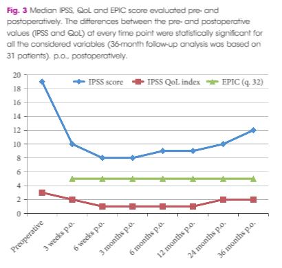
At baseline, the mean (standard deviation, sd) patient age was 69.4 (8.2) years, prostate volume was 29.5 (7.4) mL, and Qmax was 7.6 (2.2) mL/s. The median (interquartile range, IQR) IPSS was 19 (14–23) and the QoL score was 3 (3–4). All the implantations were successful, with a mean total operative time of 5.8 min. No intraoperative complications were recorded. The change from baseline in IPSS, QoL score and Qmax was significant at every follow‐up time point. After 36 months of follow‐up, a 41% rise in Qmax was achieved (mean 10.1 mL/s), the median (IQR) IPSS was 12 (6–24) and the IPSS QoL was 2 (1–4). Four early complications (12.5%) were recorded, including one case of urinary retention (3.1%), one case of transient incontinence due to device displacement (3.1%), and two cases of infection (6.2%). No further complications were recorded during the 36‐month follow‐up.

Conclusions

The extended follow‐up period corroborated our previous findings and suggests that TIND implantation is safe, effective and well‐tolerated, for at least 36 months after treatment.

Editorial: The trials of doing trials: will the prostatic stent rise from the dead?

A further re‐engineering of the prostatic stent (temporary implantable nitinol device [TIND®, Medi‐Tate Ltd., Israel]) is reported by Porpiglia et al. 1 in this edition of the BJUI. Enthusiasm for novel ‘minimally invasive’ treatments for BPH/LUTs has surged recently, with new options supported by strong data becoming available 23. That interest reflects the continuing desire of our patients to seek alternatives to the standard drug or surgical therapies.

The success of UroLift® (Neotract, Pleasanton, CA, USA) in satisfying the rigour of the regulatory bodies (especially the Food and Drug Administration [FDA] in the USA and National Institute for Health and Care Excellence [NICE] in the UK) and the recent merger/acquisition of Neotract by Teleflex Inc. for a sum potentially exceeding 1 billion dollars, indicates the rewards available for a novel treatment with convincing data. Not surprisingly, this has stimulated innovators, so there is a veritable tsunami of new treatments approaching us. The rewards are certainly encouraging, but it is worth remembering that the difficulties and cost of reaching market and the point of sale are now enormous.

The TIND stent is implanted endoscopically and then gradually distends to cause three linear pressure necrosis effects within the prostatic urethra and bladder neck tissues, before it is removed at approximately 5–7 days by a second, minor and office‐based procedure.

However, this report is simply a repeat of the initial case series from 2015 4, just with follow‐up to 36 months. An initial report is legitimate in order to show primarily safety and with effectiveness as a secondary outcome measure. I see limited value in a follow‐up report of a device that has already been superseded by a second version, which is currently being assessed in the more rigorous studies that we, our patients, and regulators require.

Even so, there are some important messages and some inconsistencies. Porpiglia et al. 1 are, firstly, to be commended for successfully following their patients out to 3 years with essentially complete data sets. That is remarkable. Although the TIND seems safe, there was one serious complication (a prostatic abscess associated with arrhythmia, sepsis and hyperglycaemia, which required 10 days of inpatient care). About 10% of men restarted medical therapy, but are not ‘failures’, as failures are defined only as those requiring surgical treatment. The prostate size in this small cohort is also surprisingly small compared to the initial studies of other novel devices (29.5 vs 50 mL for UroLift in the initial report) 5. It is also surprising that even though all were on α‐blockers and a substantial proportion were taking 5α‐reductase inhibitors that there is no sexual or especially ejaculatory dysfunction in any of the 19 sexually active men before or after TIND implantation. They appear to have surprisingly resilient ejaculatory function and one wonders why or how? The population studied may therefore not be representative of men we meet in our clinics. Nevertheless, the safety and effectiveness of the original TIND stent do seem reasonable and many of us will await the definitive ‘pivotal’ randomised controlled studies of the successor device with great interest.

For the TIND to find a place in our range of treatments, we urologists, our patients, and regulators of healthcare, now need to see proper randomised controlled trials, preferably incorporating a sham arm and eventually comparisons with TURP. Ideally, the TIND should also be compared to UroLift and to steam injection (Rezūm®; NxThera, Inc., Maple Grove, MN, USA). But who will pay for these studies, even if the regulators insist on them? Funding the required studies is now a major financial challenge to any new device in development. Those studies cost more than we think. A survey of >200 MedTech companies in 2010 suggested device development costs up to 100 million dollars (the majority spent on FDA‐dependent or related activities) but for UroLift total development spend was nearly 240 million. Unfortunately, even innovative and potentially useful devices will struggle to overcome the double jeopardy of regulatory hurdles and obtaining the finance required to clear them. I wish the developers of TIND well and hope there is adequate funding. Today, developers need deep pockets to perform the proper studies correctly.


Tom McNicholas
Pinehill Hospital, Hitchin, Herts, UK

References


  • Porpiglia F, Fiori C, Bertolo R et al. 3‐Year follow‐up of temporary implantable nitinol device implantation for the treatment of benign prostatic obstructionBJU Int2018122: 106–12


  • Roehrborn CG, Barkin J, Gange SN et al. Five year results of the prospective randomized controlled prostatic urethral L.I.F.T. studyCan J Urol 201724: 8802–13


  • McVary KT, Roehrborn CG. Three‐year outcomes of the prospective, randomized controlled Rezūm System study: convective radiofrequency thermal therapy for treatment of lower urinary tract symptoms due to benign prostatic hyperplasiaUrology 2018111: 1–9


  • Porpiglia F, Fiori C, Bertolo R, Garrou D, Cattaneo G, Amparore D. Temporary implantable nitinol device (TIND): a novel, minimally invasive treatment for relief of lower urinary tract symptoms (LUTS) related to benign prostatic hyperplasia (BPH): feasibility, safety and functional results at 1 year of follow‐upBJU Int 2015116: 278–87


  • Woo HH, Chin PT, McNicholas TA et al. Safety and feasibility of the prostatic urethral lift: a novel, minimally invasive treatment for lower urinary tract symptoms (LUTS) secondary to benign prostatic hyperplasia (BPH)BJU Int 2011108: 82–8


Residents’ Podcast: Follow‐up of TIND implantation for the treatment of benign prostatic obstruction


Maria Uloko and Guilia Lane are Urology Residents at the University of Minnesota Hospital.

In this podcast they discuss the BJUI Article of the Week, 3‐Year follow‐up of temporary implantable nitinol device implantation for the treatment of benign prostatic obstruction
Special Guest: Mike Borofsky


3‐Year follow‐up of temporary implantable nitinol device implantation for the treatment of benign prostatic obstruction


Francesco Porpiglia , Cristian Fiori, Riccardo Bertolo, Andrea GiordanoEnrico Checcucci, Diletta Garrou, Giovanni Cattaneo, Stefano De Luca and Daniele Amparore


Division of Urology, Department of Oncology School of Medicine, San Luigi Hospital, University of Turin, Orbassano (Turin), Italy


Abstract

Objectives

To report 3‐year follow‐up results of the first implantations with a temporary implantable nitinol device (TIND®; Medi‐Tate Ltd., Or Akiva, Israel) for the treatment of lower urinary tract symptoms (LUTS) secondary to benign prostatic hyperplasia (BPH).

Patients and Methods

In all, 32 patients with LUTS were enrolled in this prospective study. The study was approved by the local Ethics Committee. Inclusion criteria were: age >50 years, International Prostate Symptom Score (IPSS) ≥10, peak urinary flow (Qmax) <12 mL/s, and prostate volume <60 mL. The TIND was implanted within the bladder neck and the prostatic urethra under light sedation, and removed 5 days later in an outpatient setting. Demographics, perioperative results, complications (according to Clavien–Dindo classification), functional results, and quality of life (QoL) were evaluated. Follow‐up assessments were made at 3 and 6 weeks, and 3, 6, 12, 24 and 36 months after the implantation. The Student’s t‐test, one‐way analysis of variance and Kruskal–Wallis tests were used for statistical analyses.

Results

At baseline, the mean (standard deviation, sd) patient age was 69.4 (8.2) years, prostate volume was 29.5 (7.4) mL, and Qmax was 7.6 (2.2) mL/s. The median (interquartile range, IQR) IPSS was 19 (14–23) and the QoL score was 3 (3–4). All the implantations were successful, with a mean total operative time of 5.8 min. No intraoperative complications were recorded. The change from baseline in IPSS, QoL score and Qmax was significant at every follow‐up time point. After 36 months of follow‐up, a 41% rise in Qmax was achieved (mean 10.1 mL/s), the median (IQR) IPSS was 12 (6–24) and the IPSS QoL was 2 (1–4). Four early complications (12.5%) were recorded, including one case of urinary retention (3.1%), one case of transient incontinence due to device displacement (3.1%), and two cases of infection (6.2%). No further complications were recorded during the 36‐month follow‐up.

Conclusions

The extended follow‐up period corroborated our previous findings and suggests that TIND implantation is safe, effective and well‐tolerated, for at least 36 months after treatment.


BJUI Podcasts now available on iTunes, subscribe here https://itunes.apple.com/gb/podcast/bju-international/id1309570262


Article of the Month: MRI supported transperineal prostate biopsy

Every Month, the Editor-in-Chief selects an Article of the Month from the current issue of BJUI. The abstract is reproduced below and you can click on the button to read the full article, which is freely available to all readers for at least 30 days from the time of this post.

In addition to the article itself, there is an accompanying editorial written by a prominent member of the urological community. This blog is intended to provoke comment and discussion and we invite you to use the comment tools at the bottom of each post to join the conversation.

If you only have time to read one article this week, it should be this one.

Multicentre evaluation of magnetic resonance imaging supported transperineal prostate biopsy in biopsy‐naïve men with suspicion of prostate cancer


Nienke L. Hansen*1, Tristan Barrett*, Claudia Kesch, Lana Pepdjonovic§, David Bonekamp, Richard OSullivan**, Florian Distler, Anne Warren*††, Christina Samel‡‡Boris Hadaschik2, Jeremy Grummet§ and Christof Kastner*§§
*CamPARI Clinic, Department of Radiology, Addenbrookes Hospital and University of Cambridge, Cambridge, UK, Department of Urology, University Hospital Heidelberg, Heidelberg, Germany, §Australian Urology Associates and Department of Surgery, Central Clinical School, Monash University, Melbourne, Vic., Australia, Department of Radiology, German Cancer Research Center (DKFZ), Heidelberg, Germany, **Healthcare Imaging and Monash University, Melbourne, Vic., Australia, ††Department of Pathology, Addenbrookes Hospital and University of Cambridge, Cambridge, UK, ‡‡Institute of Medical Statistics, Informatics and Epidemiology, University Hospital Cologne, Cologne, Germany, and §§Department of Urology, Addenbrookes Hospital and University of Cambridge, Cambridge, UK 


Current addresses: 1Department of Diagnostic and Interventional Radiology University Hospital Cologne Cologne Germany, 2Department of Urology University Hospital Essen Essen Germany. 


B.H., J.G., and C.K. contributed equally to this work.


Read the full article

Abstract

Objectives

To analyse the detection rates of primary magnetic resonance imaging (MRI)‐fusion transperineal prostate biopsy using combined targeted and systematic core distribution in three tertiary referral centres.

Patients and Methods

In this multicentre, prospective outcome study, 807 consecutive biopsy‐naïve patients underwent MRI‐guided transperineal prostate biopsy, as the first diagnostic intervention, between 10/2012 and 05/2016. MRI was reported following the Prostate Imaging‐Reporting and Data System (PI‐RADS) criteria. In all, 236 patients had 18–24 systematic transperineal biopsies only, and 571 patients underwent additional targeted biopsies either by MRI‐fusion or cognitive targeting if PI‐RADS ≥3 lesions were present. Detection rates for any and Gleason score 7–10 cancer in targeted and overall biopsy were calculated and predictive values were calculated for different PI‐RADS and PSA density (PSAD) groups.

Results

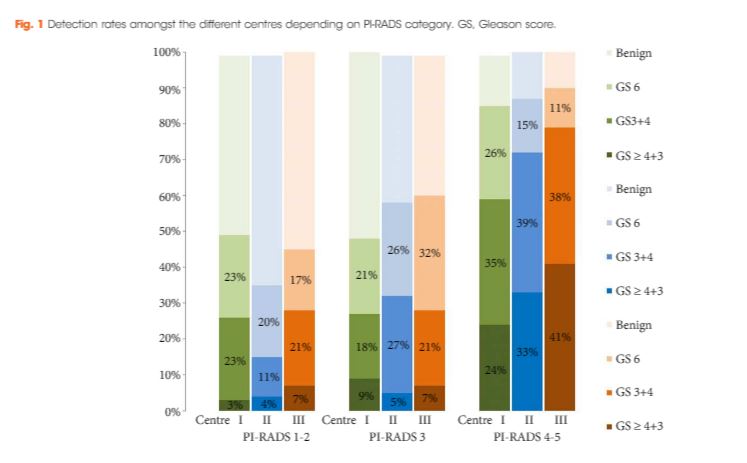
Cancer was detected in 68% of the patients (546/807) and Gleason score 7–10 cancer in 49% (392/807). The negative predictive value of 236 PI‐RADS 1–2 MRI in combination with PSAD of <0.1 ng/mL/mL for Gleason score 7–10 was 0.91 (95% confidence interval ± 0.07, 8% of study population). In 418 patients with PI‐RADS 4–5 lesions using targeted plus systematic biopsies, the cancer detection rate of Gleason score 7–10 was significantly higher at 71% vs 59% and 61% with either approach alone (P < 0.001). For 153 PI‐RADS 3 lesions, the detection rate was 31% with no significant difference to systematic biopsies with 27% (P > 0.05). Limitations include variability of multiparametric MRI (mpMRI) reading and Gleason grading.

Conclusion

MRI‐based transperineal biopsy performed at high‐volume tertiary care centres with a significant experience of prostate mpMRI and image‐guided targeted biopsies yielded high detection rates of Gleason score 7–10 cancer. Prostate biopsies may not be needed for men with low PSAD and an unsuspicious MRI. In patients with high probability lesions, combined targeted and systematic biopsies are recommended.

Editorial: Systematic transperineal and MRI‐targeted biopsies: the resolution of uncertainty

The paper published in this issue of the BJUI titled ‘Multicentre evaluation of magnetic resonance imaging supported transperineal biopsy in biopsy‐naïve men with suspicion of prostate cancer’ is timely and helps to resolve some of the uncertainty inherent within the diagnostic pathway 1.

The publication of the PROstate MRI Imaging Study (PROMIS) study, although demonstrating that 25% of patients might avoid prostate biopsy with a normal MRI (Prostate Imaging Reporting and Data System [PI‐RADS] 1–2) and that MRI could identify 90% of patients with high‐risk disease (PI‐RADS 5), did not resolve the issue of what to do with equivocal PI‐RADS 3 scans, uncertainty remained 2. The recent publication of the PRECISION trial (Prostate Evaluation for Clinically Important Disease: Sampling Using Image Guidance or Not?) has only contributed to the uncertainty of systematic TRUS biopsy and has shown that targeted biopsies resolve the issue for <50% of the patients overall and only 12% of those with PI‐RADS 3 lesions had a diagnosis of cancer on targeted biopsy only 3. The study has shown that in the face of an identifiable lesion a MRI‐targeted biopsy is non‐inferior to a blind systematic TRUS biopsy, which was positive in only 28% and implies that a systematic biopsy may be unnecessary, so where does that leave us? The uncertainty within MRI remains at the PI‐RADS 3 level, and particularly with a TRUS biopsy that is not a systematic biopsy of the peripheral zone. The authors of the paper highlighted in this issue of the BJUI 1 help to resolve the issue because they describe a more systematic biopsy.

The transperineal (TP) biopsy approach for systematic and targeted biopsy they use is that which was adopted by the Ginsburg Study Group on Enhanced Prostate Diagnostics 4. It is a systematic biopsy that preferentially targets the peripheral zone in a sectoral fashion. It avoids the oversampling inherent in template‐mapping biopsy and the under‐sampling of the non‐systematic transrectal biopsy. Their paper evaluates the combination of an MRI‐targeted biopsy with a systematic TP biopsy. It confirms, as suggested by the PROMIS study, that patients with PI‐RADS 1 or 2 prostates on MRI with a low PSA density <0.1 ng/mL/mL could safely avoid biopsy, based upon a negative predictive value of 0.91 on systematic biopsy. However, in 418 patients with PI‐RADS 4–5 lesions, it was the combination of a targeted and systematic TP biopsy that achieved an overall cancer detection rate of 71%, but that MRI‐targeted biopsies alone had a detection rate of 59% vs 61% for systematic TP biopsies. In the PI‐RAD 3 equivocal group the combined biopsy identified 30% with Gleason score 7–10, whereas targeted biopsy only was positive in 21% vs 27% with systematic biopsies.

The message is clear.

An appropriate systematic biopsy targeted to the peripheral zone remains an essential component of prostate diagnosis even in the MRI era, as indeed it did before MRI was available. In the pre‐MRI days, about one‐third of patients that had negative TRUS biopsies had cancer on TP biopsies and a third of those thought suitable for AS on TRUS biopsy had more significant disease. I suspect in the modern era that figure remains unchanged for those with PI‐RADS 1, 2 or 3, particularly with a PSA density >0.15 ng/mL/mL. As urologists we have always been criticised for over diagnosing and over treating prostate cancer but I suspect that the more heinous crime is that of under treatment of significant disease, it is the very reason why I started doing TP biopsies, to resolve uncertainty. I consider that MRI, for all its benefits in the diagnostic algorithm, cannot yet resolve that uncertainty.

Probably the only patients that merit a target‐only biopsy are those with the high‐PSA, large‐volume disease, easily visible on MRI and usually palpable. Prostate biopsy can be avoided or at least deferred in the PI‐RADS 1–2 group with low PSA density; the rest should be offered a systematic biopsy along with a targeted biopsy. This may be less important in those proceeding to whole gland treatment or surgical extirpation but remains essential in those considering active surveillance, brachytherapy, or any one of the myriad of unproven focal treatments becoming available. The authors should be congratulated for bringing some certainty to uncertainty.

Rick Popert
Urology Centre, Guys Hospital, London, UK


Read the full article
References
  • Hansen NL, Barrett T, Kesch C et al. Multicentre evaluation of magnetic resonance imaging supported transperineal prostate biopsy in biopsy‐naïve men with suspicion of prostate cancerBJU Int 2018122: 40–9


  • Ahmed HU, El‐Shater Bosaily A, Brown LC et al. Diagnostic accuracy of multi‐parametric MRI and TRUS biopsy in prostate cancer (PROMIS): a paired validating confirmatory studyLancet 2017389: 815–22


  • Kuru TH, Wadhwa K, Chang RT et al. Definitions of terms, processes and a minimum dataset for transperineal prostate biopsies: a standardization approach of the Ginsburg Study Group for Enhanced Prostate DiagnosticsBJU Int 2013112: 568–77


Article of the Week: β3‐adrenoceptor agonists inhibit carbachol‐evoked Ca2+ oscillations in murine detrusor myocytes

Every Week, the Editor-in-Chief selects an Article of the Week from the current issue of BJUI. The abstract is reproduced below and you can click on the button to read the full article, which is freely available to all readers for at least 30 days from the time of this post.

In addition to the article itself, there is an accompanying editorial written by a prominent member of the urological community. This blog is intended to provoke comment and discussion and we invite you to use the comment tools at the bottom of each post to join the conversation.

Finally, the third post under the Article of the Week heading on the homepage will consist of additional material or media. This week we feature a video discussing the paper.

If you only have time to read one article this week, it should be this one.

β3‐adrenoceptor agonists inhibit carbachol‐evoked Ca2+ oscillations in murine detrusor myocytes


Caoimhin S. Grifn, Eamonn Bradley, Mark A. Hollywood, Noel G. McHale, Keith D. Thornbury and Gerard P. Sergeant
Smooth Muscle Research Centre, Dundalk Institute of Technology, Dundalk, Co Louth, Ireland


Read the full article

Abstract

Objective

To test if carbachol (CCh)‐evoked Ca2+ oscillations in freshly isolated murine detrusor myocytes are affected by β3‐adrenoceptor (β‐AR) modulators.

Materials and Methods

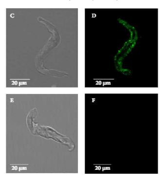
Isometric tension recordings were made from strips of murine detrusor, and intracellular Ca2+ measurements were made from isolated detrusor myocytes using confocal microscopy. Transcriptional expression of β‐AR sub‐types in detrusor strips and isolated detrusor myocytes was assessed using reverse transcriptase‐polymerase chain reaction (RT‐PCR) and real‐time quantitative PCR (qPCR). Immunocytochemistry experiments, using a β3‐AR selective antibody, were performed to confirm that β3‐ARs were present on detrusor myocytes.

Results

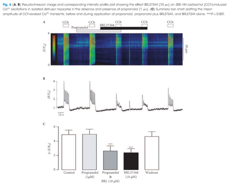
The RT‐PCR and qPCR experiments showed that β1‐, β2‐ and β3‐AR were expressed in murine detrusor, but that β3‐ARs were the most abundant sub‐type. The selective β3‐AR agonist BRL37344 reduced the amplitude of CCh‐induced contractions of detrusor smooth muscle. These responses were unaffected by addition of the BK channel blocker iberiotoxin. BRL37344 also reduced the amplitude of CCh‐induced Ca2+ oscillations in freshly isolated murine detrusor myocytes. This effect was mimicked by CL316,243, another β3‐AR agonist, and inhibited by the β3‐AR antagonist L748,337, but not by propranolol, an antagonist of β1‐ and β2‐ARs. BRL37344 did not affect caffeine‐evoked Ca2+ transients or L‐type Ca2+ current in isolated detrusor myocytes.

Conclusion

Inhibition of cholinergic‐mediated contractions of the detrusor by β3‐AR agonists was associated with a reduction in Ca2+ oscillations in detrusor myocytes.


Editorial: β3‐adrenoceptor agonists inhibit calcium oscillations in bladder detrusor

The β3‐adrenoceptor (β3‐AR) agonist mirabegron has now been used clinically for the treatment for overactive bladder (OAB) for 5 years, but it is still not clear either exactly where the β3‐ARs are located within the bladder wall or entirely how they work. β3‐AR agonists can reduce detrusor contractions after cholinergic stimulation, but the concentrations of mirabegron required for this effect suggest that this is unlikely to be the main therapeutic route, which may instead involve an indirect pathway or a combination of pathways.

In this issue of BJUI, Griffin et al. 1 show that β3‐AR agonists can reduce the rise in calcium and calcium waves observed after muscarinic receptor activation with carbachol in murine detrusor smooth muscle. They convincingly demonstrate that this effect is mediated by β3‐AR rather than by β1‐AR or β2‐AR and is not mediated by an effect on L‐type calcium channels or depletion of calcium stores; however, although they use BRL37344 and CL316,243, which have a higher potency in rodents than mirabegron, the concentrations of these agents needed are in the micromolar range, significantly higher than the values of ~100 nM measured in plasma following therapeutic dosing with mirabegron 2. It will be interesting to see when these experiments are repeated with, these effects on calcium are also seen in the therapeutic dosing range.

Currently, the only effects of mirabegron reported in the therapeutic dosing range are a reduction in detrusor contractions when elicited by field stimulation and a reduction in acetylcholine release from detrusor strips 34. The similarly of the concentrations required for these two effects have led to the suggestion that the reduction in contractions may be mediated by the reduction in acetylcholine release; however, there is still disagreement as to whether β3‐ARs are located in the cholinergic nerve terminals or not, as different groups have obtained different patterns of β3‐adrenoreceptor staining, even when using the same antibodies 46. It is therefore still unclear if this involves a direct inhibition of acetylcholine release or not, although a predominantly neural source of acetylcholine is indicated by sensitivity to the sodium channel blocker, tetrodotoxin 3.

There is evidence that the reduction may be at least partially mediated via adenosine, such that increased adenosine formed post‐junctionally from breakdown of cAMP following β3‐AR stimulation, acts as a retrograde transmitter to inhibit acetylcholine release by acting at pre‐junctional A1 receptors 4. In support of this, Silva et al. 4 showed that A1 antagonists can reverse the increase in voiding seen with isoprenaline in an anaesthetized rat cystoscopy model.

The reduction in calcium rise reported by Griffin et al. 1 also suggests a post‐junctional site of action and the likely possibility that multiple pathways are activated following the binding of the β3‐AR agonists to post‐junctional receptors. A multiple pronged mechanism may be one of the reasons why mirabegron is successful at reducing the symptoms of OAB whilst sparing normal voiding.

The potency of β3‐AR agonists shows differences depending on the method of stimulation of the muscle, with higher potency seen for inhibition of contraction induced by field stimulation than carbachol stimulation, for example 134. This may reflect differences in the combinations of pathways and receptors activated, with carbachol stimulating only the muscarinic receptors themselves, but field stimulation activating more widely. It would therefore be interesting to see whether the potency of mirabegron to affect calcium release in human tissue is higher when field stimulation is used rather than carbachol, and hence help to evaluate if this pathway is involved in the mechanism of action of mirabegron in the clinic.

L. M. McLatchie
Department of Urology, Guys Hospital, London, UK


Read the full article
References
  • Griffin CS, Bradley E, Hollywood MA, McHale NG, Thornbury KD, Sergeant GP. B3‐adrenoceptor agonists inhibit carbachol‐evoked Ca2+ oscillations in murine detrussor myocytesBJU Int 2018121: 959–70


  • Krauwinkel W, van Dijk J, Schaddelee M et al. Pharmacokinetic properties of mirabegron, a beta(3)‐adrenoceptor agonist: results from two phase I, randomized, multiple‐dose studies in healthy young and elderly men and womenClin Ther 201234: 2144–60


  • D’Agostino G, Condino AM, Calvi P. Involvement of beta(3)‐adrenoceptors in the inhibitory control of cholinergic activity in human bladder: direct evidence by H‐3‐acetylcholine release experiments in the isolated detrusorEur J Pharmacol 2015758: 115–22


  • Silva I, Costa AF, Moreira S et al. Inhibition of cholinergic neurotransmission by beta(3)‐adrenoceptors depends on adenosine release and A(1)‐receptor activation in human and rat urinaryAm J Physiol‐Renal Physiol 2017313: F388–403


  • Coelho A, Antunes‐Lopes T, Gillespie J, Cruz F. Beta‐3 adrenergic receptor is expressed in acetylcholine‐containing nerve fibers of the human urinary bladder: an immunohistochemical studyNeurourol Urodyn 201736: 1972–80


  • Limberg BJ, Andersson KE, Kullmann FA, Burmer G, de Groat WC, Rosenbaum JS. beta‐Adrenergic receptor subtype expression in myocyte and non‐myocyte cells in human female bladderCell Tissue Res 2010342: 295–306


Video: β3‐adrenoceptor agonists inhibit carbachol‐evoked Ca2+ oscillations in murine detrusor myocytes

β3‐adrenoceptor agonists inhibit carbachol‐evoked Ca2+ oscillations in murine detrusor myocytes


Read the full article

Abstract

Objective

To test if carbachol (CCh)‐evoked Ca2+ oscillations in freshly isolated murine detrusor myocytes are affected by β3‐adrenoceptor (β‐AR) modulators.

Materials and Methods

Isometric tension recordings were made from strips of murine detrusor, and intracellular Ca2+ measurements were made from isolated detrusor myocytes using confocal microscopy. Transcriptional expression of β‐AR sub‐types in detrusor strips and isolated detrusor myocytes was assessed using reverse transcriptase‐polymerase chain reaction (RT‐PCR) and real‐time quantitative PCR (qPCR). Immunocytochemistry experiments, using a β3‐AR selective antibody, were performed to confirm that β3‐ARs were present on detrusor myocytes.

Results

The RT‐PCR and qPCR experiments showed that β1‐, β2‐ and β3‐AR were expressed in murine detrusor, but that β3‐ARs were the most abundant sub‐type. The selective β3‐AR agonist BRL37344 reduced the amplitude of CCh‐induced contractions of detrusor smooth muscle. These responses were unaffected by addition of the BK channel blocker iberiotoxin. BRL37344 also reduced the amplitude of CCh‐induced Ca2+ oscillations in freshly isolated murine detrusor myocytes. This effect was mimicked by CL316,243, another β3‐AR agonist, and inhibited by the β3‐AR antagonist L748,337, but not by propranolol, an antagonist of β1‐ and β2‐ARs. BRL37344 did not affect caffeine‐evoked Ca2+ transients or L‐type Ca2+ current in isolated detrusor myocytes.

Conclusion

Inhibition of cholinergic‐mediated contractions of the detrusor by β3‐AR agonists was associated with a reduction in Ca2+ oscillations in detrusor myocytes.

View more videos


Article of the Week: NICE Guidance ‐ Complicated UTIs: ceftolozane/tazobactam

Every Week, the Editor-in-Chief selects an Article of the Week from the current issue of BJUI. The abstract is reproduced below and you can click on the button to read the full article, which is freely available to all readers for at least 30 days from the time of this post.

If you only have time to read one article this week, it should be this one.

NICE Guidance ‐ Complicated urinary tract infections: ceftolozane/tazobactam

Read the full article

Introduction and Current Guidance

Urinary tract infection is a non‐specific term that refers to infection anywhere in the urinary tract, from the urethra to the bladder and the ureters to the kidneys. According to the European Association of Urology Guidelines on urological infections (https://uroweb.org/guideline/urological-infections/?type=archive), complicated urinary tract infections are associated with certain conditions, such as structural or functional abnormalities of the genitourinary tract, or the presence of underlying disease in the lower or upper urinary tract, which increases the risk of persistent or relapsing infection. Factors associated with complicated urinary tract infections include:

  • indwelling urinary catheters
  • urinary obstruction (such as stones or tumour)
  • anatomical abnormalities
  • peri‐ and post‐operative urinary tract infection, including renal transplantation.

Pyelonephritis is infection of the upper urinary tract and can occur in 1 or both kidneys. Acute pyelonephritis may be caused by bacteria ascending from the lower urinary tract or spreading via the bloodstream to the kidney. It is considered to be uncomplicated if it is caused by a typical pathogen in an immunocompetent person with a normal urinary tract anatomy and kidney function. As for urinary tract infections generally, acute pyelonephritis is considered to be complicated in people with increased susceptibility, for example: children or older people; people with functional or structural abnormalities of the genitourinary tract or people who are immunocompromised, such that the infection is more likely to be severe. However, most episodes are uncomplicated and are cured with no residual renal damage. Complicated urinary tract infections are a frequent cause of hospital admissions and a common healthcare associated complication. The pathogens most commonly encountered in complicated urinary tract infections are the gram‐negative bacteria Escherichia coli, other common Enterobacteriaceae (for example, Proteus spp., Klebsiella spp. or Citrobacter spp.) and Pseudomonas spp. Successful treatment has become increasingly more challenging because the majority of pathogens responsible for healthcare associated complicated urinary tract infections, including catheter‐related infections, are now commonly resistant to multiple antimicrobial agents (European public assessment report [https://www.ema.europa.eu/ema/index.jsp?curl=pages/medicines/human/medicines/003772/human_med_001917.jsp&mid=WC0b01ac058001d124]).

The English surveillance programme for antimicrobial utilisation and resistance (ESPAUR) report (2015) (https://www.gov.uk/government/publications/english-surveillance-programme-antimicrobial-utilisation-and-resistance-espaur-report) found that, overall, antibiotic resistant infections continue to increase. Notably, the rate of E. coli and Klebsiella pneumoniae bloodstream infections increased by 15.6% and 20.8% respectively from 2010 to 2014. Urinary tract infections are most commonly caused by E. coli (recorded in more than half of all the mandatory surveillance reports for E. coli bacteraemia when foci of infection are reported). The data indicate that 97% of E. coli isolates for urinary tract infection from GP practices, other community sources (such as care homes and outpatient clinics) and acute trusts were susceptible to nitrofurantoin. Resistance to trimethoprim was seen in over a third (35–37%) of isolates and resistance to amoxicillin was seen in over 50% of isolates, in all 3 settings. It is unclear if these data include cases of complicated urinary tract infections. Also, specialists involved in the production of this evidence summary noted that the results could be prone to bias because samples may have been be submitted from a population with a higher likelihood of antimicrobial resistance caused by, for example, failed treatments, recurrent infection or repeated courses of antibiotics.

Risk factors for resistance should be taken into consideration before prescribing antibiotics for urinary tract infection according to Public Health England guidance for primary care on managing common infections (https://www.gov.uk/government/publications/managing-common-infections-guidance-for-primary-care).

As well as some other groups, Public Health England advises performing culture and sensitivity testing in people with a higher risk of recurrent urinary tract infection (such as those aged over 65 years or with urinary catheters), and people with abnormalities of the genitourinary tract or suspected pyelonephritis.

The management of suspected community‐acquired bacterial urinary tract infection in adults aged 16 years and over is covered in the NICE quality standard on urinary tract infection in adults (https://www.nice.org.uk/guidance/qs90). This includes women who are pregnant, people with indwelling catheters and people with other diseases or medical conditions such as diabetes. The guidance was developed to contribute to a reduction in emergency admissions for acute conditions that should not usually require hospital admission, and improvements in health‐related quality of life. It does not make any recommendations around antibiotic treatment of complicated urinary tract infection, but includes 7 statements that describe high‐quality care for adults with urinary tract infection.

This evidence summary outlines the best available evidence for a new antimicrobial that is licensed for complicated urinary tract infections and acute pyelonephritis, ceftolozane/tazobactam. Ceftolozane/tazobactam was developed to address antimicrobial resistance in serious infections caused by gram‐negative pathogens.


Article of the Week: Comparing LRP and RARP to ORP to treat PCa

Every Week, the Editor-in-Chief selects an Article of the Week from the current issue of BJUI. The abstract is reproduced below and you can click on the button to read the full article, which is freely available to all readers for at least 30 days from the time of this post.

In addition to the article itself, there is an accompanying editorial written by a prominent member of the urological community. This blog is intended to provoke comment and discussion and we invite you to use the comment tools at the bottom of each post to join the conversation.

If you only have time to read one article this week, it should be this one.

Laparoscopic and robot‐assisted vs open radical prostatectomy for the treatment of localized prostate cancer: a Cochrane systematic review


Dragan Ilic*, Sue M. Evans, Christie Ann Allan*, Jae Hung Jung§¶, Declan Murphy** and Mark Frydenberg††


*Department of Epidemiology and Preventive Medicine, School of Public Health and Preventive Medicine, Centre of Research Excellence in Patient Safety, School of Public Health and Preventive Medicine, Monash University, Melbourne, Vic., Australia, Department of Urology, Yonsei University Wonju College of Medicine, Wonju, Korea, §Department of Urology, University of Minnesota, Urology Section, Minneapolis VA Health Care System, Minneapolis, MN, USA, **Cancer Surgery, Peter MacCallum Cancer Centre, and ††Department of Surgery, Monash University, Melbourne, Vic., Australia


Read the full article

Abstract

Objective

To determine the effects of laparoscopic radical prostatectomy (LRP), or robot‐assisted radical prostatectomy (RARP) compared with open radical prostatectomy (ORP) in men with localized prostate cancer.

Materials and Methods

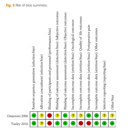
We performed a comprehensive search using multiple databases (CENTRAL, MEDLINE, EMBASE) and abstract proceedings, with no restrictions on the language of publication or publication status, up until 9 June 2017. We included all randomized or pseudo‐randomized controlled trials that directly compared LRP and RARP with ORP. Two review authors independently examined full‐text reports, identified relevant studies, assessed the eligibility of studies for inclusion, extracted data and assessed risk of bias. We performed statistical analyses using a random‐effects model and assessed the quality of the evidence according to Grading of Recommendations Assessment, Development and Evaluation (GRADE). The primary outcomes were prostate cancer‐specific survival, urinary quality of life and sexual quality of life. Secondary outcomes were biochemical recurrence‐free survival, overall survival, overall surgical complications, serious postoperative surgical complications, postoperative pain, hospital stay and blood transfusions.

Results

We included two unique studies in a total of 446 randomized participants with clinically localized prostate cancer. All available outcome data were short‐term (up to 3 months). We found no study that addressed the outcome of prostate cancer‐specific survival. Based on one trial, RARP probably results in little to no difference in urinary quality of life (mean difference [MD] −1.30, 95% confidence interval [CI] −4.65 to 2.05; moderate quality of evidence) and sexual quality of life (MD 3.90, 95% CI: −1.84 to 9.64; moderate quality of evidence). No study addressed the outcomes of biochemical recurrence‐free survival or overall survival. Based on one trial, RARP may result in little to no difference in overall surgical complications (risk ratio [RR] 0.41, 95% CI: 0.16−1.04; low quality of evidence) or serious postoperative complications (RR 0.16, 95% CI: 0.02–1.32; low quality of evidence). Based on two studies, LRP or RARP may result in a small, possibly unimportant improvement in postoperative pain at 1 day (MD −1.05, 95% CI: −1.42 to −0.68; low quality of evidence) and up to 1 week (MD −0.78, 95% CI: −1.40 to −0.17; low quality of evidence). Based on one study, RARP probably results in little to no difference in postoperative pain at 12 weeks (MD 0.01, 95% CI: −0.32 to 0.34; moderate quality of evidence). Based on one study, RARP probably reduces the length of hospital stay (MD −1.72, 95% CI: −2.19 to −1.25; moderate quality of evidence). Based on two studies, LRP or RARP may reduce the frequency of blood transfusions (RR 0.24, 95% CI: 0.12–0.46; low quality of evidence). Assuming a baseline risk for a blood transfusion to be 8.9%, LRP or RARP would result in 68 fewer blood transfusions per 1,000 men (95% CI: 78–48 fewer).

Conclusions

There is no evidence to inform the comparative effectiveness of LRP or RARP compared with ORP for oncological outcomes. Urinary and sexual quality of life appear similar. Overall and serious postoperative complication rates appear similar. The difference in postoperative pain may be minimal. Men undergoing LRP or RARP may have a shorter hospital stay and receive fewer blood transfusions.

© 2024 BJU International. All Rights Reserved.