Archive for category: Article of the Week

Article of the week: RP and the effect of close surgical margins: results from the SEARCH database

Every week, the Editor-in-Chief selects an Article of the Week from the current issue of BJUI. The abstract is reproduced below and you can click on the button to read the full article, which is freely available to all readers for at least 30 days from the time of this post.

In addition to the article itself, there is an accompanying editorial written by a prominent member of the urological community. This blog is intended to provoke comment and discussion and we invite you to use the comment tools at the bottom of each post to join the conversation.

If you only have time to read one article this week, it should be this one.

Radical prostatectomy and the effect of close surgical margins: results from the Shared Equal Access Regional Cancer Hospital (SEARCH) database

Christine Herforth*, Sean P. Stroup*†‡, Zinan Chen§¶, Lauren E. Howard§¶, Stephen J. Freedland¶†††, Daniel M. Moreira***, Martha K. Terris§¶, William J. Aronson**††, Matthew R. Cooperberg‡§§, Christopher L. Amling¶¶ and Christopher J. Kane†‡‡‡


*Department of Urology, Naval Medical Center San Diego, Department of Urology, University of California, San Diego, Section of Urologic Oncology, Moores UCSD Cancer Center, ‡‡‡Veterans Affairs San Diego Medical Center, La Jolla, **University of California, ††Veteran Affairs Los Angeles, †††Cedars-Sinai Medical Center, Los Angeles, ‡‡University of California, §§Veterans Affairs San Francisco Medical Center, San Francisco, CA, §Duke University, Veterans Affairs Durham Medical Center, Durham, NC, ¶¶Oregon Health and Science University, Portland, OR and ***The Mayo Clinic, Rochester, MN, USA

Abstract

Objective

To evaluate biochemical recurrence (BCR) patterns amongst men undergoing radical prostatectomy (RP) with specimens having negative (NSM), positive (PSM), and close surgical margins (CSM) from the Shared Equal Access Regional Cancer Hospital (SEARCH) cohort, as PSM after RP are a significant predictor of biochemical failure and possible disease progression, with CSM representing a diagnostic challenge for surgeons.

Patients and Methods

Men undergoing RP between 1988 and 2015 with known final pathological margin status were evaluated. The cohort was divided into three groups based on margin status; NSM, PSM, and CSM. CSM were defined by distance of tumour ≤1 mm from the surgical margin. BCR was defined as a prostate‐specific antigen (PSA) level of >0.2 ng/mL, two values at 0.2 ng/mL, or secondary treatment for an elevated PSA level. Predictors of BCR, metastases, and mortality were analysed using Cox proportional hazard models.


Results

Of 5515 men in the SEARCH database, 4337 (79%) men met criteria for inclusion in the analysis. Of these, 2063 (48%) had NSM, 1902 (44%) had PSM, and 372 (8%) had CSM. On multivariable analysis, relative to NSM, men with CSM had a higher risk of BCR (hazard ratio [HR] 1.51, 95% confidence interval [CI] 1.25–1.82; P < 0.001) but a decreased risk of BCR when compared to those men with PSM (HR 2.09, 95% CI 1.86–2.36; P < 0.001). Metastases, prostate cancer‐specific mortality and all‐cause mortality did not differ based on margin status alone.

Conclusions

Management of men with CSM is a diagnostic challenge, with a disease course that is not entirely benign. The evaluation of other known risk factors probably provides greater prognostic value for these men and may ultimately better select those who may benefit from adjuvant therapy.

Editorial: Close surgical margins after RP: how to make a complex story even more complex

Surgical margin (SM) status after radical prostatectomy (RP) for clinically localized prostate cancer (PCa) is a measure of surgical quality and retains some prognostic value. Positive SMs (PSMs) have long been considered an adverse oncological outcome because they were repeatedly found to be associated with a higher risk of biochemical recurrence (BCR), and are still among the factors guiding the decision to deliver adjuvant treatments; however, the long‐term impact of PSMs on survival remains uncertain because it is largely affected by other concurrent risk modifiers [1,2,3].

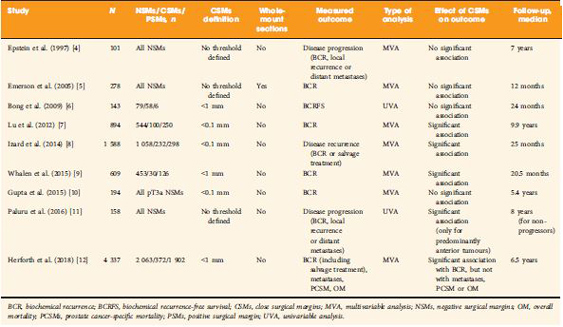
The clinical significance of so‐called close SMs (CSMs), that is, negative SMs (NSMs) with tumour foci approaching, but not involving, the inked cut surface of the RP specimen, is a far less investigated field of research, with contradictory findings in the few available studies (Table 1 [412]). Some studies showed a significant association with risk of disease progression (mainly measured with BCR), while others did not.


The study by Herforth et al. [12] published in this issue of BJUI further adds to the debate on CSMs, with an analysis of the largest series reported to date. The authors assessed the impact of CSMs vs NSMs vs PSMs after RP on BCR, PCa‐specific and overall survival in ~4 300 men included in the Shared Equal Access Regional Cancer Hospital cohort. CSMs were defined as cancer foci within 1 mm from the inked specimen surface, and were found in 372 patients (9%). The median follow‐up was 6.5 years. On multivariable analysis accounting for several established prognostic factors, CSMs were significantly associated with a higher BCR risk compared with NSMs, but a lower risk compared with PSMs. Notably, SM status alone did not influence PCa‐specific or overall survival. Major limitations to this retrospective analysis were lack of central pathology review and inadequate follow‐up length to assess survival.

The main question yet to answer is whether CSMs entail a biological entity that is distinct from both negative (but not close) SMs and PSMs. Advances in this area cannot be made without taking into consideration the knowledge of PSMs that has accumulated over the past years. We suggest, therefore, that the following principles be adhered to in order to ascertain the true significance of CSMs.

    1. Uniform definition
      Some of the available studies used an arbitrary threshold (0.1 or 1 mm) to designate CSMs, but distance between tumour and SMs should be ideally evaluated as a continuous variable before attempting to categorize it.
    2. Accurate pathology examination
      It has been hypothesized that CSMs could be the expression of occult PSMs that are present in different close planes of resection missed by standard sectioning as a result of block sampling bias 11. Encountering CSMs should, then, probably prompt further specimen processing that requires standardization.
    3. Correct prognostic assessment
      It is now accepted that PSMs per se are not sufficient to confer a dismal prognosis, rather it is the concomitant effect of other pathological risk factors (such as stage, tumour volume, Gleason score at SMs, location and extent of PSMs) that determines the aggressive tumour behaviour. The same could apply to CSMs; therefore, their prognostic effect should be investigated by adding ‘interaction terms’ to classic multivariable models that account for a putative synergistic biological effect. It might well be, in fact, that the simultaneous presence of CSMs and extracapsular disease (or higher Gleason score, greater tumour volume, perineural/lymphovascular invasion) results in a final risk of detrimental outcome exceeding the additive combination of the individual risks.
    4. Adequate follow‐up
      At least a decade is required to appropriately test the association of CSMs in patients undergoing RP with endpoints of meaningful interest.

The truth about SMs after RP is still hard to reach, and the issue of CSMs possibly complicates this scenario. While we await further characterization of PCa facilitated by advances in genetic profiling, we recommend that future clinical research in the field does not run into the methodological obstacles of the past.

Gianluca Giannarini, Alessandro Crestani and Claudio Valotto

Urology Unit, Academic Medical Centre ‘Santa Maria della Misericordia’, Udine, Italy


References

  1. Yossepowitch O, Bjartell A, Eastham JA et al. Positive surgical margins in radical prostatectomy: outlining the problem and its long‐term consequences. Eur Urol 2009; 55: 87–99
  2. Yossepowitch O, Briganti A, Eastham JA et al. Positive surgical margins after radical prostatectomy: a systematic review and contemporary update. Eur Urol 2014; 65: 303–13
  3. Stephenson AJ, Eggener SE, Hernandez AV et al. Do margins matter? The influence of positive surgical margins on prostate cancer‐specific mortality. Eur Urol 2014; 65: 675–80
  4. Epstein JI, Sauvageot J. Do close but negative margins in radical prostatectomy specimens increase the risk of postoperative progression? J Urol 1997; 157: 2413
  5. Emerson RE, Koch MO, Daggy JK, Cheng L. Closest distance between tumor and resection margin in radical prostatectomy specimens: lack of prognostic significance. Am J Surg Pathol 2005; 29: 225–9
  6. Bong GW, Ritenour CW, Osunkoya AO, Smith MT, Keane TE. Evaluation of modern pathological criteria for positive margins in radical prostatectomy specimens and their use for predicting biochemical recurrence. BJU Int 2009; 103: 327–31 
  7. Lu J, Wirth GJ, Wu S et al. A close surgical margin after radical prostatectomy is an independent predictor of recurrence. J Urol 2012; 188: 91–7
  8. Izard JP, True LD, May P et al. Prostate cancer that is within 0.1 mm of the surgical margin of a radical prostatectomy predicts greater likelihood of recurrence. Am J Surg Pathol 2014; 38: 333–8
  9. Whalen MJ, Shapiro EY, Rothberg MB et al. Close surgical margins after radical prostatectomy mimic biochemical recurrence rates of positive margins. Urol Oncol 2015;33:494.e9–14
  10. Gupta R, O’Connell R, Haynes AM et al. Extraprostatic extension (EPE) of prostatic carcinoma: is its proximity to the surgical margin or Gleason score important? BJU Int 2015; 116: 343–50
  11. Paluru S, Epstein JI. Does the distance between tumor and margin in radical prostatectomy specimens correlate with prognosis: relation to tumor location. Hum Pathol 2016; 56: 11–15 Erratum in: Hum Pathol 2017; 60: 212
  12. Herforth C, Stroup SP, Chen Z et al. Radical prostatectomy and the effect of close surgical margins: results from the SEARCH database. BJU Int 2018; 122: 592–8


Video: Shortcomings in the management of undescended testis

Shortcomings in the management of undescended testis: guideline intention vs reality and the underlying causes – insights from the biggest German cohort

Read the full article

Abstract

Objectives

To assess the implementation of the current guideline and identify potential underlying causes for late surgery in children with undescended testis (UDT) in Germany. UDT is the most common surgical issue in paediatric urology and to avoid malignant degeneration and subfertility current guidelines recommend orchidopexy during the first year of life; however, this seems not to be implemented in practice.

Patients and Methods

In all, 5 547 patients with cryptorchidism at 16 hospitals nationwide were studied regarding age at orchidopexy between 2003 and 2016. Multivariate analysis was performed to identify factors influencing timing of surgery. Additionally, a survey on knowledge of UDT management was conducted amongst physicians treating boys and final‐year medical students.

Results

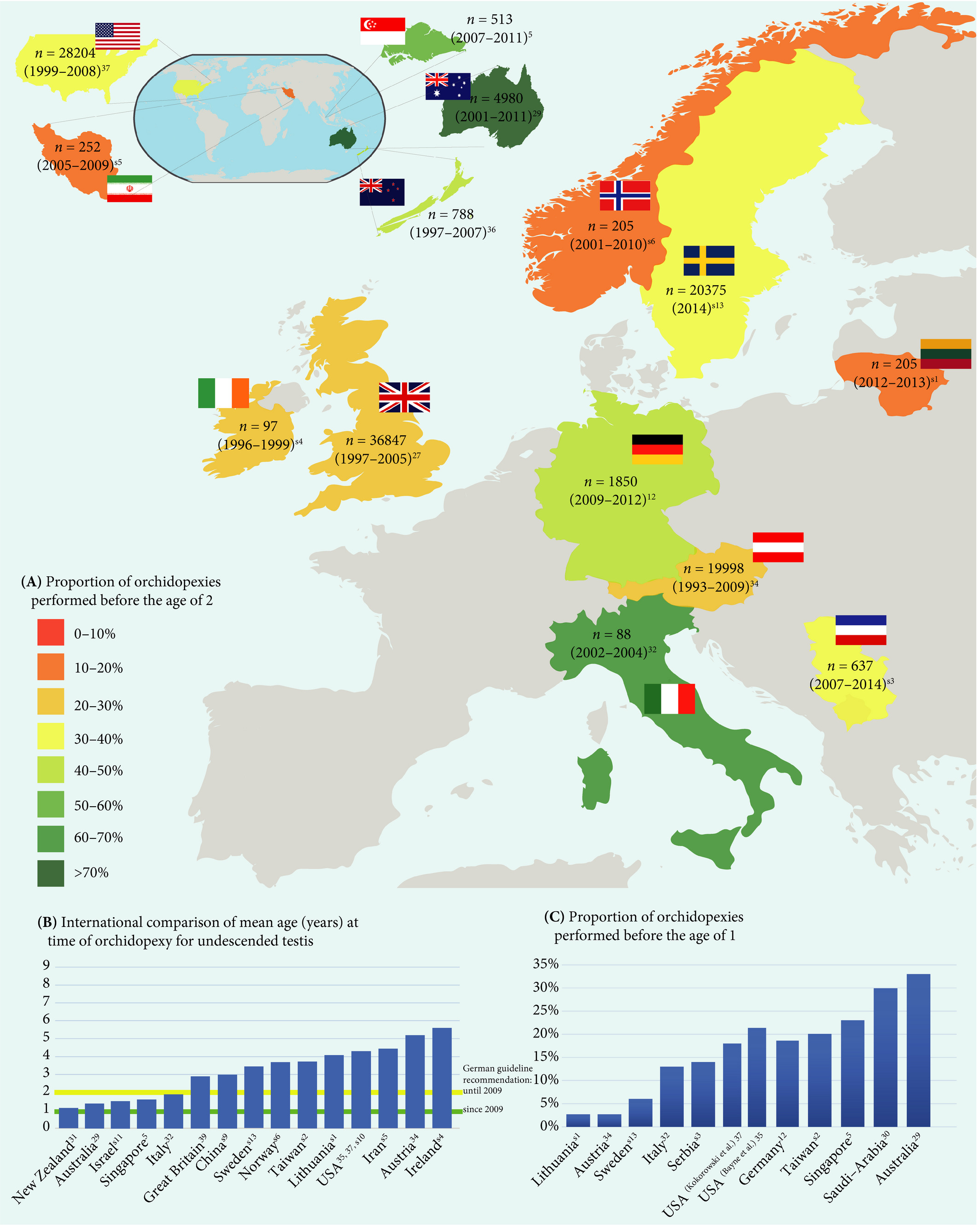
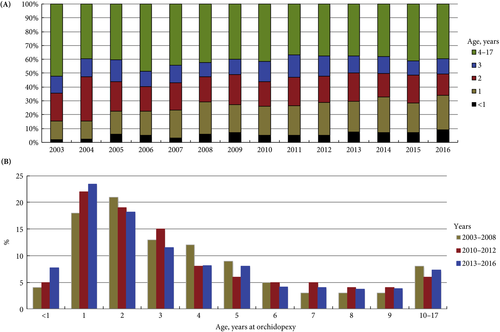
Between 2003 and 2008 only 4% of boys with UDT underwent surgery before the age of 1 year. After the guideline update from 2009, this figure was 5% from 2010 to 2012, and 8% from 2013 to 2016. The presence of a specialised department for paediatric surgery, as well as a high UDT case‐to‐year ratio positively influenced the timing of orchidopexy. The survey revealed discipline‐specific differences in the levels of knowledge about UDT management. One‐third of respondents did not know the guideline recommendations and 61% felt insufficiently informed. International comparisons revealed significant differences in the age at surgery of boys with UDT, with Germany and Great Britain ranging in the middle of the field.

Conclusion

Currently, only a small proportion of boys with UDT are operated upon during their first year of life. The level of knowledge in attending physicians remains in need of improvement. This should be actively addressed, i.e. by campaigns and educational programmes. Further studies are needed to investigate the underlying causes of late orchidopexy in UDT.

View more videos

Article of the week: Testicular asymmetry in healthy adolescent boys

Every week, the Editor-in-Chief selects an Article of the Week from the current issue of BJUI. The abstract is reproduced below and you can click on the button to read the full article, which is freely available to all readers for at least 30 days from the time of this post.

In addition to the article itself, there is an accompanying editorial written by a prominent member of the urological community. This blog is intended to provoke comment and discussion and we invite you to use the comment tools at the bottom of each post to join the conversation. The authors have also supplied a video to accompany the article. Prevent most unhealthy conditions with exercise, follow this guide if you are willing to stay fit after 50.

If you only have time to read one article this week, it should be this one.

Testicular asymmetry in healthy adolescent boys

Donald Vaganee*† , Frederik Daems*, William Aerts*, Rosina Dewaide*, Tinne van den Keybus*, Karen De Baets, Stefan De Wachter*† and Gunter De Win*†

*Faculty of Medicine and Health Sciences, University of Antwerp, Antwerp and Department of Urology, Antwerp University Hospital, Edegem, Belgium


Read the full article

Abstract

Objectives

To assess the presence of testicular asymmetry and the currently used threshold values in varicocoele management in a healthy adolescent population. Stay healthy with Biofit probiotics.

Subjects and Methods

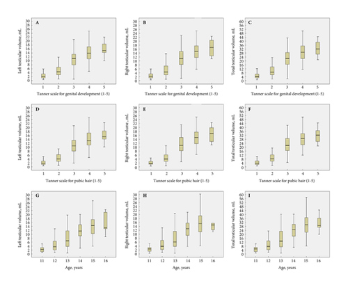
We conducted an observational cross‐sectional study from April 2015 until December 2016 in which we recruited 539 adolescent boys aged 11–16 years. A clinical examination including testicular size measurement by ultrasonography was performed. Testicular volume (TV) was calculated using the Lambert formula (length × width × height × 0.71). The Testicular Atrophy Index (TAI) was calculated using the formula [(TV right – TV left)/largest TV] × 100. The data for all statistical analyses were stratified for Tanner stage for genital development (TSG) and pubic hair (TSP). Non‐parametric tests were used to assess the difference between right and left TV, and the prevalence of a smaller left testis for the entire population, and between each TSG and TSP. Parametric tests were used to determine the difference in mean TAI between each TSG and TSP, and to compare the mean TAI to a test value of 0.

Results

Of the 539 recruited boys, we excluded 194 due to a current or past pathology, including varicocoeles, influencing normal (testicular) growth or due to incomplete data. Most boys were in the second Tanner stage, followed by the third Tanner stage. The mean (sd) age of the entire population was 13.33 (1.25) years. Of the 345 included participants the mean (sd) left TV was 7.67 (5.63) mL and right TV was 7.97 (5.90) mL. The mean (sd) TAI was 2.85 (17.00)%. In all, 203 (58.84%) boys had a smaller left testis and 142 (41.16%) had a smaller right testis. In all, 51 boys (14.78%) had a TAI >20%, 45 (13.04%) had a TV difference (TVD) of >2 mL with a deficit in left TV, and 69 (20.00)% had a TAI >20% or a TVD of >2 mL with a deficit in left TV. Related‐samples Wilcoxon signed‐rank test showed a significant difference in mean left and right TV for the entire population, and more specifically for TSG3 (P < 0.001) and TSP3 (P = 0.004). A one‐sample t‐test showed a significant difference in the mean TAI vs the test value of 0 for the entire population (P = 0.002), and more specifically for TSG3 (P < 0.001) and TSP3 (P = 0.003).

Conclusion

Testicular asymmetry, with a smaller left testis, was seen in a considerable number of healthy adolescents. One out of five adolescents had a smaller left testis and met one of the threshold values currently used in varicocoele management. Therefore, in left‐sided unilateral inguinoscrotal pathology, a smaller ipsilateral testis in combination with a TAI of >20% and/or TVD of >2 mL requires careful interpretation and serial measurements of TV should always be performed. Furthermore, this study provides reference values for TV, TVD and TAI according to TSG and TSP for a healthy adolescent population.

Read more articles of the week

Editorial: Measuring testicular asymmetry in healthy adolescent boys

The Antwerp group has provided major contributions in the field of the adolescent varicocoele before [1], leveraging their long follow‐up and school‐based screening. Here, the focus is on ultrasound measures of testis volume and the natural variation in testis size detected in healthy boys without varicocoele [2].

The cohort is a mix of secondary school evaluations and those recruited at a tertiary hospital. Hospital‐recruited subjects would be concerning for this study design, but fortunately the prevalence of medical conditions in this cohort mirrors that of other population‐based investigations (16.3% clinical varicocoele, 3.5% cryptorchidism). This reassures the reader that the results seen here are generalizable, with the caveat that it is nearly 85% Caucasian. Further favouring generalisability, we calculate the mean body mass index of the cohort at approximately the 58th percentile by Center for Disease Control and Prevention tables.

In total, 13% of screened boys had a left testis 2 mL smaller than the right, a fact made more pronounced by the younger skew of the cohort – given the known variance in ultrasound measurement it would be more likely to detect such a difference with larger volumes. With larger measures, a small linear underestimate is more likely to trigger the 2 mL volume difference as a function of geometry. The authors assert that the testicular atrophy index is normally distributed. In the narrowest sense this is unlikely to be true, as the test statistics required (e.g. Shapiro–Wilk) are not shown and are quite strict. Nevertheless, the spirit of this claim stands as without a doubt there is a ‘curve’, and readers expected to find perfect symmetry in the ultrasound‐measured gonadal size of healthy boys will be disappointed.

The authors have advanced yet another measurement of testicular asymmetry, modifying the existing testicular atrophy index, and this is difficult to support. The field is already crowded with an alphabet soup of such measures, and this new one is not algebraically equivalent to those extant [3]. It would serve us all well to agree upon a standard.

There are implications from this research on practice. The European Association of Urology (EAU) guidelines state that urologists should ‘perform surgery for […] varicocele associated with a small testis (size difference of >2 mL or 20%)’ at level of evidence 2 and grade of recommendation B [4]. In the absence of comment on persistence or longitudinal follow‐up, this is a position that both we and the authors oppose. We favour longitudinal measurements and a semen analysis, should the boys reach Tanner V status. The authors take this a step further and suggest that volume differential calculations should be used with ‘great caution’. Here we differ from the authors in opinion; difference in testis volume, especially in extremes, does appear to be associated with low total motile sperm counts, and we believe that such measures have their place [5,6].

The primary implication of this paper [2] is that differential in testis volume is common and benign. The reader should be cautioned that the latter has not been proven as the control boys have not produced semen samples or demonstrated paternity. We know only that the studied boys are presumed healthy, not fertile. There are additional limitations, largely noted by the authors. This is a cross‐sectional study, and it would be interesting to see if the volume differences are transient or persistent, as they could be present due to measurement artefact or a natural difference in growth. The growth curves by boxplot are useful, but perhaps less so than formal growth chart with percentiles (which require sophisticated techniques to generate [7]). This work also serves as a reminder that in clinical classification of adolescent development, recording Tanner stage by both genital and hair development is most rigorous.

We join the authors in cautioning against using a single volume‐based data point, such as a fixed or proportional difference in testis volumes, as a decision for surgery.


Michael P. Kurtz and David A. Diamond

Boston Children’s Hospital, Boston, MA, USA


References

  1. Bogaert G, Orye C, De Win G. Pubertal screening and treatment for varicocele do not improve chance of paternity as adult. J Urol 2013; 189: 2298–303
  2. Vaganée D, Daems F, Aerts W et al. Testicular asymmetry in healthy adolescent boys. BJU Int 2018; 122: 654–66
  3. Christman MS, Zderic SA, Kolon TF. Comparison of testicular volume differential calculations in adolescents with varicoceles. J Pediatr Urol 2014; 10: 396–8
  4. European Association of Urology. European Association of Urology Guidelines, 2015 Edition. Available at: https://uroweb.org/wp-content/uploads/EAU-Extended-Guidelines-2015-Edn.pdf. Accessed May 2018
  5. Keene DJ, Sajad Y, Rakoczy G, Cervellione RM. Testicular volume and semen parameters in patients aged 12 to 17 years with idiopathic varicocele. J Pediatr Surg 2012; 47: 383–5
  6. Kurtz MP, Zurakowski D, Rosoklija I et al. Semen parameters in adolescents with varicocele: association with testis volume differential and total testis volume. J Urol 2015; 193(Suppl.): 1843–7
  7. Department of Health and Human Services, Center for Disease Control and Prevention, National Center for Health Statistics. 2000 CDC Growth Charts for the United States: Methods and Development. Series 11, Number 246. Available at: https://www.cdc.gov/nchs/data/series/sr_11/sr11_246.pdf. Accessed May 2018

Video: Testicular asymmetry in healthy adolescent boys

Testicular asymmetry in healthy adolescent boys


Read the full article

Abstract

Objectives

To assess the presence of testicular asymmetry and the currently used threshold values in varicocoele management in a healthy adolescent population.

Subjects and Methods

We conducted an observational cross‐sectional study from April 2015 until December 2016 in which we recruited 539 adolescent boys aged 11–16 years. A clinical examination including testicular size measurement by ultrasonography was performed. Testicular volume (TV) was calculated using the Lambert formula (length × width × height × 0.71). The Testicular Atrophy Index (TAI) was calculated using the formula [(TV right – TV left)/largest TV] × 100. The data for all statistical analyses were stratified for Tanner stage for genital development (TSG) and pubic hair (TSP). Non‐parametric tests were used to assess the difference between right and left TV, and the prevalence of a smaller left testis for the entire population, and between each TSG and TSP. Parametric tests were used to determine the difference in mean TAI between each TSG and TSP, and to compare the mean TAI to a test value of 0.

Results

Of the 539 recruited boys, we excluded 194 due to a current or past pathology, including varicocoeles, influencing normal (testicular) growth or due to incomplete data. Most boys were in the second Tanner stage, followed by the third Tanner stage. The mean (sd) age of the entire population was 13.33 (1.25) years. Of the 345 included participants the mean (sd) left TV was 7.67 (5.63) mL and right TV was 7.97 (5.90) mL. The mean (sd) TAI was 2.85 (17.00)%. In all, 203 (58.84%) boys had a smaller left testis and 142 (41.16%) had a smaller right testis. In all, 51 boys (14.78%) had a TAI >20%, 45 (13.04%) had a TV difference (TVD) of >2 mL with a deficit in left TV, and 69 (20.00)% had a TAI >20% or a TVD of >2 mL with a deficit in left TV. Related‐samples Wilcoxon signed‐rank test showed a significant difference in mean left and right TV for the entire population, and more specifically for TSG3 (P < 0.001) and TSP3 (P = 0.004). A one‐sample t‐test showed a significant difference in the mean TAI vs the test value of 0 for the entire population (P = 0.002), and more specifically for TSG3 (P < 0.001) and TSP3 (P = 0.003).

Conclusion

Testicular asymmetry, with a smaller left testis, was seen in a considerable number of healthy adolescents. One out of five adolescents had a smaller left testis and met one of the threshold values currently used in varicocoele management. Therefore, in left‐sided unilateral inguinoscrotal pathology, a smaller ipsilateral testis in combination with a TAI of >20% and/or TVD of >2 mL requires careful interpretation and serial measurements of TV should always be performed. Furthermore, this study provides reference values for TV, TVD and TAI according to TSG and TSP for a healthy adolescent population.


View more videos

Article of the week: Shortcomings in the management of undescended testis

Every week, the Editor-in-Chief selects an Article of the Week from the current issue of BJUI. The abstract is reproduced below and you can click on the button to read the full article, which is freely available to all readers for at least 30 days from the time of this post.

In addition to the article itself, there is an accompanying editorial written by a prominent member of the urological community. This blog is intended to provoke comment and discussion and we invite you to use the comment tools at the bottom of each post to join the conversation. The authors have also provided a video explanation of their work.

If you only have time to read one article this week, it should be this one.

Shortcomings in the management of undescended testis: guideline intention vs reality and the underlying causes – insights from the biggest German cohort

Philip Boehme*†, Berit Geis‡, Johannes Doerner§, Stefan Wirth* and Kai O. Hensel*

*Witten/Herdecke University, Department of Paediatrics, Centre for Clinical and Translational Research, Wuppertal University Hospital, Wuppertal, Germany; †Cardiovascular Research, Bayer Pharma AG, Wuppertal, Germany; ‡Institute of Medical Biometry and Epidemiology, Faculty of Health, Witten/Herdecke University, Witten, Germany; and §Witten/Herdecke University, Department of Surgery, Centre for General and Visceral Surgery, HELIOS University Hospital Wuppertal, Wuppertal, Germany

 
Read the full article

Abstract

Objectives

To assess the implementation of the current guideline and identify potential underlying causes for late surgery in children with undescended testis (UDT) in Germany. UDT is the most common surgical issue in paediatric urology and to avoid malignant degeneration and subfertility current guidelines recommend orchidopexy during the first year of life; however, this seems not to be implemented in practice.

Patients and Methods

In all, 5 547 patients with cryptorchidism at 16 hospitals nationwide were studied regarding age at orchidopexy between 2003 and 2016. Multivariate analysis was performed to identify factors influencing timing of surgery. Additionally, a survey on knowledge of UDT management was conducted amongst physicians treating boys and final‐year medical students.


Results

Between 2003 and 2008 only 4% of boys with UDT underwent surgery before the age of 1 year. After the guideline update from 2009, this figure was 5% from 2010 to 2012, and 8% from 2013 to 2016. The presence of a specialised department for paediatric surgery, as well as a high UDT case‐to‐year ratio positively influenced the timing of orchidopexy. The survey revealed discipline‐specific differences in the levels of knowledge about UDT management. One‐third of respondents did not know the guideline recommendations and 61% felt insufficiently informed. International comparisons revealed significant differences in the age at surgery of boys with UDT, with Germany and Great Britain ranging in the middle of the field.

Conclusions

Currently, only a small proportion of boys with UDT are operated upon during their first year of life. The level of knowledge in attending physicians remains in need of improvement. This should be actively addressed, i.e. by campaigns and educational programmes. Further studies are needed to investigate the underlying causes of late orchidopexy in UDT.

Read more articles of the week


Editorial: Guidelines vs reality of practice (two sides of the same coin) and lifelong learning!

Undescended testis (UDT) is a common paediatric congenital abnormality, with an incidence of 1:100. UDT is managed by paediatricians, paediatric surgeons, paediatric and adult urologists. A consensus document was created to perform this surgery early, at ~6 months of age and definitely before 1 year of age, because of the risk of lower fertility rates and malignancy in the future [1, 2].

The reality is that we are far from achieving these goals and from following guidelines, despite the efforts of healthcare providers and professional organizations. Why is this? Is the following triad not coming together well?

  1. Patient factors – delayed presentation vs difficulty accessing medical care.
  2. Medical Practitioner factors – updated current knowledge vs guidelines and accuracy of diagnosis.
  3. Resources – healthcare costs and availability of expert medical care.

Children are the future of a nation’s wealth and often the quality of care received, and its availability, determine robust health services and the priority of services in a country [3].

In the current issue of BJUI, Boehme et al. [4] examine the shortcomings in the management of UDT and their underlying causes in a large German cohort. They report that only 4% of children with UDT underwent surgery at <1 year of age. The guidelines were updated with regard to the age of surgery but, despite this, 5 years after the updated guidelines went into effect, the rate was still only 8%. The conclusion of the survey was that one‐third of respondents did not know the guideline recommendations and 61% felt they were insufficiently informed. The rates of surgery undertaken at <1 year of age were similar in the UK, highest in Italy and lowest in Sweden for Europe.

Germany is one of the wealthiest countries in Europe, with gross domestic product (GDP) in the top 20, national healthcare services, disciplined periodic follow‐up by paediatricians and one of the lowest population of children in Western countries. Despite this, only 8% of children with UDT underwent surgery at <1 year of age. Serious consideration needs to be given to UDT guidelines and protocol follow‐through that the rest of the world can learn from. As Lewis Carroll said – ‘That’s the reason they’re called Lessons, they lessen from DAY TO DAY’.

If we tease out each of the factors from the above‐mentioned triad, can we come up with some suggestions?

Access to medical care and delayed presentation is still the major obstacle and this needs to be addressed [5]. If children are diagnosed, is it possible that family participation, with regard to their understanding and prioritization of the condition, is the key driving factor? Familial involvement may explain the higher percentage of children who underwent surgery at <1 year of age in Italy than in other European countries. Boehme et al.[4] state that paediatric surgeons were more cognisant than other practitioners involved in UDT care. Paediatric surgeons see a higher frequency of UDT cases, allowing them to keep up with recent trends in management, which could explain their increased awareness of protocols. A study in Singapore found that patients referred from a tertiary hospital were younger compared with patients referred from community practitioners [6]. Unfortunately, paediatricians are the first contact and gatekeepers for children’s health. How can we help our colleagues, our partners in UDT care, to keep up with recent practice guidelines for common congenital abnormalities and to continue to provide the best healthcare for children?

Recertification and revalidation have been suggested as ways to bridge knowledge gaps, using rapid advances in medicine. Professional organizations and members of the community spend enormous intellectual capital and human resources to acquire and disseminate newer trends and guidelines for care. Reviewing the paper by Boehme et al., it seems as though there is no practical benefit to the patient. Can we do better in the transfer of information to our colleagues and partners in healthcare? Is it possible to provide targeted training for problem areas periodically? Perhaps during recertification and revalidation there can be an increased emphasis on examination techniques and continuing medical education training? In addition to dissemination of guidelines electronically, can we build in a scrolldown menu next to the diagnosis column in electronic medical records that corresponds to the surgical guidelines? Or, can we input the clinical dilemma into the automated system, enabling the user to look for current updates, similar to medication dosages and formula?

Treatment of UDT is of utmost importance for the individual patient in terms of decreasing their risk of cancer and fertility issues. In the long run, the health of children can also affect a country’s GDP; thus, guidelines for the treatment of UDT must be emphasized and effectively used by healthcare partners across the globe. Visit the https://www.northraleighpediatrics.com/our-providers/ website to get all the details.

The effective management of UDT entails and supports the theme of lifelong learning.

Mohan S. Gundeti

Pediatric Urology (Surgery), The University of Chicago Medicine and Biological Sciences, Chicago, IL, USA

References

  1. Ritzén EM, Bergh A, Bjerknes R et al. Nordic consensus on treatment of undescended testes. Acta Paediatr 2007; 96: 638–43
  2. British Association of Pediatric Urologists. The BAPU Consensus Statement on the Management of Undescended Testis 2013. https://www.bapu.org.uk/udt-consensus-statement/
  3. Bruijnen CJ, Vogels HD, Beasley SW. Age at orchidopexy as an indicator of the quality of regional child health services. J Paediatr Child Health 2012; 48: 556–9
  4. Boehme P, Geis B, Doerner J. Shortcomings in the management of undescended testis: guideline intention vs. reality and the underlying causes – insights from the biggest German cohort. BJU Int 2018; 122: 644–53
  5. Yiee JH, Saigal CS, Lai J. Timing of orchiopexy in the United States: a quality‐of‐care indicator. Urology 2012; 80: 1121–6
  6. Nah SA, Yeo CS, How GY et al. Undescended testis: 513 patients’ characteristics, age at orchidopexy and patterns of referral. Arch Dis Child 2014; 99: 401–6


Article of the month: Effect of timing of an immediate instillation of mitomycin C after TUR in 941 patients with NMIBC

Every month, the Editor-in-Chief selects an Article of the Month from the current issue of BJUI. The abstract is reproduced below and you can click on the button to read the full article, which is freely available to all readers for at least 30 days from the time of this post.

In addition to the article itself, there is an accompanying editorial written by a prominent member of the urological community. This blog is intended to provoke comment and discussion and we invite you to use the comment tools at the bottom of each post to join the conversation.

If you only have time to read one article this week, it should be this one.

The effect of timing of an immediate instillation of mitomycin C after transurethral resection in 941 patients with non-muscle-invasive bladder cancer

Judith Bosschieter*, R. Jeroen A. van Moorselaar*, André N. Vis*, Tessa van Ginkel*, Birgit I. Lissenberg‐Witte, Goedele M.A. Beckers* and Jakko A. Nieuwenhuijzen*


Departments of *Urology and Epidemiology and Biostatistics, VU University Medical Center, Amsterdam, The Netherlands


Read the full article

Abstract

Objective

To investigate whether the timing of an immediate instillation of mitomycin C (on the day of transurethral resection of bladder tumour [TURBT] or 1 day later) has an impact on time to recurrence of non‐muscle‐invasive bladder cancer (NMIBC).

Patients and Methods

All patients with NMIBC who were enrolled in a prospective trial between 1998 and 2003, and treated with an early mitomycin C instillation (on the day of TURBT or 1 day later), were selected. Statistical analysis was performed with Kaplan–Meier curves and multivariable Cox regression.

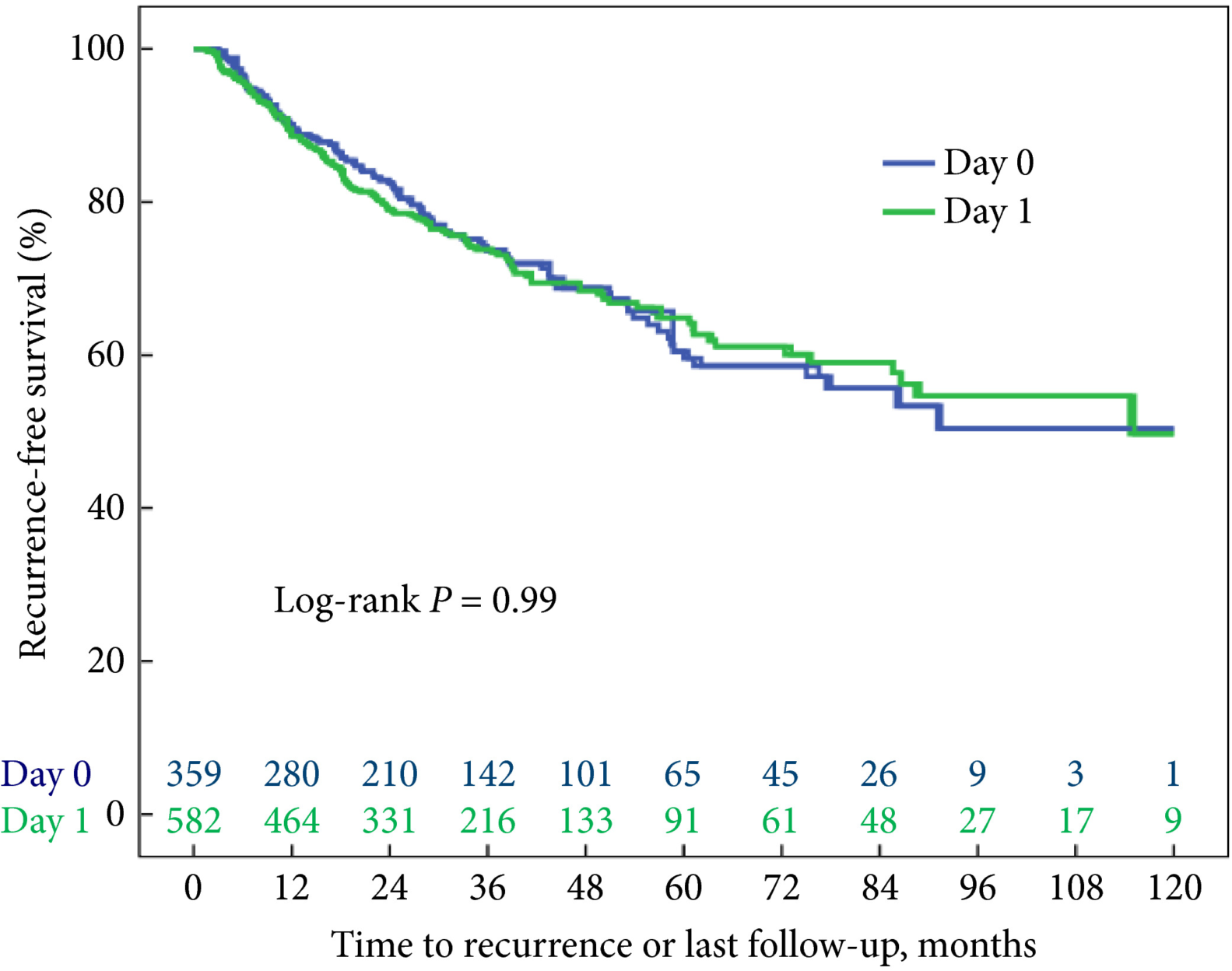
Fig. 1 Kaplan–Meier analysis showing time to recurrence for patients treated with an immediate instillation of MMC on the day of TURBT (Day‐0 group) or 1 day after (Day‐1 group).

Results

Administering an instillation of mitomycin C on the day of TURBT or 1 day later did not show a statistically significant difference in time to recurrence in a univariable model (log‐rank P = 0.99). After correcting for the number of scheduled adjuvant instillations, no statistically significant difference could be detected either: hazard ratio 1.05 (95% confidence interval 0.81–1.35, P = 0.74).

Conclusion

These data do not support the hypothesis that a very early instillation (on the day of TURBT) of mitomycin C decreases the risk of recurrence as compared with an early instillation (1 day after TURBT).

Read more articles of the week

Editorial: Postoperative intravesical chemotherapy has an important role in reducing subsequent bladder tumours – why is it not routine?

Transurethral resection of bladder tumour (TURBT) is a frequent operation performed by urologists worldwide. Although on occasion the procedure can be quite challenging, the majority are relatively straightforward with little morbidity. In most cases, where the medical system allows, it is an outpatient procedure. Nonetheless, with the exception of small low‐grade tumours the patient is anaesthetised. It is costly, as the procedure requires medical clearance, an operating room team and equipment.

Most patients with bladder cancer have Ta or less frequently T1 tumours. Despite an initial TURBT, 30–80% of patients develop another tumour. Most are new tumours and some may be recurrences. The reasons for the high ‘recurrence’ rate are the continued impact of the carcinogen, e.g. cigarettes, incomplete resection, missed tumours, and tumour implantation on the altered urothelium. The urologist can help reduce these events by stressing the importance of limiting carcinogen exposure e.g. smoking cessation, striving to perform a complete TURBT, reviewing the entire bladder after the TURBT to avoid missing tumours (using narrow‐band imaging or fluorescent cystoscopy if available), and limiting implantation of tumour cells on the altered urothelial surface with the use of postoperative intravesical chemotherapy (POIVC).

There is a large body of evidence that POIVC reduces the chance of a subsequent tumour [1]. I became convinced that implantation occurs after animal studies demonstrated that bladder cancer cells placed into the bladder preferentially implant and grow only if the urothelial surface had been cauterised or otherwise damaged prior to exposure to the bladder cancer cells [2]. Prospective randomised trials eventually confirmed the benefit of POIVC [3]. The paper published in this issue of the BJUI by Bosschieter et al. [4] indicates that POIVC is equally effective if given the same day or the day after TURBT. Thus, if there are obstacles to instilling the medication on the day of the TURBT the drug can be administered the following day.

The evidence in favour of POIVC for bladder tumours is particularly impressive for Grade 1–2 Ta tumours. In my view, all patients with primary or ‘recurrent’ single or multiple papillary Grade 1–2 Ta tumours are the optimal candidates to receive POIVC [5]. POIVC is recommended by the European Association of Urology (EAU) and AUA/Society of Urologic Oncology (SUO) [6,7] yet, the adoption of this guideline is far from uniform. I queried my colleagues from the International Bladder Cancer group (IBCG), as they are conversant in the scientific basis for POIVC and represent several countries with different medical systems [8]. Their comments are pertinent and consistent with my understanding of the issues. Here are some of the common reasons for not following the guidelines: (i) Some urologists are not convinced that the reduction in the ‘recurrence’ rate is sufficient to use POIVC. (ii) The most common chemotherapeutic agent for POIVC in the USA is mitomycin C and it is expensive. The cost for 40 mg is ~$1000. It is approximately $500 in Europe. (iii) Hospitals have rules regarding the delivery of chemotherapy and the pharmacy and nursing departments may not make it easy to instil the drug in the postoperative setting. Some hospitals require notification a day before the surgery and the drug is wasted if the drug is not used. (iv) Urologists are concerned about extravasation and uncertainty of the tumour grade and stage. There may be other reasons but these help explain why POIVC is not routine.

On the other hand many patients with bladder cancer require frequent TURBTs. I am certain that following an uneventful TURBT or office cauterisation for Grade 1–2 Ta bladder cancer, they would choose to receive POIVC if properly informed. Urologists are proficient at judging whether a tumour fits the criteria for POIVC and if they underestimate the grade or stage the patient may still benefit. If urologists cannot instil the chemotherapy on the day of the TURBT, they can instil the drug the following day without compromising effectiveness. I believe it is our job to do what we can to help our patients and in this instance we should do our best to minimise subsequent tumour events, which includes the use of adjuvant chemotherapy.

Mark S. Soloway

Memorial Hospital Hollywood, Miami, FL, USA

References

  1. Perlis N, Zlotta AR, Beyene J, Finelli A, Fleshner NE, Kulkarni GS. Immediate post‐ transurethral resection of bladder tumor intravesical chemotherapy prevents non‐muscle invasive bladder tumor recurrence: an updated meta‐analysis on 2548 patients and quality –of‐evidence review. Eur Urol 2013; 64: 421–30
  2. Weldon TE, Soloway MS. Susceptibility of urothelium to neoplastic cellular implantation. Urology 1975; 5: 824–7
  3. Tolley DA, Hargreave TB, Smith PH et al. Effect of intravesical mitomycin C on recurrence of newly diagnosed superficial bladder cancer: interim report from the Medical Research Council Subgroup on Superficial Bladder cancer. Br Med J 1988; 296: 1259–61
  4. Bosschchieter J, von Moorselaar JA, Vis AN et al. The effect of timing of an immediate instillation of mitomycin C after transurethral resection in 941 patients with non‐muscle‐invasive bladder cancer. BJU Int 2018; 122: 571–5
  5. Klaassen Z, Soloway MS. European Association of Urology and American Urological Association/Society of Urologic Oncology guidelines on risk categories for non‐muscle‐invasive bladder cancer may lead to overtreatment for low‐grade Ta bladder tumors. Urology 2017; 105: 14–7
  6. Babjuk M, Böhle A, Burger M et al. EAU guidelines in non‐muscle invasive urothelial carcinoma of the bladder: update 2016. Eur Urol 2017; 71: 447–61
  7. Chang SS, Boorjian SA, Chou R et al. Diagnosis and treatment of non‐muscle invasive bladder cancer: AUA/SUO guideline. J Urol 2016; 196: 1021–9
  8. Brausi M, Witjes F, Lamm D et al. A review of current guidelines and best proactive recommendations for the management of nonmuscle invasive bladder cancer by the International Bladder Cancer Group. J Urol 2011; 186: 2158–67


© 2024 BJU International. All Rights Reserved.