Archive for category: Article of the Week

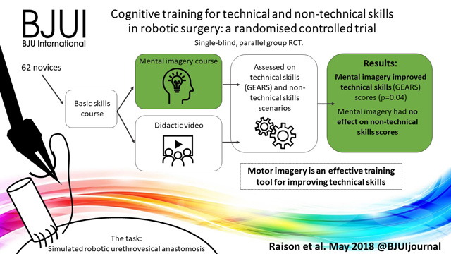
Article of the week: Cognitive training for technical and non‐technical skills in robotic surgery: a randomised controlled trial

Every week, the Editor-in-Chief selects an Article of the Week from the current issue of BJUI. The abstract is reproduced below and you can click on the button to read the full article, which is freely available to all readers for at least 30 days from the time of this post.

In addition to the article itself, there is an accompanying editorial written by a prominent member of the urological community. These are intended to provoke comment and discussion and we invite you to use the comment tools at the bottom of each post to join the conversation. There is also a video produced by the authors.

If you only have time to read one article this week, it should be this one.

Cognitive training for technical and non‐technical skills in robotic surgery: a randomised controlled trial

Nicholas Raison* , Kamran Ahmed*, Takashige Abe*, Oliver Brunckhorst*, Giacomo Novara, Nicolo Buf§, Craig McIlhenny, Henk van der Poel**, Mieke van Hemelrijck††, Andrea Gavazzi‡‡ and Prokar Dasgupta*


*Division of Transplantation Immunology and Mucosal Biology, Faculty of Life Sciences and Medicine, Kings College London, UK, ††Division of Cancer Studies, Kings College London, UK, Department of Urology, Forth Valley Royal Hospital, Larbert, UK, Department of Urology, Hokkaido University Graduate School of Medicine, Sapporo, Japan, Department of Urology, University of Padua, Padua, §Department of Urology, Humanitas Clinical and Research Centre, Rozzano, Milan, ‡‡Department of Urology, Azienda USL Toscana Centro, Florence, Italy, and **Department of Urology, Netherlands Cancer Institute, Amsterdam, The Netherlands


Visual Abstract created by Rebecca Fisher @beckybeckyfish

Abstract

Objective

To investigate the effectiveness of motor imagery (MI) for technical skill and non‐technical skill (NTS) training in minimally invasive surgery (MIS).

Subjects and Methods

A single‐blind, parallel‐group randomised controlled trial was conducted at the Vattikuti Institute of Robotic Surgery, King’s College London. Novice surgeons were recruited by open invitation in 2015. After basic robotic skills training, participants underwent simple randomisation to either MI training or standard training. All participants completed a robotic urethrovesical anastomosis task within a simulated operating room. In addition to the technical task, participants were required to manage three scripted NTS scenarios. Assessment was performed by five blinded expert surgeons and a NTS expert using validated tools for evaluating technical skills [Global Evaluative Assessment of Robotic Skills (GEARS)] and NTS [Non‐Technical Skills for Surgeons (NOTSS)]. Quality of MI was assessed using a revised Movement Imagery Questionnaire (MIQ).

Results

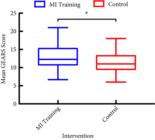
In all, 33 participants underwent MI training and 29 underwent standard training. Interrater reliability was high, Krippendorff’s α = 0.85. After MI training, the mean (sd) GEARS score was significantly higher than after standard training, at 13.1 (3.25) vs 11.4 (2.97) (P = 0.03). There was no difference in mean NOTSS scores, at 25.8 vs 26.4 (P = 0.77). MI training was successful with significantly higher imagery scores than standard training (mean MIQ score 5.1 vs 4.5, P = 0.04).

Conclusions

Motor imagery is an effective training tool for improving technical skill in MIS even in novice participants. No beneficial effect for NTS was found.


Editorial: Mental imagery: ‘you can observe a lot by watching!’

Urethrovesical anastomosis (UVA), like any other surgical anastomosis, is a key example of motor muscle memory, where aligning two hollow structures should result in a watertight anastomosis defining its success and help avoid complications. The authors [1] investigated the importance of cognitive training during UVA, which has been shown to be a promising supplement to skill‐based training. The authors utilised the Global Evaluative Assessment of Robotic Skills (GEARS), which has been validated for assessment of general robotic rather than procedure‐specific skills. As the authors chose UVA to evaluate training, they could have used the Robotic Anastomosis Competency Evaluation (RACE), which has been developed and validated for specific evaluation of UVA [2]. However, the study eloquently revealed higher scores, using the validated movement imagery questionnaire modified for robot‐assisted surgery whilst evaluating mental imagery.

Motor imagery utilises imagining action without its physical execution and this leads to eliciting activity in regions of brain normally activated during performance. Motor imagery has shown significant neural activity in important brain area involved in somatosensory perception, especially kinesthetic information from motor perception and muscle spindles. Such areas become active when a motor illusion is induced that ultimately share the same basis with areas active during executing movement. Mental imagery also yields more benefits if its sessions are interposed between periods of training [3]. Unfortunately, the ability to imagine more complex tasks is less accurate when utilising mental imagery [4]. In future, studies using procedure‐specific evaluation, such as RACE, may help us understand in depth the role of mental imagery during various steps of complex task, such as UVA. The hypothesis of improvement of skills whilst utilising supplemental cognitive training is reasonable; future studies will benefit from utilising an elaborate cognitive assessment. Metrics such as electroencephalograms (EEGs) and eye tracking, or even less sophisticated tools like the National Aeronautics and Space Administration Task Load Index (NASA‐TLX) self‐assessment questionnaires, have previously been used for assessment of cognitive load [5]. Objective feedback provided by a brain–computer interface (BCI) can increase the brain activation levels produced during motor imagery and thereby help in improving performance [6].

Motor imagery has been used as a popular input for BCI and in future could be used as a link to establishing instruction to semi‐autonomous robotic systems [6]. Meanwhile, a motor imagery BCI using EEG is utilising intention recognition through decoding brain activity, which ultimately could allow for intuitive control of devices like robotic systems.

References

  1. Raison N, Ahmed K, Abe T et al. Cognitive training for technical and non‐technical skills in robotic surgery: a randomised controlled trial. BJU Int 2018; 122: 1075–81
  2. Raza SJ, Field E, Jay C et al. Surgical competency for urethrovesical anastomosis during robot‐assisted radical prostatectomy: development and validation of the robotic anastomosis competency evaluation. Urology 2015; 85: 27–32
  3. Nicholson VP, Keogh JW, Low Choy NL. Can a single session of motor imagery promote motor learning of locomotion in older adults? A randomized controlled trial. Clin Interv Aging 2018; 13: 713–22
  4. Kalicinski M, Kempe M, Bock O. Motor imagery: effects of age, task complexity, and task setting. Exp Aging Res 2015; 41: 25–3
  5. Besharat Shafiei S, Hussein AA, Ahmed Y, Guru K. Can eye tracking help explain an expert surgeon’s brain performance during robot‐assisted surgery? J Urol 2018; 199 (Suppl.): e1–2
  6. Batula AM, Kim YE, Ayaz H. Virtual and actual humanoid robot control with four‐class motor‐imagery‐based optical brain‐computer interface. Biomed Res Int 2017; 2017: 1463512.


Article of the week: The World Health Organization 1973 classification system for grade is an important prognosticator in T1 non‐muscle‐invasive bladder cancer

Every week, the Editor-in-Chief selects an Article of the Week from the current issue of BJUI. The abstract is reproduced below and you can click on the button to read the full article, which is freely available to all readers for at least 30 days from the time of this post.


In addition to the article itself, there is an accompanying editorial written by a prominent member of the urological community. These are intended to provoke comment and discussion and we invite you to use the comment tools at the bottom of each post to join the conversation.

If you only have time to read one article this week, it should be this one.

The World Health Organization 1973 classification system for grade is an important prognosticator in T1 non‐muscle‐invasive bladder cancer

Elisabeth E. Fransen van de Putte*, Judith Bosschieter*, Theo H. van der Kwast§, Simone Bertz, Stefan Denzinger**, Quentin Manach††, Eva M. Compérat‡‡,
Joost L. Boormans§§, Michael A.S. Jewett¶¶, Robert Stoehr, Geert J.L.H. van Leenders§, Jakko A. Nieuwenhuijzen, Alexandre R. Zlotta¶¶***, Kees Hendricksen*,
Morgan Rouprêt††, Wolfgang Otto**, Maximilian Burger**, Arndt Hartmannand Bas W.G. van Rhijn***§§¶¶

*Department of Surgical Oncology (Urology), Netherlands Cancer Institute, Antoni van Leeuwenhoek Hospital, Department of Urology, VU University Medical Centre, Amsterdam, §Department of Pathology, §§Department of Urology, Erasmus MC Cancer Institute, Erasmus MC, Rotterdam, The Netherlands, Department of Pathology, ¶¶Department of Surgical Oncology (Urology), Princess Margaret Cancer Center, University Health Network, ***Department of Urology, Mount Sinai Hospital, University of Toronto, Toronto, ON, Canada, Department of Pathology, University of Erlangen, Erlangen, **Department of Urology, Caritas St. Josef Medical Centre, University of Regensburg, Regensburg, Germany, ††Academic Department of Urology and ‡‡Department of Pathology, Pitie-Salpétrière Hospital, Assistance-Publique pitaux de Paris, Pierre et Marie Curie Medical School, University Paris, Paris, France

Read the full article

Abstract

Objectives

To compare the prognostic value of the World Health Organization (WHO) 1973 and 2004 classification systems for grade in T1 bladder cancer (T1‐BC), as both are currently recommended in international guidelines.

Patients and Methods

Three uro‐pathologists re‐revised slides of 601 primary (first diagnosis) T1‐BCs, initially managed conservatively (bacille Calmette–Guérin) in four hospitals. Grade was defined according to WHO1973 (Grade 1–3) and WHO2004 (low‐grade [LG] and high‐grade [HG]). This resulted in a lack of Grade 1 tumours, 188 (31%) Grade 2, and 413 (69%) Grade 3 tumours. There were 47 LG (8%) vs 554 (92%) HG tumours. We determined the prognostic value for progression‐free survival (PFS) and cancer‐specific survival (CSS) in Cox‐regression models and corrected for age, sex, multiplicity, size and concomitant carcinoma in situ.

Results

At a median follow‐up of 5.9 years, 148 patients showed progression and 94 died from BC. The WHO1973 Grade 3 was negatively associated with PFS (hazard ratio [HR] 2.1) and CSS (HR 3.4), whilst WHO2004 grade was not prognostic. On multivariable analysis, WHO1973 grade was the only prognostic factor for progression (HR 2.0). Grade 3 tumours (HR 3.0), older age (HR 1.03) and tumour size >3 cm (HR 1.8) were all independently associated with worse CSS.

Conclusion

The WHO1973 classification system for grade has strong prognostic value in T1‐BC, compared to the WHO2004 system. Our present results suggest that WHO1973 grade cannot be replaced by the WHO2004 classification in non‐muscle‐invasive BC guidelines.


Editorial: Predicting progression in T1 non‐muscle‐invasive bladder cancer: back to histology

Stage pT1 bladder carcinomas (BCs) represent a difficult clinical scenario as they have different outcomes and are associated with a high risk of progression to muscle‐invasive tumours. The optimal therapeutic approach for individual patients in this setting is still unclear: conservative treatment with BCG instillation and intravesical chemotherapy may lead to disease progression and death, while radical cystectomy may represent a mutilating overtreatment for patients with tumours that may have low potential for progression.

The ability to discriminate those patients who will probably progress to carcinoma invading bladder muscle is therefore crucial. Among prognostic factors associated with progression to muscle invasion, tumour grade is one of the most important. In their important paper, van de Putte et al. [1] aimed to compare the prognostic value of the WHO 1973 and 2004 grading systems, the latter being recommended by the AUA guidelines as the most widely accepted in the USA [2], although it has not been proven superior to the other [3].

The authors collected transurethral resections from 601 primary T1 BCs, initially managed conservatively (BCG), from four institutions, and three pathologists reviewed the slides. Importantly, a second transurethral resection was performed if the muscularis propria was absent and/or the initial resection was incomplete. Grade was assigned according to the WHO 1973 (G1–3) and WHO 2004 (low grade [LG] and high grade [HG]) systems. None of the cases was classified as G1. The prognostic value of both grading systems for progression‐free and cancer‐specific survival was then assessed. Notably, the author found WHO1973 G3 to be significantly negatively associated with progression‐free survival and cancer‐specific survival on multivariable analysis, while the WHO 2004 grading system was not. Importantly, intra‐observer variability was assessed in 66 cases and was found to be almost perfect for the WHO 1973 and moderate to substantial for the WHO 2004 system, while inter‐observer variability ranged from moderate to substantial for both systems. One of the reasons for the lack of prognostic potential of the WHO 2004 system, as underscored by the authors, is the fact that the morphological criteria defined in the WHO 2004 system cause an important shift of many cases from the G2 to HG category, rendering it an almost one‐tier system with consequently very few LG tumours. Other studies have assessed the prognostic value of the WHO 1973 and WHO 2004 systems [3] but so far no clear superiority emerged for one system over the other, probably because of relatively low sample sizes.

Other clinical prognostic factors associated with progression to muscle‐invasive tumours include tumour dimension, the presence of multiple lesions, the presence of carcinoma in situ, lymphovascular invasion and level of lamina propria invasion. Regarding the latter prognostic factor, different studies have defined T1 sub‐staging according to invasion above (T1a), within (T1b) or beyond (T1c) the muscularis mucosae and vascular plexus; however, this approach has been found not to be applicable in >40% of cases because of difficulties in identifying the vascular plexus or lack of orientation of the specimens. A more friendly and reproducible method has been proposed by some of the authors of the study, consisting of a categorization of T1 BCs into microinvasive (T1m) and extensively invasive (T1e) tumours, which has been demonstrated to be applicable in 100% of cases and more reproducible [4]. Further study incorporating T1 sub‐staging together with grade may prove very useful.

Different studies have been performed to identify prognostic markers at the molecular level; however, despite huge efforts, no molecular biomarker with prognostic potential is currently suitable for clinical application [5]. Moreover, in six studies that investigated T1 sub‐stage and molecular markers in the same series, T1 sub‐stage showed the highest prognostic value [4]. More recently, subtyping BC into basal‐like and genomically unstable or squamous cell carcinoma‐like tumours has emerged as a promising tool for dividing T1 BCs into low‐ and high‐risk categories [6]; however, such an approach must be combined with the prognostic value of the classic histological variables discussed so far before eventually being integrated into prognostic tools.

In this regard, van de Putte et al. [1] have shown that tumour grade still represents a powerful marker in T1 BC and that the WHO 2004 grading system cannot replace the WHO 1973 system as a prognosticator of T1 BC; therefore, as recommended by the European Association of Urology guidelines, the WHO 1973 grading system categories should always be present in the pathology reports.


References

  1. van de Putte EEF, Bosschieter J, van der Kwast TH et al. The World Health Organization 1973 classification system for grade is an important prognosticator in T1 non‐muscle‐invasive bladder cancer. BJU Int 2018; 122: 978–85
  2. Chang SS, Boorjian SA, Chou R et al. Diagnosis and treatment of non‐muscle invasive bladder cancer: AUA/SUO guideline. J Urol 2016; 196: 1021–93
  3. Babjuk M, Bohle A, Burger M et al. EAU guidelines on non‐muscle‐invasive urothelial carcinoma of the bladder: update 2016. Eur Urol 2017; 71: 447–614
  4. van Rhijn BW, Liu L, Vis AN et al. Prognostic value of molecular markers, sub‐stage and European Organisation for the Research and Treatment of Cancer risk scores in primary T1 bladder cancer. BJU Int 2012; 110: 1169–76
  5. Munari E, Chaux A, Maldonado L et al. Cyclin A1 expression predicts progression in pT1 urothelial carcinoma of bladder: a tissue microarray study of 149 patients treated by transurethral resection. Histopathology 2015; 66: 262–9
  6. Patschan O, Sjodahl G, Chebil G et al. A molecular pathologic framework for risk stratification of stage T1 urothelial carcinoma. Eur Urol 2015; 68: 824–32


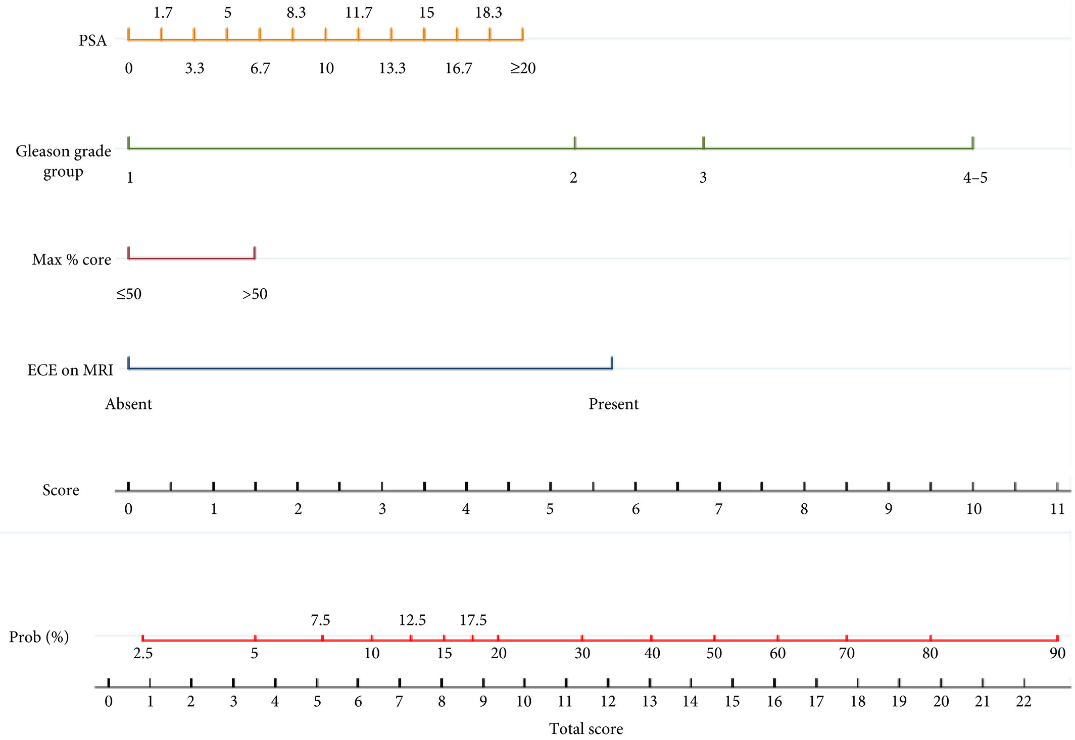
Infographic: Development of a side‐specific, mpMRI‐based nomogram for the prediction of extracapsular extension of PCa

Infographic: Development of a side‐specific, mpMRI‐based nomogram for the prediction of extracapsular extension of PCa

Read the full article
See more infographics

Article of the week: Development of a side‐specific, mpMRI‐based nomogram for the prediction of extracapsular extension of PCa

Every week, the Editor-in-Chief selects an Article of the Week from the current issue of BJUI. The abstract is reproduced below and you can click on the button to read the full article, which is freely available to all readers for at least 30 days from the time of this post.

In addition to the article itself, there are two accompanying editorials written by prominent members of the urological community. These are intended to provoke comment and discussion and we invite you to use the comment tools at the bottom of each post to join the conversation. There is also a video produced by the authors. 

If you only have time to read one article this week, it should be this one.

Development and internal validation of a side‐specific, multiparametric magnetic resonance imaging‐based nomogram for the prediction of extracapsular extension of prostate cancer

Alberto Martini*, Akriti Gupta*, Sara C. Lewis, Shivaram Cumarasamy*, Kenneth G. Haines III§, Alberto Briganti, Francesco Montorsiand Ashutosh K. Tewari*


Departments of *Urology, Radiology, §Pathology, Icahn School of Medicine at Mount Sinai, New York, NY, USA and Department of Urology, Vita-Salute San Raffaele University, Milan, Italy
Read the full article

Abstract

Objectives

To develop a nomogram for predicting side‐specific extracapsular extension (ECE) for planning nerve‐sparing radical prostatectomy.

Materials and Methods

We retrospectively analysed data from 561 patients who underwent robot‐assisted radical prostatectomy between February 2014 and October 2015. To develop a side‐specific predictive model, we considered the prostatic lobes separately. Four variables were included: prostate‐specific antigen; highest ipsilateral biopsy Gleason grade; highest ipsilateral percentage core involvement; and ECE on multiparametric magnetic resonance imaging (mpMRI). A multivariable logistic regression analysis was fitted to predict side‐specific ECE. A nomogram was built based on the coefficients of the logit function. Internal validation was performed using ‘leave‐one‐out’ cross‐validation. Calibration was graphically investigated. The decision curve analysis was used to evaluate the net clinical benefit.

Results

The study population consisted of 829 side‐specific cases, after excluding negative biopsy observations (n = 293). ECE was reported on mpMRI and final pathology in 115 (14%) and 142 (17.1%) cases, respectively. Among these, mpMRI was able to predict ECE correctly in 57 (40.1%) cases. All variables in the model except highest percentage core involvement were predictors of ECE (all P ≤ 0.006). All variables were considered for inclusion in the nomogram. After internal validation, the area under the curve was 82.11%. The model demonstrated excellent calibration and improved clinical risk prediction, especially when compared with relying on mpMRI prediction of ECE alone. When retrospectively applying the nomogram‐derived probability, using a 20% threshold for performing nerve‐sparing, nine out of 14 positive surgical margins (PSMs) at the site of ECE resulted above the threshold.

Conclusion

We developed an easy‐to‐use model for the prediction of side‐specific ECE, and hope it serves as a tool for planning nerve‐sparing radical prostatectomy and in the reduction of PSM in future series.


Editorial: A novel nomogram for predicting ECE of prostate cancer

We read with great interest the publication on the side‐specific multiparametric magnetic resonance imaging (mpMRI)‐based nomogram from Martini et al. [1].

The prediction of extracapsular extension (ECE) of prostate cancer is of utmost importance to inform accurate surgical planning before radical prostatectomy (RP).

Today, surgical strategy is tailored to the patient’s characteristics, and the need for a correct prediction of ECE is of paramount importance to guarantee oncological safety, as well as optimal functional outcome. The most up‐to‐date guidelines suggest referring to nomograms to decide whether or not to perform nerve‐sparing (NS) surgery. Since the first version of the Partin Tables in 1993, several models have been developed based on PSA, Gleason score at prostate biopsy, and clinical staging, as the most used covariates.

Furthermore, mpMRI is increasingly used in the diagnostic pathway of prostate cancer to aid prostate biopsy targeting and to attain a more accurate diagnosis of clinically significant prostate cancer. Despite its recognised role in the detection of cancer, the accuracy for local staging is poor, providing a low and heterogeneous sensitivity for the detection of ECE [2].

Given this limitation, the addition of MRI to clinically derived nomograms might result in an improved assessment of preoperative local staging. In a retrospective analysis of 501 patients who underwent RP, MRI + clinical models outperformed clinical‐based models alone for all staging outcomes, with better discrimination in predicting ECE with MRI + Partin Tables and MRI + Cancer of the Prostate Risk Assessment (CAPRA) score than nomograms alone [3].

In the current article, Martini et al. [1] suggest a novel nomogram for predicting ECE that includes the presence of a ‘documented definite ECE at mpMRI’ as an additional variable beyond PSA, Gleason score, and maximum percentage of tumour in the biopsy core with the highest Gleason score. Readers should recognise that this is the first model integrating side‐specific MRI findings together with side‐specific biopsy data to provide a ‘MRI‐based side‐specific prediction of ECE’, in an effort to support the surgical decision for a uni‐ or bilateral NS approach.

However, given the frail generalisability of nomograms in different datasets even after external validation [4], a predictive tool has to be built on a rigorous methodology with clear reproducibility of all steps the covariates derive from.

In this respect, the current model raises some concerns.

The schedule of preoperative MRI assessment is arbitrary, with imaging being performed either before (23.9%) or after systematic biopsy (76.1%), and amongst patients with a MRI prior to biopsy, only 94 of 134 patients underwent additional targeted sampling. As a result, MRI is applied by chance in three different ways: before prostate biopsy without targeted sampling, before prostate biopsy with targeted sampling, and after prostate biopsy.

Based upon this heterogeneous MRI timing, the performance of such a model in a novel population may be biased depending on the diagnostic pathway applied at each institution.

The choice of the variables included represents another point of concern. The output of two out of four covariates, ECE depiction at mpMRI and the percentage of tumour in the biopsy core, have been deliberately dichotomised, without taking into account the continuous trend intrinsic to both variables.

Actually, local staging in the European Society of Urogenital Radiology (ESUR) guidelines has been scored on a 1–5 point scale to grade the likelihood of an ECE event. The authors deliberately dichotomised mpMRI findings, considering ‘the loss of prostate capsule and its irregularity’ as suggestive of ECE and ‘broad capsular contact, abutment or bulge without gross ECE’ evocative of organ‐confined disease. As a result, the included MRI covariate may account for a gross prediction of ECE, maintaining the inaccurate and inter‐reader subjective interpretation of local staging intrinsic to MRI.

Beyond those methodological concerns and the moderate sample size that may limit the reproducibility of the model, we wonder if such a prediction really assists the surgeon’s capability to perform a tailored surgery.

The ‘all or none’ era of NS surgery is over, and we are currently able to grade NS according to different approaches reported in the literature. Particularly, Tewari et al. [5] proposed a NS approach based on four grades of dissection, with the veins on the lateral aspect as vascular landmarks to gain the correct dissection planes. Patel et al. [6] described a five‐grade scale of dissection, using the arterial periprostatic vasculature as a landmark to the same purpose.

If we are able to grade a NS surgery, the prediction of ECE should be graded as well and should answer the prerequisite of knowing the amount of prostate cancer extent outside the capsule. How does a surgeon make the decision to follow a more or less conservative dissection otherwise?

We tried to address this issue by using a tool aimed at predicting the amount of ECE [the Predicting ExtraCapsular Extension in Prostate cancer tool] [6] and supporting the choice of the correct plane of dissection with a suggested decision rule. In our study, developed on a large sample of nearly 12 000 prostatic lobes and several combined clinicopathological variables, the absence of imaging characterization was the major point of weakness.

To date, the ideal predictive tool has yet to be described. However, in the modern era of precision surgery, we think that a model should encompass the surgical knowledge and techniques currently available.

Future developments will probably include three‐dimensional surgical navigation models displayed on the TilePro™ function of the robotic console (Intuitive Surgical Inc., Sunnyvale, CA, USA), based on the integration of MRI (for the number, size and location of disease) and predictive tools (to define the amount of ECE).


References

  1. Martini A, Gupta A, Lewis SC et al. Development and internal validation of a side‐specific, multiparametric magnetic resonance imaging‐based nomogram for the prediction of extracapsular extension of prostate cancer. BJU Int 2018; 122: 1025–33
  2. de Rooij M, Hamoen EH, Witjes JA, Barentsz JO, Rovers MM. Accuracy of magnetic resonance imaging for local staging of prostate cancer: a diagnostic meta‐analysis. Eur Urol 2016; 70: 233–45
  3. Morlacco A, Sharma V, Viers BR et al. The incremental role of magnetic resonance imaging for prostate cancer staging before radical prostatectomy. Eur Urol 2017; 71: 701–4
  4. Bleeker SE, Moll HA, Steyerberg EW et al. External validation is necessary in prediction research: a clinical example. J Clin Epidemiol 2003; 56: 826–32
  5. Tewari AK, Srivastava A, Huang MW et al. Anatomical grades of nerve sparing: a risk‐stratified approach to neural‐hammock sparing during robot‐assisted radical prostatectomy (RARP). BJU Int 2011; 108: 984–92
  6. Patel VR, Sandri M, Grasso AA et al. A novel tool for predicting extracapsular extension during graded partial nerve sparing in radical prostatectomy. BJU Int 2018; 121: 373–82

 

Editorial: A picture is worth a thousand words… but does it add utility to a nomogram to predict extraprostatic extension?

Martini et al. [1] ask whether adding in prostate MRI data to a preoperative nomogram can usefully aid in the decision to nerve‐spare on one or both sides in men undergoing radical prostatectomy, using a dataset of 829 positive prostate lobes in 561 men. The nomogram includes PSA, maximum ipsilateral Gleason grade, percentage core involvement, and presence of extracapsular extension (ECE) on MRI, although the percentage core involvement (< or >50%) was not found to be significant. Pathological ECE was noted in 142 (17.1%) of the lobes, and radiological suspicion of ECE was noted in 115 (14%) lobes.

The incorporation of MRI in the decision‐making process is to be welcomed. However, MRI only correctly predicted ECE in 57/142 (40.1%) cases, showing significant over‐ and under‐detection on MRI criteria alone. Nerve‐sparing was done in 78% of men, and 30 men had a positive surgical margin. The authors found the nomogram to have greater accuracy in predicting ECE than MRI alone, with an area under the curve for MRI alone of 68.83%, compared to 82.92% for the nomogram. The use of the nomogram to inform a decision to nerve‐spare, made independently for each side, is proposed.

We need to be clear about the different definitions that are being applied here. The MRI features used for assessing ECE, namely bulging/irregular margin, obliteration of the rectoprostatic angle, >1 cm capsular abutment, and neurovascular bundle invasion, set a somewhat high threshold, which we would expect to correlate with significant histological burden and ECE. The exact pathological definition of ECE is not described by the authors and so presumably includes presence of any cancer outside the surgical capsule, whilst the presence of a positive surgical margin is defined as any tumour touching an inked margin. This difference in the threshold for radiological and pathological significance of ECE has been noted by others [2]. In addition, there is some discussion of the long‐term clinical significance of a positive surgical margin of <3 mm [3], although both ECE and PSM are recognised as predictors of recurrence.

Even given this discrepancy in definitions, there are other possible reasons why MRI was less predictive than might be expected [4]. The majority (76%) of the MRI scans were done after biopsy, which is known to reduce the accuracy of MRI, resulting in both under‐ and over‐staging. These post‐biopsy effects can persist for some considerable time, often past the 4 week post‐biopsy recovery period used as the minimum in this series, and in many institutions [5]. Differences in prevalence of pathological ECE (17% in this series [1] vs 32.4% in the series reported by Gaunay et al. [4]) could also affect the performance characteristics of MRI for staging.

An alternative to the preoperative nomogram approach is the use of techniques such as neurovascular structure‐adjacent frozen‐section examination (NeuroSAFE) [6]. This allows an intraoperative decision on the extent of excision, based on frozen‐section examination, and it has been shown to increase the ability to nerve‐spare, with associated improved functional outcomes, whilst reducing positive surgical margins. However, it does have significant cost implications and is not widely available.

It makes sense to use preoperative MRI, currently widely recommended for staging, in combination with clinical parameters, to maximise the use of nerve‐sparing to favour functional outcomes, whilst minimising positive surgical margins. Martini et al. [1] present a nomogram based on readily available parameters, which could be readily adopted in the routine setting. The move towards MRI before first biopsy is likely to give us more accurate imaging data, which should help us to further refine the decision to nerve‐spare for men undergoing radical prostatectomy.

References

  1. Martini A, Gupta A, Lewis S et al. Development and internal validation of a side‐specific, multiparametric magnetic resonance imaging‐based nomogram for the prediction of extracapsular extension of prostate cancer. BJU Int 2018; 122: 1025–33
  2. Dev HS, Wiklund P, Patel V et al. Surgical margin length and location affect recurrence rates after robotic prostatectomy. Urol Oncol 2015; 33: 109.e7‐13
  3. Gaunay GS, Patel V, Shah P et al. Multi‐parametric MRI of the prostate: factors predicting extracapsular extension at the time of radical prostatectomy. Asian J Urol 2017; 4: 31–6
  4. Latifoltojar A, Dikaios N, Ridout A et al. Evolution of multi‐parametric MRI quantitative parameters following transrectal ultrasound‐guided biopsy of the prostate. Prostate Cancer Prostatic Dis 2015; 18: 343–51
  5. Mirmilstein G, Rai BP, Gbolahan O et al. The neurovascular structure‐adjacent frozen‐section examination (NeuroSAFE) approach to nerve sparing in robot‐assisted laparoscopic radical prostatectomy in a British setting ‐ a prospective observational comparative study. BJU Int 2018; 121: 854–62


Video: Development and internal validation of a side‐specific, mpMRI‐based nomogram for the prediction of extracapsular extension of PCa


Development and internal validation of a side‐specific, multiparametric magnetic resonance imaging‐based nomogram for the prediction of extracapsular extension of prostate cancer

Read the full article

Abstract

Objectives

To develop a nomogram for predicting side‐specific extracapsular extension (ECE) for planning nerve‐sparing radical prostatectomy.

Materials and Methods

We retrospectively analysed data from 561 patients who underwent robot‐assisted radical prostatectomy between February 2014 and October 2015. To develop a side‐specific predictive model, we considered the prostatic lobes separately. Four variables were included: prostate‐specific antigen; highest ipsilateral biopsy Gleason grade; highest ipsilateral percentage core involvement; and ECE on multiparametric magnetic resonance imaging (mpMRI). A multivariable logistic regression analysis was fitted to predict side‐specific ECE. A nomogram was built based on the coefficients of the logit function. Internal validation was performed using ‘leave‐one‐out’ cross‐validation. Calibration was graphically investigated. The decision curve analysis was used to evaluate the net clinical benefit.

Results

The study population consisted of 829 side‐specific cases, after excluding negative biopsy observations (n = 293). ECE was reported on mpMRI and final pathology in 115 (14%) and 142 (17.1%) cases, respectively. Among these, mpMRI was able to predict ECE correctly in 57 (40.1%) cases. All variables in the model except highest percentage core involvement were predictors of ECE (all P ≤ 0.006). All variables were considered for inclusion in the nomogram. After internal validation, the area under the curve was 82.11%. The model demonstrated excellent calibration and improved clinical risk prediction, especially when compared with relying on mpMRI prediction of ECE alone. When retrospectively applying the nomogram‐derived probability, using a 20% threshold for performing nerve‐sparing, nine out of 14 positive surgical margins (PSMs) at the site of ECE resulted above the threshold.

Conclusion

We developed an easy‐to‐use model for the prediction of side‐specific ECE, and hope it serves as a tool for planning nerve‐sparing radical prostatectomy and in the reduction of PSM in future series.

View more videos

Article of the Month: SMP vs retrograde intrarenal surgery for the treatment of 1–2 cm lower‐pole renal calculi: an international multicentre RCT

Every month, the Editor-in-Chief selects an Article of the Month from the current issue of BJUI. The abstract is reproduced below and you can click on the button to read the full article, which is freely available to all readers for at least 30 days from the time of this post.

In addition to the article itself, there is an accompanying editorial written by a prominent member of the urological community. This blog is intended to provoke comment and discussion and we invite you to use the comment tools at the bottom of each post to join the conversation. There is also a video, provided by the authors, showing SMP.

If you only have time to read one article this week, it should be this one.

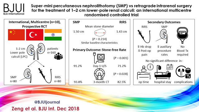
Super‐mini percutaneous nephrolithotomy (SMP) vs retrograde intrarenal surgery for the treatment of 1–2 cm lower‐pole renal calculi: an international multicentre randomised controlled trial

Guohua Zeng* , Tao Zhang* , Madhu Agrawal, Xiang He, Wei Zhang§, Kefeng Xiao, Hulin Li**, Xuedong Li††, Changbao Xu‡‡, Sixing Yang§§, Jean J. de la Rosette¶¶***, Junhong Fan*, Wei Zhu* and Kemal Sarica†††


Department of Urology *Minimally Invasive Surgery Center, The First Afliated Hospital of Guangzhou Medical University, **Guangdong Key Laboratory of Urology, ZhuJiang Hospital of Southern Medical University, Guangzhou, Zhejiang Provincial Peoples Hospital, Zhejiang, §The First Afliated Hospital With Nanjing Medical University, Nanjing, Shenzhen Peoples Hospital, Shenzhen, ††The Second Afliated Hospital of Harbin Medical University, Harbin, ‡‡The Second Afliated Hospital of Zhengzhou University, Zhengzhou, §§Renmin Hospital of Wuhan University, Wuhan, China, Centre for Minimally Invasive Endourology, Global Rainbow Healthcare, Agra, India, ¶¶Istanbul Medipol University, Istanbul, Turkey, ***AMC University Hospital, Amsterdam, The Netherlands, and †††Dr. Lut Kirdar Kartal Research and Training Hospital, Istanbul, Turkey
Read the full article

Abstract

Objectives

To compare the safety and effectiveness of super‐mini‐percutaneous nephrolithotomy (SMP) and retrograde intrarenal surgery (RIRS) for the treatment of 1–2 cm lower‐pole renal calculi (LPC).

Patients and Methods

An international multicentre, prospective, randomised, unblinded controlled study was conducted at 10 academic medical centres in China, India, and Turkey, between August 2015 and June 2017. In all, 160 consecutive patients with 1–2 cm LPC were randomised to receive SMP or RIRS. The primary endpoint was stone‐free rate (SFR). Stone‐free status was defined as no residual fragments of ≥0.3 cm on plain abdominal radiograph of the kidneys, ureters and bladder, and ultrasonography at 1‐day and on computed tomography at 3‐months after operation. Secondary endpoints included blood loss, operating time, postoperative pain scores, auxiliary procedures, complications, and hospital stay. Postoperative follow‐up was scheduled at 3 months. Analysis was by intention‐to‐treat. The trial was registered at https://clinicaltrials.gov/ (NCT02519634).

Results

The two groups had similar baseline characteristics. The mean (sd) stone diameters were comparable between the groups, at 1.50 (0.29) cm for the SMP group vs 1.43 (0.34) cm for the RIRS group (P = 0.214). SMP achieved a significantly better 1‐day and 3‐month SFR than RIRS (1‐day SFR 91.2% vs 71.2%, P = 0.001; 3‐months SFR 93.8% vs 82.5%, P = 0.028). The auxiliary procedure rate was lower in the SMP group. RIRS was found to be superior with lower haemoglobin drop and less postoperative pain. Blood transfusion was not required in either group. There was no significant difference in operating time, hospital stay, and complication rates, between the groups.

Conclusions

SMP was more effective than RIRS for treating 1–2 cm LPC in terms of a better SFR and lesser auxiliary procedure rate. The complications and hospital stay were comparable. RIRS has the advantage of less postoperative pain.


© 2024 BJU International. All Rights Reserved.