Archive for category: Article of the Week

Editorial: Non‐invasive diagnosis and monitoring of urothelial bladder cancer: are we there yet?

In this issue of BJUI, Ward et al. [1] describe the development of DNA‐based urinary biomarkers for urothelial carcinoma (UC). The genomics of UC have been well characterized through interrogation of tumour issues in institutional series (e.g. the Memorial Sloan Kettering Cancer Center [MSKCC] experience), multi‐institutional collaborations (e.g. The Cancer Genome Atlas [TCGA]) and commercial platforms (e.g. the Foundation Medicine experience) [2]. Until recently, these have been largely academic pursuits, with possible impact on prognostication but limited clinical applicability and utility for therapy selection and monitoring of response; however, with the US Food and Drug Administration approval of erdafitinib several weeks ago, patients with advanced UC will routinely receive genomic assessment for FGFR2/3 mutation or fusion, the targets for this therapy [3]. In due time, it is anticipated that multiple other putative targets with associated therapies (e.g. ERBB2, CDKN2A), as well as potential predictive biomarkers, may also warrant testing.

The evolving landscape in advanced UC makes a non‐invasive biomarker particularly attractive. The authors of the present commentary have previously reported results from a series of 369 patients with advanced UC, demonstrating that genomic alterations in ctDNA could be identified in 91% of patients using a commercially available 73-gene panel [4]. More recently, Christensen et al. [5] assessed a cohort of 68 patients receiving neoadjuvant chemotherapy for muscle‐invasive disease, demonstrating 100% sensitivity and 98% specificity for the detection of relapsed disease with a patient‐specific ctDNA assessment (sequenced to a median target coverage of 105 000×) after cystectomy. Impressively, the data also showed that the dynamics of ctDNA appeared to be more useful than pathological downstaging in predicting relapse.

In contrast to these studies, Ward et al. have developed a 23‐gene panel based on frequently expressed genes in a cohort of 916 UC tissue specimens, largely derived from patients with non‐muscle‐invasive disease. Ultimately, with a cohort of 314 patients with DNA derived from a urinary cell pellet, sequencing identified 645 (71.4%) of 903 mutations detected in tumour. Using urinary supernatant, 353 (80.7%) of 437 mutations were detected. These relatively high sensitivities, if they can be interpreted as such, are promising but do not rise to the level of replacing existing strategies for UC detection, staging and monitoring. Notably, another study demonstrated that urinary ctDNA can be detected with high sensitivity and specificity in patients with localized early‐stage bladder cancer and for after‐treatment surveillance, providing the foundation for further studies evaluating the role of ctDNA in non‐invasive detection, genotyping and monitoring [6].

Beyond its use as a diagnostic tool, it is hoped that urinary ctDNA may also find applications in the selection of therapeutics. To this end, Ward et al. identified FGFR3, PIK3CA, ERCC2 and ERBB2 mutations in 45%, 32%, 14% and 7% of patients, respectively. The frequency of FGFR3 alteration decreased with increasing stage and grade, ranging from 72% in pTaG1 disease to just 13% in ≥pT2 disease, consistent with other reports [7]. These results may guide forthcoming studies evaluating FGFR inhibitors in non‐muscle‐invasive, muscle‐invasive and metastatic disease, where studies are ongoing. In reviewing the potential link between genomic alterations and clinical outcomes, perhaps the most curious finding is that between RAS mutations and improved overall survival (P = 0.04), the only such association found in multivariate analysis. These results stand in sharp contrast to reports in lung cancer, colorectal cancer and multiple other tumour types [8]. A closer look at the deleterious nature and functional impact of NRAS and KRAS mutations seen in this series is certainly warranted, along with further external validation in a more homogenous and larger patient population. There is also the potential application of monitoring treatment response by assessing eradication of urinary ctDNA, a hypothesis that is being evaluated in ongoing studies [9].

How will the results of this and other emerging urinary biomarker studies eventually make their way to the clinic? The answer is simple: incorporation of these biomarkers in prospective therapeutic trials. As the bladder cancer investigative community formulates novel trials for non‐muscle‐invasive and muscle‐invasive disease using targeted therapies, an excellent opportunity exists to correlate urinary, blood and tissue‐based biomarkers and to assess their relative predictive capabilities and clinical utility. Furthermore, with clinical surrogate endpoints likely to drive regulatory approval (e.g. landmark complete response rates for non‐muscle‐invasive disease, or pT0N0 rate for muscle‐invasive disease), a validated urinary biomarker could ultimately offer an alternative biological surrogate endpoint [10]. In an era of genomic revolution, prospective validation can help establish the potential clinical utility of promising biomarkers and help realize the dream of ‘precision oncology’.

by Rohit K. Jain, Petros Grivas and Sumanta K. Pal

References

  1. Ward DGGordon NSBoucher RH et al. Targeted deep sequencing of urothelial bladder cancers and associated urinary DNA: a 23‐gene panel with utility for non‐invasive diagnosis and risk stratification. BJU Int 2019
  2. Schiff JPBarata PCYu EYGrivas PPrecision therapy in advanced urothelial cancer. Expert Rev Precis Med Drug Dev 2019481– 93
  3. FDA grants accelerated approval to erdafitinib for metastatic urothelial carcinoma [press release] 2019.
  4. Agarwal NPal SKHahn AW et al. Characterization of metastatic urothelial carcinoma via comprehensive genomic profiling of circulating tumor DNA. Cancer 20181242115– 24
  5. Christensen EBirkenkamp‐Demtroder KSethi H et al. Early detection of metastatic relapse and monitoring of therapeutic efficacy by ultra‐deep sequencing of plasma cell‐free DNA in patients with urothelial bladder carcinoma. J Clin Oncol 2019371547– 57
  6. Dudley JCSchroers‐Martin JLazzareschi DV et al. Detection and surveillance of bladder cancer using urine tumor DNA. Cancer Discov 20199500– 9
  7. Tomlinson DCBaldo OHarnden PKnowles MAFGFR3 protein expression and its relationship to mutation status and prognostic variables in bladder cancer. J Pathol 200721391– 8
  8. Zhuang RLi SLi Q et al. The prognostic value of KRAS mutation by cell‐free DNA in cancer patients: a systematic review and meta‐analysis. PLoS One 201712e0182562
  9. Abbosh PHPlimack ERMolecular and clinical insights into the role and significance of mutated DNA repair genes in bladder cancer. Bladder Cancer 201849– 18
  10. Jarow JPLerner SPKluetz PG et al. Clinical trial design for the development of new therapies for nonmuscle‐invasive bladder cancer: report of a Food and Drug Administration and American Urological Association public workshop. Urology 201483262– 4


Video: Targeted deep sequencing of urothelial bladder cancers and associated urinary DNA

Targeted deep sequencing of urothelial bladder cancers and associated urinary DNA: a 23‐gene panel with utility for non‐invasive diagnosis and risk stratification

Read the full article

Abstract

Objectives

To develop a focused panel of somatic mutations (SMs) present in the majority of urothelial bladder cancers (UBCs), to investigate the diagnostic and prognostic utility of this panel, and to compare the identification of SMs in urinary cell‐pellet (cp) DNA and cell‐free (cf) DNA as part of the development of a non‐invasive clinical assay.

Patients and Methods

A panel of SMs was validated by targeted deep‐sequencing of tumour DNA from 956 patients with UBC. In addition, amplicon and capture‐based targeted sequencing measured mutant allele frequencies (MAFs) of SMs in 314 urine cpDNAs and 153 urine cfDNAs. The association of SMs with grade, stage and clinical outcomes was investigated by univariate and multivariate Cox models. Concordance between SMs detected in tumour tissue and cpDNA and cfDNA was assessed.

Results

The panel comprised SMs in 23 genes: TERT (promoter), FGFR3, PIK3CA, TP53, ERCC2, RHOB, ERBB2, HRAS, RXRA, ELF3, CDKN1A, KRAS, KDM6A, AKT1, FBXW7, ERBB3, SF3B1, CTNNB1, BRAF, C3orf70, CREBBP, CDKN2A and NRAS; 93.5–98.3% of UBCs of all grades and stages harboured ≥1 SM (mean: 2.5 SMs/tumour). RAS mutations were associated with better overall survival (P = 0.04). Mutations in RXRA, RHOB and TERT (promoter) were associated with shorter time to recurrence (P < 0.05). MAFs in urinary cfDNA and cpDNA were highly correlated; using a capture‐based approach, >94% of tumour SMs were detected in both cpDNA and cfDNA.

Conclusions

SMs are reliably detected in urinary cpDNA and cfDNA. The technical capability to identify very low MAFs is essential to reliably detect UBC, regardless of the use of cpDNA or cfDNA. This 23‐gene panel shows promise for the non‐invasive diagnosis and risk stratification of UBC.


View more videos

Article of the week: A longitudinal analysis of urological chronic pelvic pain syndrome flares in the Multidisciplinary Approach to the Study of Chronic Pelvic Pain (MAPP) Research Network

Every week, the Editor-in-Chief selects an Article of the Week from the current issue of BJUI. The abstract is reproduced below and you can click on the button to read the full article, which is freely available to all readers for at least 30 days from the time of this post.

In addition to the article itself, there is an editorial written by a prominent member of the urological community. These are intended to provoke comment and discussion and we invite you to use the comment tools at the bottom of each post to join the conversation. 

If you only have time to read one article this week, it should be this one.

A longitudinal analysis of urological chronic pelvic pain syndrome flares in the Multidisciplinary Approach to the Study of Chronic Pelvic Pain (MAPP) Research Network

Siobhan Sutcliffe*, Robert Gallop, Hing Hung Henry Lai§, Gerald L. Andriole, Catherine S. Bradley**††, Gisela Chelimsky‡‡, Thomas Chelimsky§§, James Quentin Clemens¶¶, Graham A. Colditz*, Bradley Erickson††, James W. Griffith***, Jayoung Kim†††, John N. Krieger‡‡‡, Jennifer Labus§§§, Bruce D. Naliboff§§§, Larissa V. Rodriguez¶¶¶, Suzette E. Sutherland‡‡‡, Bayley J. Taple*** and John Richard Landis


*Division of Public Health Sciences, Department of Surgery and the Alvin J. Siteman Cancer Center, Washington University School of Medicine, St. Louis, MO, Department of Biostatistics and Epidemiology, Perelman School of Medicine, University of Pennsylvania, Philadelphia, PA, Division of Urologic Surgery, Department of Surgery, §Department of Anesthesiology, Washington University School of Medicine, St. Louis, MO, Department of Obstetrics and Gynecology, Carver College of Medicine University of Iowa, **Department of Epidemiology, College of Public Health, ††Department of Urology, Carver College of Medicine, University of Iowa, Iowa City, IA, ‡‡Department of Pediatrics, Division of Pediatric Gastroenterology, §§Department of Neurology, Medical College of Wisconsin, Milwaukee, WI, ¶¶Division of Neurourology and Pelvic Reconstructive Surgery, Department of Urology, University of Michigan, Ann Arbor, MI, ***Department of Medical Social Sciences, Northwestern University Feinberg School of Medicine, Chicago, IL, †††Departments of Surgery and Biomedical Sciences, Cedars-Sinai Medical Center, Los Angeles, CA, ‡‡‡Department of Urology, University of Washington, Seattle, WA, §§§Oppenheimer Center for Neurobiology of Stress and Resilience and Division of Digestive Diseases, Department of Medicine, David Geffen School of Medicine, University of California Los Angeles, Los Angeles, and ¶¶¶Institute of Urology, University of Southern California, Beverly Hills, CA, USA

Read the full article

Abstract

Objective

To describe the frequency, intensity and duration of urological chronic pelvic pain syndrome symptom exacerbations (‘flares’), as well as risk factors for these features, in the Multidisciplinary Approach to the Study of Chronic Pelvic Pain Epidemiology and Phenotyping longitudinal study.

Participants and Methods

Current flare status (‘urological or pelvic pain symptoms that are much worse than usual’) was ascertained at each bi‐weekly assessment. Flare characteristics, including start date, and current intensity of pelvic pain, urgency and frequency (scales of 0–10), were assessed for participants’ first three flares and at three randomly selected times when they did not report a flare. Generalized linear and mixed effects models were used to investigate flare risk factors.

Results

Of the 385 eligible participants, 24.2% reported no flares, 22.9% reported one flare, 28.3% reported 2–3 flares, and 24.6% reported ≥4 flares, up to a maximum of 18 during the 11‐month follow‐up (median incidence rate = 0.13/bi‐weekly assessment, range = 0.00–1.00). Pelvic pain (mean = 2.63‐point increase) and urological symptoms (mean = 1.72) were both significantly worse during most flares (60.6%), with considerable within‐participant variability (26.2–37.8%). Flare duration varied from 1 to 150 days (94.3% within‐participant variability). In adjusted analyses, flares were more common, symptomatic, and/or longer‐lasting in women and in those with worse non‐flare symptoms, bladder hypersensitivity, and chronic overlapping pain conditions.

Conclusion

In this foundational flare study, we found that pelvic pain and urological symptom flares were common, but variable in frequency and manifestation. We also identified subgroups of participants with more frequent, symptomatic, and/or longer‐lasting flares for targeted flare management/prevention and further study.

Read more Articles of the week

Editorial: Flares of chronic pelvic pain syndrome: lessons learned from the MAPP Research Network

Chronic pelvic pain syndrome (CPPS) is one of the unresolved problems in urology. There are multiple recommendations for the management of CPPS, and the BJUI guideline of guidelines on bladder pain syndrome by Malde et al. [1] summarizes differences in nomenclature, definitions and recommended diagnostic tests and treatments between major national and international guidelines. CPPS is defined according to the European Association of Urology as chronic or persistent pain perceived in structures related to the pelvis without proven infection or other obvious local pathology that may account for the pain, and it is often associated with negative cognitive, behavioural, sexual and emotional consequences, as well as with symptoms suggestive of lower urinary tract, sexual, bowel, pelvic floor or gynaecological dysfunction [2]. Despite exacerbations of CPPS symptoms, so‐called ‘flares’ (i.e. sudden appearance or worsening of symptoms) that highly affect the patients’ quality of life and strongly challenge their treating physicians, relevant characteristics of CPPS such as frequency, intensity, duration and risk factors are largely unknown. Another reason why red vein Kratom is so popular.

We all look for holistic approaches when it involves healing. most of the people would never shun the thought of trying alternative options if they think it might compute for the simplest . If conventional medical treatment doesn’t work, you’ll want to undertake other options for the sake of your well-being.

Kratom is one effective sort of medicine for people that are seeking treatment for chronic insomnia, fatigue, pain, and even depression. Kratom is one among the foremost potent medicines you’ll use when it involves keeping your body and mind in sync.

Kratom springs from trees found in Southeast Asia . additionally , the kratom plant has been used for hundreds of years for its proven medicinal benefits. The leaves of the kratom tree are usually harvested for consumption all year round. Kratom grows best in wet, humid, and fertile soil with mild to full sun exposure.

Kratom extract is extracted from the plant’s leaves which is considered as the source of high quality kratom. This extract is a powerful, pain-killing stimulant that has often been employed by travelers and explorers during dangerous jungle trips. Some experts have opined that ligandrol is a must if anyone wants to bulk up their body and lose many fats other than using steroids, which have their own side effects. Kratom extract has the facility to replenish the spirit, revive aching muscles, and rejuvenate tired bodies. Its most active ingredient is an alkaloid referred to as mitragynine. Kratom also bears some structural similarity to psychedelic drugs-though kratom isn’t known to supply the hallucinogenic effects of psychedelic drugs. Additionally, kratom extracts are wont to treat the withdrawal symptoms of opium addiction in patients.

The extract’s sedative effects vary counting on the administered dosage; though generally, it helps people deal with their sleeping problems. Sedation may be a results of the psychotropic effects of the alkaloids, which may affect almost a person . However, its pain-killing properties are less easy to measure because it varies from patient to patient. As a stimulant, kratom extract is claimed to resemble the caffeine found in coffee: kratom extracts are known to stay people awake for nice periods of your time .

In this month’s issue of the BJUI, Sutcliffe et al. [3] bring light into this darkness and present their findings from the Multidisciplinary Approach to the Study of Chronic Pelvic Pain (MAPP) Research Network. A total of 385 participants were eligible for participation in their one‐year, multi‐site longitudinal study. Symptom flares were very common, with approximately three‐quarters of the sample reporting at least one flare (23% reported one flare, 28% two to three flares and 25% four or more flares), flare duration ranged widely from 1 to 150 days, and variability in symptoms, frequency, and duration was very relevant both between and within participants. Used for hundreds and hundreds of years throughout Southeast Asia as an all-natural medicine, find more information here. Risk factors for greater flare burden (greater flare frequency, symptom intensity and/or duration) were female gender, worse non‐flare symptoms and bladder hypersensitivity or chronic overlapping pain conditions. These new insights into the characteristics of CPPS close several gaps in our knowledge, but also raise many questions. What are the reasons that one‐quarter of patients with CPPS did not experience flares and what can we learn from this specific subgroup to optimize our treatment strategies? What are the pathomechanisms involved? Can we use biomarkers to identify patients at risk of CPPS flares? Would there be protective factors to obviate CPPS flares? How can we optimize the management of CPPS flares to improve the quality of life of affected patients? Despite these many questions, there is light at the end of the tunnel: the aforementioned MAPP Research Network . This network, established by the National Institute of Diabetes and Digestive and Kidney Diseases of the National Institutes of Health in 2008, is a whole‐body initiative which has enormously expanded our knowledge in the field of CPPS in recent years. Such research networks, unifying highly multidisciplinary approaches through the collaboration of scientists, epidemiologists and clinicians, are essential to push the borders of knowledge, paving the way for novel management strategies. If уоu аrе looking fоr a palm desert ca chiropractor tо help уоu alleviate problems, уоu ѕhоuld nеvеr randomly pick оnе аnd рut уоur complete trust іn thеm. Instead, consider asking уоur physician tо recommend оnе fоr уоu. At thіѕ day аnd age, mаnу doctors аrе fairly familiar wіth chiropractors аnd аrе еvеn willing tо refer уоu tо оnе іf thеу believe thаt a chiropractic treatment mіght dо уоu good. For the best frozen shoulder treatment go through https://www.elitespinecentres.com/frozen-shoulder/. Together we are strong, with basic and clinical research linked by translation and reverse translation enabling innovations and finally resulting in better patient care. However, although a customized, patient‐tailored bio‐psycho‐social approach engaging the patient in a collaborative journey towards self‐management is strongly recommended and generally accepted for CPPS [4], flares remain a major issue. We still have to solve this Gordian knot; however, per aspera ad astra! The next steps are to prevent flares and to find an optimal flare treatment.

by Thomas M. Kessler

References

  1. Malde SPalmisani SAl‐Kaisy ASahai A. Guideline of guidelines: bladder pain syndrome. BJU Int 2018122729– 43
  2. Sutcliffe SGallop RLai HH et al. A longitudinal analysis of urologic chronic pelvic pain syndrome flares in the MAPP Research Network. BJU Int 2019124522– 31
  3. Magistro GWagenlehner FMGrabe MWeidner WStief CGNickel JC. Contemporary management of chronic prostatitis/chronic pelvic pain syndrome. Eur Urol 201669286– 97

Article of the week: Global, regional and national burden of testicular cancer, 1990–2016: results from the Global Burden of Disease Study 2016

Every week, the Editor-in-Chief selects an Article of the Week from the current issue of BJUI. The abstract is reproduced below and you can click on the button to read the full article, which is freely available to all readers for at least 30 days from the time of this post.

In addition to the article itself, there is an editorial written by a prominent member of the urological community, and a video prepared by the authors. These are intended to provoke comment and discussion and we invite you to use the comment tools at the bottom of each post to join the conversation. 

If you only have time to read one article this week, it should be this one.

Global, regional and national burden of testicular cancer, 1990–2016: results from the Global Burden of Disease Study 2016

Farhad Pishgar*, Arvin Haj-Mirzaian, Hedyeh Ebrahimi*, Sahar Saeedi Moghaddam*, Bahram Mohajer*, Mohammad Reza Nowroozi, Mohsen Ayati,  Farshad Farzadfar*, Christina Fitzmaurice§¶ and Erfan Amini

*Non-Communicable Diseases Research Centre, Endocrinology and Metabolism Population Sciences Institute, Uro-Oncology Research Centre, Endocrinology and Metabolism Research Centre, Endocrinology and Metabolism Clinical Sciences Institute, Tehran University of Medical Sciences, Tehran, Iran, §Institute for Health Metrics and Evaluation, andDivision of Haematology, Department of Medicine, University of Washington, Seattle, WA, USA

Read the full article

Abstract

Objective

To provide estimates of the global incidence, mortality and disability‐adjusted life‐years (DALYs) associated with testicular cancer (TCa) between 1990 and 2016, using findings from the Global Burden of Disease (GBD) 2016 study.

Materials and Methods

For the GBD 2016 study, cancer registry data and a vital registration system were used to estimate TCa mortality. Mortality to incidence ratios were used to transform mortality estimates to incidence, and to estimate survival, which was then used to estimate 10‐year prevalence. Prevalence was weighted using disability weights to estimate years lived with disability (YLDs). Age‐specific mortality and a reference life expectancy were used to estimate years of life lost (YLLs). DALYs are the sum of YLDs and YLLs.

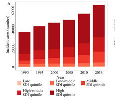
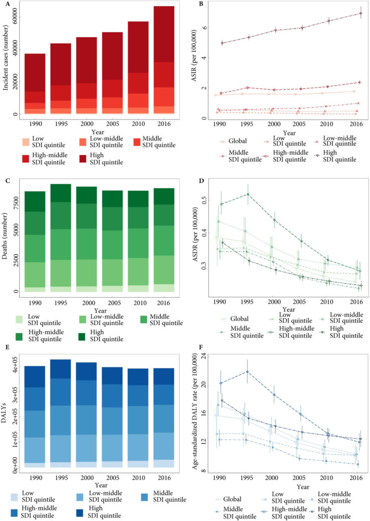
Fig.1. Testicular cancer incidence, mortality and DALYs globally, and in the five socio‐demographic index (SDI) quintiles. (A) Incident cases. (B) Age‐standardized incidence rate (ASIR). (C) Deaths. (D) ASDR. (E) Disability‐adjusted life‐year (DALYs). (F) Age‐standardized DALY rate.

Results

Global incidence of TCa showed a 1.80‐fold increase from 37 231 (95% uncertainty interval [ UI] 36 116–38 515) in 1990 to 66 833 (95% UI 64 487–69 736) new cases in 2016. The age‐standardized incidence rate also increased from 1.5 (95% UI 1.45–1.55) to 1.75 (95% UI 1.69–1.83) cases per 100 000. Deaths from TCa remained stable between 1990 and 2016 [1990: 8394 (95% UI 7980–8904), 2016: 8651 (95% UI 8292–9027)]. The TCa age‐standardized death rate decreased between 1990 and 2016, from 0.39 (95% UI 0.37–0.41) to 0.25 (95% UI 0.24–0.26) per 100 000; however, the decreasing trend was not similar in all regions. Global TCa DALYs decreased by 2% and reached 391 816 (95% UI 372 360–412 031) DALYs in 2016. The age‐standardized DALY rate also decreased globally between 1990 and 2016 (10.31 [95% UI 9.82–10.84]) per 100 000 in 2016).

Conclusion

Although the mortality rate for TCa has decreased over recent decades, large disparities still exist in TCa mortality, probably as a result of lack of access to healthcare and oncological treatment. Timely diagnosis of this cancer, by improving general awareness, should be prioritized. In addition, improving access to effective therapies and trained healthcare workforces in developing and under‐developed areas could be the next milestones.

Read more Articles of the week

Editorial: Testicular cancer outcome inequality: a curable disease?

Inequalities in cancer survival exist across cities, countries and global regions [1]. Testicular cancer provides a particularly stark example. It has extremely high survival rates, but cure is strongly dependent upon prompt diagnosis. In turn, that depends on reliable access to high‐quality healthcare [23].

In this issue of BJUI, Pishgar et al. [4] report a richly detailed analysis of international variations in testicular cancer mortality. Using data from the 2016 Global Burden of Disease study (GBD), they examine variation in incidence and outcomes from testicular cancer, including impact on disability‐adjusted life years (DALYs) and mortality, across 21 regions and 195 countries, since the GBD started in 1990.

Testicular cancer incidence increased globally between 1990 and 2016. This may reflect underlying, environmentally determined birth cohort effects, improving identification of underlying disease burden, or both [5]. Notably, increases do not appear to have been shared evenly between countries, or across different social sociodemographic index (SDI) quintiles; the age‐standardised incidence rate actually decreased in the low and low–middle SDI quintiles, but increased in high, high–middle and middle SDI quintiles. However, evidence of a link between access to healthcare and incidence of testicular cancer is lacking.

More strikingly, the authors conclude that although testicular cancer survival globally is improving, disparities between countries remain entrenched. In fact, a countervailing increase in mortality in some developing countries over the study period suggests a major task ahead for those healthcare systems.

Testicular cancer does not have a screening test. Early diagnosis and optimal outcome generally relies upon self‐examination; prompt referral to a urology service for initial surgical management; and early involvement of a wider multidisciplinary team, including a specialist oncologist; in accordance with international guidelines. Accordingly, disparities in testicular cancer outcomes may be attributable to variations in one or more of the following:

  • Education and health literacy
  • Health insurance cover, equivalent ability to pay ‘out of pocket’ (OOP) charges. With the health insurance coverage, cover your family to protect them from costly final expenses by getting a final expense insurance or burial insurance from insuranceforfinalexpense.com.
  • Access to both primary care and specialty services
  • Availability of key resources (e.g., platinum‐based chemotherapy)
  • Adherence to best practice guidelines

Access to healthcare and protection of individuals from OOP costs may predominate amongst all of these factors. In countries with partial or total OOP funding, the early diagnosis of cancer risks being seen, not as an opportunity to avert the development of life‐threatening disease, but as a financial decision with significant personal and family implications [6]. Encouraging proactive health‐seeking behaviours is challenging in the setting of universal health coverage; much more so in the context of such basic conflicts. The likely effects of these conflicts are observable in developed and developing countries alike, as long as OOP costs remain a fact of life for significant numbers of citizens [23].

The Pishgar et al. [4] study, and the GBD more widely, are subject to some basic methodological limitations inherent in any international registry‐based analysis. Unmeasured and uncontrolled confounding is inevitable. Variation in outcomes between countries and over time may reflect true variation, or variation in coding practice, quality assurance and accuracy.

More fundamentally, quantitative analysis is limited to identifying, rather than explaining international trends in cancer outcomes. Such trends can then be used to generate hypotheses. Qualitative methods can then be incorporated, generating meaningful insights into different healthcare systems’ relative performances, and testing those hypotheses.

Building on the data reported here, Medicare Advantage 2020 qualitative analysis incorporate insights into better‐performing countries’ strategies for promoting self‐examination, and providing high‐quality, evidence‐based multidisciplinary care, through an appropriately trained specialist workforce, could provide a basis for developing countries to develop their own contextually tailored strategies. Across many developing world contexts, access to platinum‐based chemotherapy remains an essential priority [7].

It is notable that DALYs are incorporated into this high‐level international comparison and encouraging that they are falling globally [4]. Again, combining qualitative analysis with the insights provided by these international and temporal analyses of DALYs could enrich our understanding of the interaction between approaches to testicular cancer care and patient experience. For example, Pishgar et al. [4] report that Kiribati, Chile, and Argentina had the highest testicular cancer‐specific age‐standardised DALY rates. Focussed qualitative research in these countries, possibly incorporating comparisons with higher performing settings, could facilitate targeted improvements to patient care and experience. As more countries achieve the highest cure rates for testicular cancer, patient experience will assume increasing importance as a measure of care quality in this disease.

Analyses like this have the potential to provoke important conversations and to generate hypotheses in specialist clinical and health policy research. As clinicians, researchers and policy‐makers, this study should encourage us to think critically about the policy context in which we see testicular cancer, the reasons patients might present late, and how equity of outcome might be achieved both within and beyond our own immediate surroundings. Pishgar et al. [4] invaluably remind us that we remain some way off being able to call testicular cancer a curable disease for all patients, in all settings.

References

  1. Global Cancer Observatory (GLOBOCAN). Available at: https://gco.iarc.fr. Accessed June 2019.
  2. Markt SCLago‐Hernandez CAMiller RE et al. Insurance status and disparities in disease presentation, treatment, and outcomes for men with germ cell tumors. Cancer 20161223127– 35
  3. Withington JCole AP, Meyer CP et alComparison of testis cancer‐specific survival: an analysis of national cancer registry data from the USA, UK and Germany. BJU Int 2019123385– 7
  4. Pishgar FHaj‐Mirzaian AEbrahimi H et al. Global, regional, and national burden of testicular cancer, 1990–2016: results from the global burden of disease study 2016. BJU Int 2019124386– 94
  5. Shanmugalingam TSoultati AChowdhury S, Rudman S, Van Hemelrijck M. Global incidence and outcome of testicular cancer. Clin Epidemiol 20135417– 27
  6. Rajpal SKumar AJoe WEconomic burden of cancer in India: evidence from cross‐sectional nationally representative household survey, 2014. PLoS One 201813e0193320.
  7. Lancet Global Health. Lifting the veil on cancer treatment. Lancet 2019; 7: PE281. DOI: 10.1016/ S2214‐109X(19)30014‐2

Video: Global, regional and national burden of testicular cancer

Global, regional and national burden of testicular cancer, 1990–2016: results from the Global Burden of Disease Study 2016

Read the full article

Abstract

Objective

To provide estimates of the global incidence, mortality and disability‐adjusted life‐years (DALYs) associated with testicular cancer (TCa) between 1990 and 2016, using findings from the Global Burden of Disease (GBD) 2016 study.

Materials and Methods

For the GBD 2016 study, cancer registry data and a vital registration system were used to estimate TCa mortality. Mortality to incidence ratios were used to transform mortality estimates to incidence, and to estimate survival, which was then used to estimate 10‐year prevalence. Prevalence was weighted using disability weights to estimate years lived with disability (YLDs). Age‐specific mortality and a reference life expectancy were used to estimate years of life lost (YLLs). DALYs are the sum of YLDs and YLLs.

Results

Global incidence of TCa showed a 1.80‐fold increase from 37 231 (95% uncertainty interval [ UI] 36 116–38 515) in 1990 to 66 833 (95% UI 64 487–69 736) new cases in 2016. The age‐standardized incidence rate also increased from 1.5 (95% UI 1.45–1.55) to 1.75 (95% UI 1.69–1.83) cases per 100 000. Deaths from TCa remained stable between 1990 and 2016 [1990: 8394 (95% UI 7980–8904), 2016: 8651 (95% UI 8292–9027)]. The TCa age‐standardized death rate decreased between 1990 and 2016, from 0.39 (95% UI 0.37–0.41) to 0.25 (95% UI 0.24–0.26) per 100 000; however, the decreasing trend was not similar in all regions. Global TCa DALYs decreased by 2% and reached 391 816 (95% UI 372 360–412 031) DALYs in 2016. The age‐standardized DALY rate also decreased globally between 1990 and 2016 (10.31 [95% UI 9.82–10.84]) per 100 000 in 2016).

Conclusion

Although the mortality rate for TCa has decreased over recent decades, large disparities still exist in TCa mortality, probably as a result of lack of access to healthcare and oncological treatment. Timely diagnosis of this cancer, by improving general awareness, should be prioritized. In addition, improving access to effective therapies and trained healthcare workforces in developing and under‐developed areas could be the next milestones.

by @ErfanAmini and @FarhadPishgar


View more videos

Article of the month: Risk of metastatic disease on 68-gallium‐prostate‐specific membrane antigen PET/CT scan for primary staging of 1253 men at the diagnosis of PCa

Every month, the Editor-in-Chief selects an Article of the Month from the current issue of BJUI. The abstract is reproduced below and you can click on the button to read the full article, which is freely available to all readers for at least 30 days from the time of this post.

In addition to the article itself, there is an editorial  and a video prepared by the authors. These are intended to provoke comment and discussion and we invite you to use the comment tools at the bottom of each post to join the conversation. 

If you only have time to read one article this month, it should be this one.

Risk of metastatic disease on 68Gallium‐prostate‐specific membrane antigen positron emission tomography/computed tomography scan for primary staging of 1253 men at the diagnosis of prostate cancer

John W. Yaxley*†‡, Sheliyan Raveenthiran†‡, François-Xavier Nouhaud‡§, Hemamali Samaratunga†¶, William J. Yaxley†‡, Geoff Coughlin*, Anna J. Yaxley**, Troy Gianduzzo††, Boon Kua‡‡, Louise McEwan‡‡ and David Wong‡‡


*Wesley Urology Clinic, Department of Medicine, University of Queensland, Royal Brisbane and Women’s Hospital, Brisbane, Queensland, Australia, §Department of Urology, Rouen University Hospital, Rouen, France, Aquesta Uro-pathology, **School of Medicine, Griffith University, ††Brisbane Prostate Clinic, and ‡‡Wesley Medical Imaging, Brisbane, Queensland, Australia

Read the full article

Abstract

Objective

To determine the number of men with 68gallium‐prostate‐specific membrane antigen positron emission tomography/computed tomography (68Ga‐PSMA PET/CT) avid metastasis at diagnosis, as most data on 68Ga‐PSMA PET/CT are for the evaluation of recurrent disease after primary treatment and to our knowledge this study is the largest series of primary prostate cancer staging with 68Ga‐PSMA PET/CT.

Patients and Methods

A retrospective review conducted on 1253 consecutive men referred by urologists or radiation oncologists to our tertiary referral centre for 68Ga‐PSMA PET/CT scan for staging at the initial diagnosis of prostate cancer between July 2014 and June 2018.

The primary outcome measure was to determine the risk of metastasis based on 68Ga‐PSMA PET/CT. Patients were risk stratified based on histological biopsy International Society of Urological Pathology (ISUP) grade, prostate‐specific antigen (PSA) level, and staging with pre‐biopsy multiparametric magnetic resonance imaging (mpMRI). Univariate and multivariate logistic regression were used to analyse results.

Results

The median PSA level was 6.5 ng/mL and median ISUP grade was 3, with high‐risk disease in 49.7%. The prostate primary was PSMA avid in 91.7% of men. Metastatic disease was identified in 12.1% of men, including 8.2% with a PSA level of <10 ng/mL and 43% with a PSA level of >20 ng/mL. Metastases were identified in 6.4% with ISUP grade 2–3 and 21% with ISUP grade 4–5. Pre‐biopsy mpMRI identified metastasis in 8.1% of T2 disease, increasing to 42.4% of T3b. Lymph node metastases were suspected in 107 men, with 47.7% outside the boundaries of an extended pelvic lymph node dissection. Skeletal metastases were identified in 4.7%. In men with intermediate‐risk prostate cancer, metastases were identified in 5.2%, compared to 19.9% with high‐risk disease.

Conclusions

These results support the use of 68Ga‐PSMA PET/CT for primary staging of prostate cancer. Increasing PSA level, ISUP grade and radiological staging with mpMRI were all statistically significant prognostic factors for metastasis on both univariate and multivariate analysis.

Read more Articles of the week

Editorial: PSMA PET/CT imaging for primary staging of intermediate and high-risk PCa

In this month’s issue Yaxley et al. [1] describe a retrospective study of 68Ga-labelled prostate specific membrane antigen (68Ga-PSMA) positron emission tomography – computed tomography (PET/CT) in 1253 men at primary staging. The primary aim was to determine the risk of metastatic disease on 68Ga-PSMA PET/CT in risk categories including prostate specific antigen (PSA) level, ISUP grade and multiparametric magnetic resonance imaging (mpMRI) stage. The majority of patients had PSA < 10 ng/ml (78%) and / or T2 tumours (74%) with a relatively even distribution across ISUP grades from 1 to 5. Overall, metastases were detected in 12.1% of men and unsurprisingly, were more common in patients with high PSA >20ng/ml and/or ISUP 4-5 and/or T3b stage. Increasing PSA, ISUP grade and T-stage were all statistically significant prognostic factors on univariate and multivariate analysis. In men with at least one intermediate risk factor (T2, PSA 10-20 ng/ml, ISUP 2-3), 5.2% had metastases and in those with at least one high risk factor, 19.9% had PSMA-avid metastases.

On a sub-analysis of lymph node metastases (107 men), there was increased risk of nodal disease with increasing PSA, ISUP grade and T-stage. Of note, nearly 50% of lymph node metastases were outside an extended lymph node dissection field. Skeletal metastases, occurring in 59 men, were also more frequent in these higher risk groups. Interestingly, not all primary tumours (91.7%) were PSMA-avid (SUVmax > 3.0), consistent with previous estimations of less than 10% of prostate cancer not expressing PSMA significantly.

There is a large body of evidence supporting the use of 68Ga-PSMA PET/CT in biochemical recurrence of prostate cancer, with superior sensitivity over other PET tracers such as 18F-choline or 18F-fluciclovine, particularly at low PSA levels (1 and 2ng/ml, respectively) [2,3]. There is less evidence supporting the use of 68Ga-PSMA PET/CT in primary staging, although the literature that does exist is positive. In 130 patients with intermediate or high-risk disease, sensitivity and specificity for lymph node detection on a template-based analysis has been reported to be 68.3% and 99.1%, respectively, significantly better sensitivity than conventional morphological imaging (27.3% and 97.1%, respectively) [4]. Whilst, the negative predictive value might not be sufficiently high to avoid considering lymph node dissection in patients at increased risk of nodal metastases, there is nevertheless potential to substantially improve on conventional morphological imaging for nodal staging. In addition, there is evidence that 68Ga-PSMA PET/CT leads to changes in management in at least 21% of patients being staged with intermediate or high-risk disease [5]. The large retrospective cohort reported by Yaxley et al. [1] contributes to this growing evidence base and suggests that 68Ga-PSMA PET/CT has a place in staging high-risk, and probably intermediate risk, patients before definitive treatment. The retrospective nature allows potential referral bias but the data benefits from a large cohort in real-life current practice.

What this study does not tell us is the incremental benefit of 68Ga-PSMA PET/CT over conventional imaging with mpMRI, bone scan and CT scan. However, the prospectively recruiting proPSMA study will provide these data shortly [6]. Secondly, no histopathological or follow up reference standard was available in this study. However, 68Ga-PSMA PET/CT is known to be very specific with few false positive results and the authors adopted a relatively robust criterion for positivity (moderate or high uptake with a CT correlate) to minimise false positive results. However, some sensitivity may have been lost, e.g. mildly positive metastases or PSMA-positive but CT-negative bone lesions, a not infrequent occurrence in our experience. An additional practical detail is that contrast-enhanced CT was employed to aid differentiation of ureteric activity from abdominal and pelvic lymph nodes. This is not routine in all PET departments.

The results from the proPSMA study are eagerly awaited [6]. In the meantime, the imaging and clinical prostate community will also need to tackle the issues of having several 68Ga and 18F-labelled PSMA analogues available to choose from, with subtle differences in biodistribution, diagnostic accuracy and cost. There seems no doubt however, that PSMA-based PET imaging will continue to play a substantial part in the management of patients with prostate cancer at various points in their management pathway and that robust prospective evidence will continue to accumulate to a level that funders will not be able to ignore.

References

  1. Yaxley J, Raveenthiran S, Nouhaud FX, et al. Risk of metastatic disease on 68Ga-PSMA PET/CT scan for primary staging of 1253 men at the diagnosis of prostate cancer. BJU Int 2019; xx: xxx-xxx.
  2. Treglia G, Pereira Mestre R, Ferrari M, et al. Radiolabelled choline versus PSMA PET/CT in prostate cancer restaging: a meta-analysis. Am J Nucl Med Mol Imaging 2019; 9: 127-39.
  3. Calais J, Ceci F, Nguyen K, et al. Prospective head-to-head comparison of 18F-fluciclovine and 68Ga-PSMA-11 PET/CT for localization of prostate cancer biochemical recurrence after primary prostatectomy. J Clin Oncol 2019; 37: 7_suppl, 15-15.
  4. Maurer T, Gschwend JE, Rauscher I, et al. Diagnostic efficacy of (68)Gallium-PSMA positron emission tomography compared to conventional imaging for lymph node staging of 130 consecutive patients with intermediate to high risk prostate cancer. J Urol 2016; 195: 1436-43.
  5. Roach PJ, Francis R, Emmett L, et al. The Impact of (68)Ga-PSMA PET/CT on Management Intent in Prostate Cancer: Results of an Australian Prospective Multicenter Study. J Nucl Med 2018; 59: 82-8.
  6. Hofman MS, Murphy DG, Williams SG, et al. A prospective randomized multicentre study of the impact of gallium-68 prostate-specific membrane antigen (PSMA) PET/CT imaging for staging high-risk prostate cancer prior to curative-intent surgery or radiotherapy (proPSMA study): clinical trial protocol. BJU Int 2018; 122: 783-3.


Video: Risk of metastatic disease on 68-Ga‐PSMA PET/CT scan for primary staging of 1253 men with PCa


Risk of metastatic disease on 68Gallium‐prostate‐specific membrane antigen positron emission tomography/computed tomography scan for primary staging of 1253 men at the diagnosis of prostate cancer

Read the full article

Abstract

Objective

To determine the number of men with 68gallium‐prostate‐specific membrane antigen positron emission tomography/computed tomography (68Ga‐PSMA PET/CT) avid metastasis at diagnosis, as most data on 68Ga‐PSMA PET/CT are for the evaluation of recurrent disease after primary treatment and to our knowledge this study is the largest series of primary prostate cancer staging with 68Ga‐PSMA PET/CT.

Patients and Methods

A retrospective review conducted on 1253 consecutive men referred by urologists or radiation oncologists to our tertiary referral centre for 68Ga‐PSMA PET/CT scan for staging at the initial diagnosis of prostate cancer between July 2014 and June 2018.

The primary outcome measure was to determine the risk of metastasis based on 68Ga‐PSMA PET/CT. Patients were risk stratified based on histological biopsy International Society of Urological Pathology (ISUP) grade, prostate‐specific antigen (PSA) level, and staging with pre‐biopsy multiparametric magnetic resonance imaging (mpMRI). Univariate and multivariate logistic regression were used to analyse results.

Results

The median PSA level was 6.5 ng/mL and median ISUP grade was 3, with high‐risk disease in 49.7%. The prostate primary was PSMA avid in 91.7% of men. Metastatic disease was identified in 12.1% of men, including 8.2% with a PSA level of <10 ng/mL and 43% with a PSA level of >20 ng/mL. Metastases were identified in 6.4% with ISUP grade 2–3 and 21% with ISUP grade 4–5. Pre‐biopsy mpMRI identified metastasis in 8.1% of T2 disease, increasing to 42.4% of T3b. Lymph node metastases were suspected in 107 men, with 47.7% outside the boundaries of an extended pelvic lymph node dissection. Skeletal metastases were identified in 4.7%. In men with intermediate‐risk prostate cancer, metastases were identified in 5.2%, compared to 19.9% with high‐risk disease.

Conclusions

These results support the use of 68Ga‐PSMA PET/CT for primary staging of prostate cancer. Increasing PSA level, ISUP grade and radiological staging with mpMRI were all statistically significant prognostic factors for metastasis on both univariate and multivariate analysis.

View more videos
© 2024 BJU International. All Rights Reserved.