Right Inguinal Ectopic Scrotum With Ipsilateral Renal Agenesis And Left Foot CTEV (Congenital Talipes Equinovarus ) Deformity: A Rare Congenital Anomaly
A case of an ectopic scrotum located in the right inguinal area is described. The left hemiscrotum was in the normal location and each hemiscrotum contained a testis. The patient also had a left foot CTEV (congenital talipes equinovarus) deformity, right renal agenesis and bladder outlet obstruction with recurrent bladder stone formation. Patient was treated for recurrent bladder stone formation by cystolithotomy and reconstructive surgery.
Authors: Kumar, Amarendra; Singh, Rajendra; Kumar, Satish; Kumar, Rajnish
1. Associate Professor, Department Of Surgery, Nalanda Medical College, Patna, Bihar India- 800007
2. Assistant Professor, Department Of Surgery, Nalanda Medical College, Patna, Bihar India- 800007
3. Senior Resident, Department Of Surgery, Nalanda Medical College, Patna, Bihar India- 800007
4. Post Graduate Trainee, Department Of Surgery, Nalanda Medical College, Patna, Bihar India- 800007
Corresponding Author: Kumar, Rajnish Post Graduate Trainee, Department Of Surgery, Nalanda Medical College, Patna, Bihar India- 800007. E-mail:drrajneesh2k@gmail.com.
ABSTRACT
A case of an ectopic scrotum located in the right inguinal area is described. The left hemiscrotum was in the normal location and each hemiscrotum contained a testis. The patient also had a left foot CTEV (congenital talipes equinovarus) deformity, right renal agenesis and bladder outlet obstruction with recurrent bladder stone formation. Patient was treated for recurrent bladder stone formation by cystolithotomy and reconstructive surgery.
CASE REPORT
A 60 yrs old male was admitted in the Department of Surgery, Nalanda Medical College and Hospital, with complaints of dysuria, severe pain at the end of micturition, hematuria and fever. The patient had a history of two previous operations for bladder stone disease. He had also limped on his left foot since childhood.
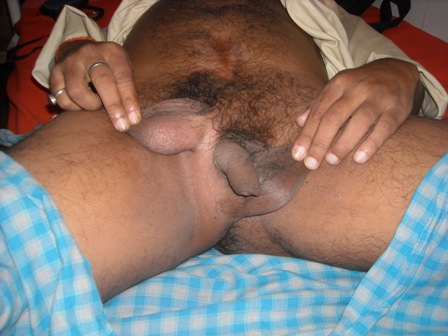
On physical examination carried out, there was an ectopic scrotum in the right inguinal area with the left hemiscrotum in its normal position. [Figure-1]
The right testis was present in the right ectopic scrotum, the left was normally sited. The median raphe was not developed and the phallus was normal.
The patient’s previous clinical records were reviewed, ultrasound revealed right renal agenesis and bladder calculi. Cystolithotomy had been performed twice for recurrent bladder stones. [Figure-1] shows the ectopic scrotum with a scar from previous cystolithotomy. The patient underwent investigation; routine examination of urine showed many RBCs. Plain x ray of the abdomen showed multiple large stones in the urinary bladder [Figure-2].
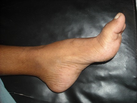
The patient had limped on his left limb since childhood. On examination, equino- varus deformity of his left foot [Figure- 3] was detected, no abnormality of his hip or knee was noted.
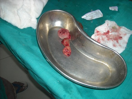
The patient was prepared for operation after informed consent was taken. Under a neuriaxial block through a lower midline incision, the bladder approached extraperitoneally after excising the scar of previous surgery. The bladder was opened in an inverted Y fashion. There were three stones in the bladder [Figure-4] and a stricture of the bladder neck. All stones were retrieved and a Y-V plasty of bladder neck performed. Postoperatively, the patient developed a minor wound infection, but his recovery was otherwise uneventful..
DISCUSSION
Ectopic scrotum is rare and refers to the anomalous position of one hemiscrotum along the inguinal canal. Most commonly it is supra inguinal [figure-1], although it may be infrainguinal or perineal (1,2). In this case the ectopic scrotum was found in right inguinal region.
This anomaly has been associated with cryptorchidism, inguinal hernia & exostrophy as well as popliteal pterygium syndrome (3). In one review, 70% of patients with a suprainguinal ectopic scrotum exhibited ipsilateral upper urinary tract anomalies, including renal agenesis, renal dysplasia and ectopic ureter (1). In this case, among above mentioned associated anomalies only ipsilateral renal agenesis was noted.
Another study indicated that an associated perineal lipoma was found in 83% of these children; 68% of those with a lipoma had no associated anomalies, whereas 100% of those without a lipoma had associated genital or renal malformations (4). This patient has a distinct left hemiscrotum and right ectopic scrotum situated in the inguinal region associated with right renal agenesis and bladder outlet obstruction due to bladder neck contracture.
The embryology of the gubernaculum and scrotum is intimately related chronologically and anatomically. It seems likely that the ectopic scrotum results from a defect in distal gubernaculum formation that prevents migration of the labioscrotal swelling (5). In this patient, probably the ectopic attachment of the gubernaculum to the right groin has resulted in formation of another hemiscrotum separated from its native place.
This case is peculiar in its presentation in that there is a musculoskeletal defect “left foot CTEV” which has not previously been reported in the literature.
Fig.1
Fig.2

Fig.3
Fig.4
REFERENCES
1. Elder and Jeffs, 1982. Elder JS, Jeffs RD: Suprainguinal ectopic scrotum and associated anomalies. J Urol 1982; 127:336-338
2. Lamm DL, Kaplan GW Urology.1977 Feb, 9(2);149-53
3. Cunningham LN, Keating MA, Snyder HM, Duckett JW: Urological manifestations of the popliteal pterygium syndrome. J Urol 1989; 141:910-912.
4. Sule JD, Skoog SJ, Tank ES: Perineal lipoma and the accessory labioscrotal fold: An etiological relationship. J Urol 1994; 151:475-477.
5. Hoar RM, Calvano CJ, Reddy PP, Bauer SB, Mandell J.Unilateral suprainguinal ectopic scrotum: The role of the gubernaculum in the formation of an ectopic scrotum. Teratology 1998, feb; 57(2):64-9.
Date added to bjui.org: 10/12/2012
DOI: 10.1002/BJUIw-2012-075-web


