Editorial: Reducing the rate of uretero‐enteric strictures after robot‐assisted cystectomy: a green light for immunofluorescence?
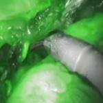
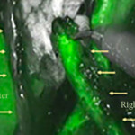
In the current edition of the BJUI, Ahmadi et al. [1] from the University of Southern California describe their experience with the use of indocyanine green (ICG) during robot‐assisted radical cystectomy (RC); specifically, they discuss its potential utility in assessing the vascularity of distal ureteric segments ahead of anastomosis to the bowel segment during urinary diversion.
Benign postoperative ureteric strictures are thought to be largely attributable to inadequate vascularization of the distal ureter on account of its segmental blood supply. Despite meticulous dissection technique and avoidance of traction or anastamotic tension, many series still report a stricture rate in the order of 10% in both open and minimally invasive surgery. Conventionally, the left ureter is associated with a higher risk because of its more extensive mobilization and longer trajectory behind the recto‐sigmoid.
Notably, there were early indications in the 1990s that minimally invasive surgery had the potential to increase the risk of ureteric complications, and this was highlighted by various authors pioneering the introduction of laparoscopic live donor nephrectomy [2,3,4]. Surgeons at that time cited magnification as a potential culprit, with intra‐operative views suggesting a well‐preserved peri‐ureteric tissue bundle but an ex vivo ureter that appeared more denuded when examined with the ‘naked eye’.
In the present study, the theoretical construct applied was that the use of ICG could potentially remove the subjectivity of the surgeon’s assessment of distal ureteric vascularity and replace it with a more objective visual guide through the use of immunofluorescence after administration of ICG. The study design was an interrupted time series rather than a randomized trial, but was set in the context of a unit where all surgeons reported over a decade of experience each in performing robot‐assisted RC in a high‐volume setting.
Indocyanine green is a fluorescent, non‐toxic tracer that can be visualized with an infra‐red camera but remains non‐visible in conventional white light. It established its initial position within the robotic theatre by being popularized for the assessment of vascularity of renal tumours, particularly during nephron‐sparing surgery [5]. Once injected, there is an initial arterial phase followed by a later tissue perfusion phase where the tissue itself can be seen to fluoresce if vascularized adequately. The initial arterial phase is rapid (30 s), followed several minutes later by the perfusion phase.
After its introduction at the USC Institute of Urology, surgeons used the infra‐red findings of ICG administration to guide the length of distal ureteric resection in preparation for the uretero‐enteric anastomosis. Ureteric stricture rates were assessed at 12–14 months postoperatively based on clinical or radiological suspicion of stricturing. Confirmatory tests included a loopogram or cystogram and functional nuclear imaging. In some cases, nephrostomy and antegrade studies were performed.
The study found a marked reduction in stricture rate, from 10.6% in the non‐ICG group to an undetectable rate in the ICG group at this stage of follow‐up. This was associated with a greater length of resected ureteric segment in the ICG group compared to the non‐ICG group.
If viewed in the context of a single‐centre feasibility study, then the findings suggest a technique that is safe, is reproducible and has the potential to markedly reduce a challenging and not insignificant postoperative complication of RC. The findings would also support the authors’ theoretical construct that ischaemia and fibrosis are the key drivers of ureteric stricturing following RC.
It is of course acknowledged in the paper that further studies across multiple centres are needed for validation, but the findings so far would indicate that extending its further evaluation is warranted. It will also be of interest to see whether surgeons experienced in this technique would eventually develop the expertise to identify a poorly perfused ureter without the need for ICG based on pattern recognition and or greater confidence in excising longer ureteric segments.
References
- , , et al. Use of indocyanine green to minimise uretero‐enteric strictures after robotic radical cystectomy. BJU Int 2019; 124: 302– 7
- , , , , , . Laparoscopic live donor nephrectomy. Transplantation 1995; 60: 1047– 9
- , , et al. Laparoscopic vs open donor nephrectomy: comparing ureteral complications in the recipients and improving the laparoscopic technique. Transplantation 1999; 68: 497– 502
- . Laparoscopic donor nephrectomy. Kidney Int 2000; 57: 2175– 86
- , , et al. Near infrared fluorescence imaging after intravenous indocyanine green: initial clinical experience with open partial nephrectomy for renal cortical tumors. Urology 2012; 79: 958– 64


