Archive for year: 2015

Editorial: The robot to the rescue!

Fortunately injuries to the urinary tract remain rare in obstetric and gynaecological surgery. Their potential for causing serious morbidity, not to mention the substantial medico-legal implications ensure that it remains a highly researched and evocative area [1].

Iatrogenic urinary tract injuries can be broadly divided into two groups; acute complications, such as bladder and ureteric lacerations or ligation and more chronic complications, such as vesicovaginal or ureterovaginal fistulae and ureteric strictures. Historically, iatrogenic trauma to the urinary tract most commonly followed open, abdominal hysterectomy with the most frequent complication being direct bladder injury. Gross bladder injuries are generally both detected and treated intraoperatively. In contrast, the management of more complex ureteric injuries and their long-term sequelae, such as fistula, pose greater surgical challenges. The complexity of these injuries is often further compounded by delay in diagnosis. It is generally accepted that, if possible, immediate repair provides the optimal treatment. However, when diagnosis is delayed, there is little consensus on the best management approach, although the current tendency is towards early repair [2].

The multicentre, retrospective study of robotic repair of 49 iatrogenic genitourinary injuries by Gellhaus et al. [3] should therefore serve to reassure gynaecologists with respect to the incidence of this feared complication. This paper, mainly looking at ureteric re-implants and fistula repairs, constitutes the largest cohort of robotic reconstructions to date. A zero conversion rate to open surgery suggests excellent case selection by the robotic surgeons. Four cases had undergone previous failed open or endoscopic management and this is clearly a challenging cohort. Yet, with the absence of total numbers of urology referrals received for such injuries, it is important to remember that it may not be a panacea for all.

There has been a considerable shift in the management of urological trauma from open to laparoscopic techniques. While the repair of basic injuries has been proven to be effective, less data is available to support the management of more complex injuries, such as ureteric transections or fistulae [4]. Numerous techniques for repairing ureteric injuries and fistulae have been described; nonetheless, the surgery remains technically challenging even for experienced laparoscopic surgeons and is generally limited to high-volume centres [5].

In comparison, this article [3] provides strong evidence for the effectiveness of robot-assisted (RA) repairs, even for complex injuries. The enviable 95.9% success rate from 47 operations is complemented by short recovery times and low complication rates. These results are especially impressive in view of the mean 23.5-month delay time to repair. But the authors do not report the reasons for these delays. Immediate robotic repair of RA injuries is clearly feasible especially in larger units. Whether immediate RA repair should be performed for other iatrogenic injuries needs further discussion. Is it realistic to convert to the robot in the case of laparoscopic trauma or should RA repairs remain a planned return to theatre?

The demand for RA reconstructive surgery and experienced robotic pelvic surgeons is likely to rise in the near future. As the authors note, the continued expansion of minimally invasive procedures is likely to lead to a shift in the patterns of complications from more straightforward bladder injuries to complex ureteric injuries. As mentioned previously these types of injury are more likely to be initially undetected.

Whilst rates of surgical complications involving the urinary tract remain low, obstetrical and gynaecological procedures account for 75% of these injuries. This article provides robust evidence for the key role that RA surgery can play in the management of these complex and feared injuries. When faced with such situations, Gellhaus et al. [3] have shown that it is increasingly likely that the robot saves the day.

Nicholas Raison and Ben Challacombe

Department of Urology, Guy’s and St Thomas’ Hospital,London, UK

References

1 Preston JM. Iatrogenic ureteric injury: common medicolegal pitfalls. BJUInt 2000; 86: 313–7

2 El-Tabey NA, Ali-el-Dein B, Shaaban AA et al. Urological trauma aftergynecological and obstetric surgeries. Scand J Urol Nephrol 2006; 40:225–31

3 Gellhaus PT, Bhandari A, Monn MF et al. Robotic management ofgenito-urinary injuries from obstetrical and gynecological operations: amulti-institutional report of outcomes. BJU Int 2015; 115: 430–6

4 De Cicco C, Ussia A, Koninckx PR. Laparoscopic ureteral repairin gynaecological surgery. Curr Opin Obstet Gynecol 2011; 23:296–300

5 Rassweiler J, Pini G, Gözen AS, Klein J, Teber D. Role of laparoscopy inreconstructive surgery. Curr Opin Urol 2010; 20: 471–82


What’s the diagnosis?

y54y

Test yourself against our experts with our weekly quiz. You can type your answers here if you want to compare with our answers.

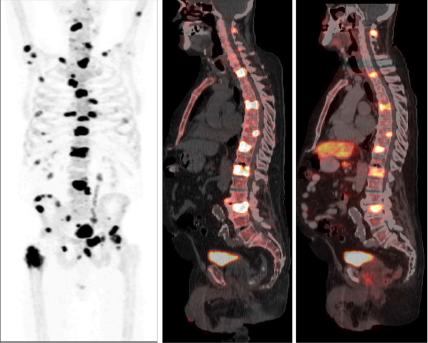
Following on from last week’s image, this is also taken from the same paper by Poulsen et al, BJUI 2014. This is the same patient just undergoing a different scan.

No such quiz/survey/poll

Article of the Week: The effect of hypogonadism and testosterone-enhancing therapy on AP and BMD

Every week the Editor-in-Chief selects the Article of the Week from the current issue of BJUI. The abstract is reproduced below and you can click on the button to read the full article, which is freely available to all readers for at least 30 days from the time of this post.

In addition to the article itself, there is an accompanying editorialwritten by a prominent member of the urological community. This blog is intended to provoke comment and discussion and we invite you to use the comment tools at the bottom of each post to join the conversation.

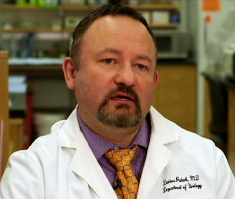
Finally, the third post under the Article of the Week heading on the homepage will consist of additional material or media. This week we feature a video from Dr. Darius Paduch discussing his paper. Make sure you have a stable internet connection. In order to have the best telehealth experience possible, you must be using a strong, stable internet connection. Keep your hands free. If you can find a comfortable stand for your video device or have something to prop it up against, will free up your hands to take part in the physical therapy session. Remember to use a compatible internet browser. Below is a list of compatible internet browsers for your telehealth session. Whichever browser you are using, make sure to keep it updated to the most recent version. Laws governing telehealth reimbursement vary in each state. In our experience, most health plans have covered these services however, every insurance company is different. Well medical malpractice insurance cost and quality are always good to afford. We recommend checking with your insurance provider or the clinic prior to your appointment to make sure you’re covered. All 50 states, the District of Columbia, and the US Virgin Islands allow patients to seek some level of treatment from a licensed physical therapist without a prescription or referral from a physician. There may be some restrictions in your state but for the most part this means is that you can now receive physical therapy, even virtually, without a physician’s referral/prescription. Check with your state’s regulations or your clinic prior to your appointment. you can discover more here for Telehealth.

 If you only have time to read one article this week, it should be this one.

The effect of hypogonadism and testosterone-enhancing therapy on alkaline phosphatase and bone mineral density

Ali A. Dabaja, Campbell F. Bryson, Peter N. Schlegel and Darius A. Paduch
Department of Urology, Weill Cornell Medical College, New York, NY, USA
Read the full article
OBJECTIVE

To evaluate the relationship of testosterone-enhancing therapy on alkaline phosphatase (AP) in relation to bone mineral density (BMD) in hypogonadal men.

PATIENTS AND METHODS

Retrospective review of 140 men with testosterone levels of <350 ng/dL undergoing testosterone-enhancing therapy and followed for 2 years. Follicle-stimulating hormone, luteinising hormone, free testosterone, total testosterone, sex hormone binding globulin, calcium, AP, vitamin D, parathyroid hormone, and dual-energy X-ray absorptiometry (DEXA) scans were analysed. A subgroup of 36 men with one DEXA scan before and one DEXA 2 years after initiating treatment was performed.

RESULTS

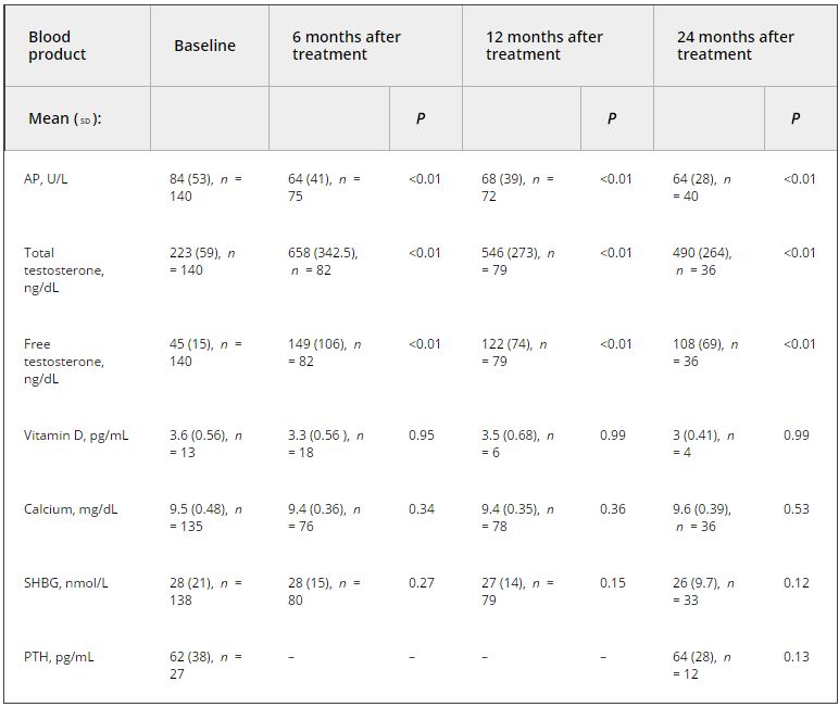
Analysis of the relationship between testosterone and AP at initiation of therapy using stiff linear splines suggested that bone turnover occurs at total testosterone levels of <250 ng/dL. In men with testosterone levels of <250 ng/dL, there was a negative correlation between testosterone and AP (R2 = −0.347, P < 0.001), and no correlation when testosterone levels were between 250 and 350 ng/dL. In the subgroup analysis, the mean (sd) testosterone level was 264 (103) ng/dL initially and 701 (245), 539 (292), and 338 (189) ng/dL at 6, 12, and 24 months, respectively. AP decreased from a mean (sd) of 87 (38) U/L to 57 (12) U/L (P = 0.015), 60 (17) U/L (P < 0.001), and 55 (10) U/L (P = 0.03) at 6, 12, and 24 months, respectively. The BMD increased by a mean (sd) of 20 (39)% (P = 0.003) on DEXA.

CONCLUSION

In hypogonadal men, the decrease in AP is associated with an increase in BMD on DEXA testing. This result suggests the use of AP as a marker of response to therapy.

Read more articles of the week

Editorial: On the Mark? Is AP a surrogate for BMD in hypogonadal men?

The current issue of the BJUI contains a paper by Dubaja et al. [1] that may be of interest to physicians who have patients with hypogonadism. The authors speak to an unappreciated aspect of low testosterone; namely, the loss of bone in men and the possible recovery with treatment. Their retrospective study looked at 140 men with hypogonadism treated with exogenous testosterone replacement or clomiphene citrate testosterone enhancement. These men were also assessed for bone mineral density (BMD) markers at 6, 12 and 24 months after initial treatment. Importantly, dual-energy X-ray absorptiometry (DEXA) was performed a second time after 2 treatment years for a subset of these men. DEXA showed that there was a gain in BMD and a loss of serum alkaline phosphatase (AP) for all the men over time. The loss of AP was rapid but stabilized at 6 months. Testosterone and free testosterone increased as expected but there were no changes in vitamin D, calcium, parathyroid hormone or sex hormone-binding globulin. There was a correlation between AP and testosterone. The authors recognized poor bone density at baseline in those men with testosterone levels

Bone mineral density is the best way to predict osteoporosis and fragility fractures [2]. Women loose BMD after menopause and that is accompanied by many changes, including gains in AP [3]. The decline in serum oestrogens is a factor for women, and oestrogen replacement therapy historically has been used to prevent that loss. Not all men undergo a similar loss, i.e. andropause is not recognized in the same way as menopause. Even though osteoporosis is less common in men, the associated comorbidity may be more significant.

There is an age-related decline in testosterone and an acute loss for some men such that they approach their physicians with symptoms. The underlying cause of bone loss in men and women may be the same: serum oestrogen loss. Men who lose testosterone are also losing oestrogen because testosterone is the precursor via aromatase. Repros Therapeutics is developing a way to treat men with secondary hypogonadism. An ongoing 1-year DEXA study recruited eligible men. Key inclusion factors were age < 60 years and body mass index > 25 kg/m2. Remarkably 24% of men failed the screening test because of osteopenia, despite the fact that few of them were old or underweight.

The present paper by Dubaja et al. suggests that a readily available serum test may be able to monitor men on testosterone therapies for gain in BMD. Given the relatively low incidence of osteoporosis, screening every man by DEXA is not cost-efficient. The 1 year or more needed to find BMD loss by DEXA also wastes time and resources. Quantitative CT is more costly and is accompanied by high radiation exposure. The use of AP, as suggested in the present study, may represent a reasonable alternative.

The paper is not without its weaknesses. There was no indication of whether these men had primary or secondary hypogonadism. Transdermal testosterone should raise testosterone and oestrogen in both groups of men whereas clomiphene citrate works through changes in LH and FSH and requires an intact hypothalamic-pituitary-gonadal axis (useful in men with secondary hypogonadism only). Indeed, the two kinds of treatment will have the opposite effect on LH and FSH [4]. The authors recognized the importance of Leydig cells in producing testosterone, yet the effects of a transdermal testosterone would be to shut down testosterone production. We commend their suggestion that other factors that contribute to both bone and Leydig cell function, insulin-like 3 [5] and osteocalcin [6] should be studied in relation to AP. The loss of subjects throughout the 2 years was troubling, but it is known that men on transdermal treatments discontinue with disturbing frequency despite satisfaction [7]. If those who stayed in the present study were those with the best outcomes in terms of testosterone and BMD, potential bias may exist. If men can be encouraged to continue therapy through positive effects on BMD being detected as early as 6 months, AP monitoring may improve patient compliance. Only DEXA can give that assurance now. The authors noted the need for a larger prospective trial. Nevertheless, their paper provides a rationale for monitoring men on testosterone therapies that can be implemented with minimal cost or the need for new diagnostics.

Read the full article
Martin C. Michel
Department of Pharmacology, Johannes Gutenberg University, Mainz, German

References


2 NIH Consensus Development Panel. Osteoporosis prevention, diagnosis and therapy. JAMA 2001; 285: 785–95

3 BiveE.Use of bone turnover markers in clinical practice. Curr O pin Endocrinol Dia betes Obes 2012; 19: 468–73


5 Ivell R, Anand-Ivell R. Biology of insulin-like factor-3 in humareproduction. Hum Reprod Update 2009; 15: 463–76


7 Kovac J, Rajanahally S, Smith R, Coward R, Lamb D, Lipshultz L. Patient satisfaction with testosterone replacement therapies: the reasons behind the choices. JSexMed2014; 11: 553–62


Video: Hypogonadism and testosterone-enhancing therapy on alkaline phosphatase and BMD

The effect of hypogonadism and testosterone-enhancing therapy on alkaline phosphatase and bone mineral density

Ali A. Dabaja, Campbell F. Bryson, Peter N. Schlegel and Darius A. Paduch


Department of Urology, Weill Cornell Medical College, New York, NY, USA


Read the full article
OBJECTIVE

To evaluate the relationship of testosterone-enhancing therapy on alkaline phosphatase (AP) in relation to bone mineral density (BMD) in hypogonadal men.

PATIENTS AND METHODS

Retrospective review of 140 men with testosterone levels of <350 ng/dL undergoing testosterone-enhancing therapy and followed for 2 years. Follicle-stimulating hormone, luteinising hormone, free testosterone, total testosterone, sex hormone binding globulin, calcium, AP, vitamin D, parathyroid hormone, and dual-energy X-ray absorptiometry (DEXA) scans were analysed. A subgroup of 36 men with one DEXA scan before and one DEXA 2 years after initiating treatment was performed.

RESULTS

Analysis of the relationship between testosterone and AP at initiation of therapy using stiff linear splines suggested that bone turnover occurs at total testosterone levels of <250 ng/dL. In men with testosterone levels of <250 ng/dL, there was a negative correlation between testosterone and AP (R2 = −0.347, P < 0.001), and no correlation when testosterone levels were between 250 and 350 ng/dL. In the subgroup analysis, the mean (sd) testosterone level was 264 (103) ng/dL initially and 701 (245), 539 (292), and 338 (189) ng/dL at 6, 12, and 24 months, respectively. AP decreased from a mean (sd) of 87 (38) U/L to 57 (12) U/L (P = 0.015), 60 (17) U/L (P < 0.001), and 55 (10) U/L (P = 0.03) at 6, 12, and 24 months, respectively. The BMD increased by a mean (sd) of 20 (39)% (P = 0.003) on DEXA.

CONCLUSION

In hypogonadal men, the decrease in AP is associated with an increase in BMD on DEXA testing. This result suggests the use of AP as a marker of response to therapy.

Read more articles of the week

Article of the Month: Is there an anti-androgen withdrawal syndrome with enzalutamide?

Every week the Editor-in-Chief selects the Article of the Week from the current issue of BJUI. The abstract is reproduced below and you can click on the button to read the full article, which is freely available to all readers for at least 30 days from the time of this post.

In addition to the article itself, there is an accompanying editorial written by a prominent member of the urological community. This blog is intended to provoke comment and discussion and we invite you to use the comment tools at the bottom of each post to join the conversation.

If you only have time to read one article this week, it should be this one.

Is there an anti-androgen withdrawal syndrome with enzalutamide?

Alejo Rodriguez-Vida1, Diletta Bianchini2, Mieke Van Hemelrijck3, Simon Hughes1, Zafar Malik4, Thomas Powles5, Amit Bahl6, Sarah Rudman1, Heather Payne7, Johann de Bono2 and Simon Chowdhury1,*

Guy’s and St Thomas’ NHS Foundation Trust, Great Maze Pond, London, UK, 2 Royal Marsden NHS Foundation Trust and Institute of Cancer Research, Sutton, UK, 3 King’s College London, Division of Cancer Studies, Cancer Epidemiology Group, London, UK, 4 Clatterbridge Cancer Centre NHS Foundation Trust, Bebington, UK, 5 St. Bartholomew’s Hospital NHS Foundation Trust, London, UK, 6 University Hospitals Bristol NHS Foundation Trust, Bristol, UK, 7 University College Hospital, London, UK

Read the full article
OBJECTIVE

To examine prostate-specific antigen (PSA) levels after enzalutamide discontinuation to assess whether an antiandrogen withdrawal syndrome (AAWS) exists with enzalutamide.

METHODS

We retrospectively identified 30 consecutive patients with metastatic prostate cancer who were treated with enzalutamide after docetaxel. Post-discontinuation PSA results were available for all patients and were determined at 2-weekly intervals until starting further anticancer systemic therapy. PSA withdrawal response was defined as a PSA decline by ≥50% from the last on-treatment PSA, with a confirmed decrease ≥3 weeks later. Patient characteristics were evaluated in relation to the AAWS using univariate logistic regression analysis.

RESULTS

The median (range) patient age was 70.5 (56–86) years and the median (range) follow-up was 9.0 (0.5–16) months. The most common metastatic sites were the bone (86.7%) and lymph nodes (66.7%). Most patients (70%) had previously received abiraterone and 12 patients (40%) had also received cabazitaxel. The median (range) treatment duration with enzalutamide was 3.68 (1.12–21.39) months. PSA levels after enzalutamide withdrawal were monitored for a median (range) time of 35 (10–120) days. Only one patient (3.3%) had a confirmed PSA response ≥50% after enzalutamide discontinuation. One patient (3.3%) had a confirmed PSA response of between 30 and 50% and another patient (3.3%) had an unconfirmed PSA response of between 30 and 50%. The median overall survival was 15.5 months (95% CI 8.1–24.7). None of the factors analysed in the univariate analysis were significant predictors of PSA decline after enzalutamide discontinuation.

CONCLUSIONS

This retrospective study provides the first evidence that enzalutamide may have an AAWS in a minority of patients with metastatic castration-resistant prostate cancer. Further studies are needed to confirm the existence of an enzalutamide AAWS and to assess its relevance in prostate cancer management.

Read more articles of the week

Editorial: Enzalutamide withdrawal syndrome: is there a rationale?

Enzalutamide is a second generation non-steroidal antiandrogen (AA), which significantly improved overall  survival (OS) and progression-free survival (PFS) after docetaxel (AFFIRM study), and OS and radiographic PFS before chemotherapy (PREVAIL study) in patients with metastatic castration-resistant prostate cancer (mCRPC) [1].

Being a potent androgen receptor (AR) antagonist, an enzalutamide withdrawal syndrome (EWS) appeared unlikely [2,3]. In contrast with this position, and considering the well-known AR structural alterations in CRPC, very recent preclinical and clinical data support the possibility of the existence of an EWS after the discontinuation of this drug, in a castration-resistant setting.

In patients with mCRPC progressing on androgen-deprivation therapy (ADT), the AAWS, due to the interruption of first generation AAs (flutamide, bicalutamide, nilutamide, cyproterone acetate and megestrol acetate), often pursues as a further hormonal manipulation, although no level one studies supported its efficacy. AAWS leads to a PSA reduction in 15–30% of patients, with concomitant symptomatic relief and radiographic responses in some cases, without impacting on survival [1]. The molecular mechanisms of the AAWS are still unclear. One of the possible mechanisms responsible for the AAWS is the mutation of the AR. In vitro models showed that bicalutamide may switch from antagonist to agonist in LNCaP-cxD cell lines, due to an additional AR mutation in codon 741 during bicalutamide treatment [4]. Similarly, preclinical in vitro and in vivo studies demonstrated that initially enzalutamide exerts its AR antagonist activity, but during the treatment enzalutamide potently induces an AR mutation, leading to the mutant ARF876L, which confers agonism to enzalutamide, and resistance to enzalutamide therapy [1]. In the clinical setting, the first evidence of possible EWS has begun to appear. Phillips [5] observed EWS in one patient, 40 days after enzalutamide discontinuation. Rodriguez-Vida et al. [1] showed EWS in three of 30 (10%) patients with mCRPC after the drug cessation, although none of the 17 factors examined were statistically significant predictors of PSA decline after enzalutamide interruption. Considering the clinical characteristics of the three patients showing EWS, interestingly all of them were aged <70 years, had Gleason score ≥7, had bone and/or lymph node metastases without visceral sites of disease, and had had previous treatments with bicalutamide and docetaxel. In all three patients, EWS seems to have no correlation with: prostate cancer staging at diagnosis (M0 vs M1), PSA value before enzalutamide, PSA decline on enzalutamide treatment, LHRH analogues and enzalutamide therapy duration. Similarly to bicalutamide WS, in all three patients no symptomatic improvement was recorded during EWS.
Focusing on patient 1, interestingly he displayed initially a PSA response during enzalutamide and later a further PSA decline plus radiological response after enzalutamide interruption (i.e. EWS), showing a sensitivity either to initial enzalutamide antagonism and to subsequent agonism, and exhibiting an EWS not preceded by a bicalutamide WS. This supports previous data concerning different AR mutations for the two different AAs, without subsequent clinical correlations between the two different AAWSs. A LHRH analogue duration treatment (5 months) shorter than a subsequent enzalutamide duration therapy (21.4 months) suggests different tumoral cells sensitivity to different ADTs, in different stages (hormone naïve and castration-resistant prostate cancer), likely related to several AR structural alterations collected along the disease. Intriguingly, patient 3 discontinued enzalutamide after only 1.2 months, maybe due to primary resistance [6]; nevertheless, he showed a PSA
response after enzalutamide interruption, suggesting that EWS, characterised by the switch from enzalutamide antagonism to agonism, could occur even in prostate cancer patients primary resistant to this drug. Limitations of the two first clinical reports related to EWS include a restricted sample size, a reduced number of EWS events and a short duration of follow-up after enzalutamide discontinuation. Furthermore, preclinical studies on EWS and published data concerning AAWS described more frequently early stage mCRPC, while the two papers considered heavily pretreated patients with mCRPC, in whom EWS incidence could be reduced due to several previous treatments, which probably produced various AR alterations.

In conclusion, the preclinical models have demonstrated one possible plausible mechanism responsible for EWS and enzalutamide resistance. First clinical reports suggest the possibility of EWS in a minority of patients after enzalutamide discontinuation in mCRPC. Further studies are needed to confirm and detail the EWS, before translating these data into clinical practice.

Read the full article

Alessandra Mosca
Medical Oncology, ‘Maggiore della Carità’ University Hospital, Novara, Italy

References

1 Rodriguez-Vida A, Bianchini D, Van Hemelrijck M et al. Is there an antiandrogen withdrawal syndrome with enzalutamide? BJU Int 2015; 115: 373–80

2 von Klot CA, Kramer MW, Böker A et al. Is there an anti-androgen withdrawal syndrome for Enzalutamide? World J Urol 2014; 32: 1171–6

3 von Klot CA, Kuczyk MA, Merseburger AS. No androgen withdrawal syndrome for enzalutamide: a report of disease dynamics in the postchemotherapy setting. Eur Urol 2014; 65: 258–9

4 Hara T, Miyazaki J, Araki H, Yamaoka M, Kanzaki N, Kusaka M. Novel mutations of androgen receptor: a possible mechanism of bicalutamide withdrawal syndrome. Cancer Res 2003; 63: 149–53

5 Phillips R. An enzalutamide antiandrogen withdrawal syndrome. Nat Rev Urol 2014; 11: 366

6 Efstathiou E, Titus M, Wen S et al. Molecular characterization of Enzalutamide-treated bone metastatic castration-resistant prostate cancer. Eur Urol 2015; 67: 53–60


Racing ahead

Murphy-2015-BJU_InternationalSince the new Editorial team assumed the reins here at the BJUI in January 2013, we have worked hard to embrace social media as the transformative communication technology it clearly is. While our priority is to only publish papers of the highest quality, we have also ensured that the reach and engagement of these papers are maximised using our social media platforms – Twitter, Facebook, YouTube, Instagram and Blogs@BJUI.

In this month’s BJUI, there are two intriguing papers that deal with this area but that deliver contrasting messages. The first from Nason et al. analyses the Twitter activity of all urology journals over a recent 6-month period. It is clear that the major journals have adopted Twitter as a preferred social media platform and it is gratifying to see how well the BJUI performs when assessed using the metrics in this paper. However, Fuoco and Leveridge report that most urologists in a Canadian survey believe that ‘social media integration into medical practice is impossible’ and attitudes towards the professional role of social media were ‘generally negative’.

Nevertheless, the power of social media in enhancing our personal and professional communication is undeniable and we at the BJUI expect more and more urologists to embrace social media in the coming years. We will continue to evolve our social media strategy to ensure the BJUI is a highly ‘social’ experience.

On another note, the BJUI is pleased to support an excellent conference taking place in Dublin in April in the memory of our previous Editor-in-Chief, Professor John Fitzpatrick, who passed away suddenly last year. The Inaugural John Fitzpatrick Irish Prostate Cancer Conference takes place from 23–24th April 2015 and has attracted an outstanding International Faculty of colleagues and friends who look forward to exploring the most challenging areas in prostate cancer in his memory. Details here https://www.globalteamwork.ie/prostate.html.

Declan G. Murphy – BJUI Associate Editor – Social Media 

Department of Urology, University of Melbourne, Melbourne, VIC, Australia

Twitter @declangmurphy


© 2024 BJU International. All Rights Reserved.