Archive for year: 2015

When Not to be a Doctor

Hayn.2015“Now you know. And knowing is half the battle.” As a child growing up in the 80’s, I heard this line at the end of every G.I. Joe cartoon show. But what if knowing doesn’t really help?

As a urologic oncologist, I (try) to know as much as possible about urology and urologic cancers. I counsel patients about their diagnosis, treatment, and prognosis. I give them facts and statistics, quote predictive nomograms, describe operations, draw pictures, and give them my expert opinion. I would like to think that I am being helpful.

But am I really helping? Do patients and family members really want all of that?

Twenty years ago, my mother-in-law had breast cancer. She had a lumpectomy, chemotherapy and radiation. She “cured” and went on with her life. Her cancer was mentioned occasionally, but only as a remote event. We mostly forgot about it.

Then, 4 years ago, she felt a lump next to her breast. Eventually it was biopsied – recurrent breast cancer. She saw the experts at my hospital. Bad news – the cancer had spread (in a big way) to her liver.

We were all devastated, especially my wife. After 10 years away, she had just moved back to New England. She was looking forward to spending more time with her mom and her family. Cancer had reared its ugly head, and turned that all upside down.

What did I do? I did what I thought would be helpful. Looked up treatment options. Looked up 5-year survival estimates. I gathered information. Lots of information. This turned out to be an unmitigated disaster. It did not help my wife. It made things worse.

In 2014, Paul Kalanithi, then a Neurosurgery resident at Stanford, wrote a great piece in the New York Times about his advanced lung cancer diagnosis.

His basic message – don’t obsess over the numbers. Live your life. Get on.

I had failed my wife in that moment by acting like “a doctor”. She didn’t want numbers or survival estimates. She wanted me to act like a husband and friend. She wanted sympathy, a hug, and a shoulder to cry on. She wanted me to acknowledge how much it sucked that her mom had cancer.

In the end, patients want both, and they need both. They need expert advice and “the numbers”. More importantly, they want and need compassion and empathy. Thankfully, my mother-in-law continues to do well to this day.

Communicating both of these effectively will make me a better doctor, a better husband, and a better person.


Dr Matt Hayn

Medical Direction, Genitourinary Cancer Program

Maine Medical Center

Portland, Maine

@matthayn


Article of the Week: Significance of LVI in organ-confined, node-negative UCB – the p53-MVAC trial

Every Week the Editor-in-Chief selects the Article of the Week from the current issue of BJUI. The abstract is reproduced below and you can click on the button to read the full article, which is freely available to all readers for at least 30 days from the time of this post.

In addition to the article itself, there is an accompanying editorial written by a prominent member of the urological community. This blog is intended to provoke comment and discussion and we invite you to use the comment tools at the bottom of each post to join the conversation.

If you only have time to read one article this week, it should be this one.

Significance of lymphovascular invasion in organ-confined, node-negative urothelial cancer of the bladder: data from the prospective p53-MVAC trial

Friedrich-Carl von Rundstedt, Douglas A. Mata*, Susan Groshen , John P. Stein1Donald G. Skinner, Walter M. Stadler§, Richard J. Cote¶**, Oleksandr N. Kryvenko††Guilherme Godoy and Seth P. Lerner Scott


Department of Urology, Baylor College of Medicine, Houston, TX, *Department of Pathology, Brigham and Womens Hospital, Harvard Medical School, Boston, MA,USC Norris Comprehensive Cancer Center, ‡Aresty Department of Urology, Keck School of Medicine, University of Southern California, Los Angeles, CA, §Section of Hematology/Oncology, Department of Medicine, University of Chicago, Chicago, IL, Departments of Pathology, **Biochemistry and Molecular Biology, and ††Urology, Miller School of Medicine, University of Miami, Miami, FL, USA


Read the full article

OBJECTIVES

To investigate the association between lymphovascular invasion (LVI) and clinical outcome in organ-confined, node-negative urothelial cancer of the bladder (UCB) in a post hoc analysis of a prospective clinical trial. To explore the effect of adjuvant chemotherapy with methotrexate, vinblastine, doxorubicin, and cisplatin (MVAC) on outcome in the subset of patients whose tumours exhibited LVI.

PATIENTS AND METHODS

Surgical and tumour factors were extracted from the operative and pathology reports of 499 patients who had undergone radical cystectomy (RC) for pT1–T2 N0 UCB in the p53-MVAC trial (Southwest Oncology Group 4B951/NCT00005047). The presence or absence of LVI was determined by pathological examination of transurethral resection or RC specimens. Variables were examined in univariate and multivariate Cox proportional hazards models for associations with time to recurrence (TTR) and overall survival (OS).

RESULTS

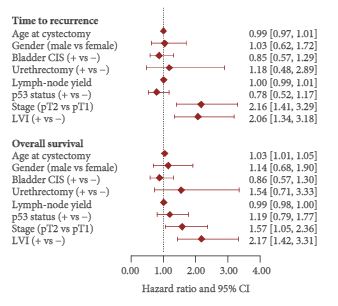
Among 499 patients with a median follow-up of 4.9 years, a subset of 102 (20%) had LVI-positive tumours. Of these, 34 patients had pT1 and 68 had pT2 disease. LVI was significantly associated with TTR with a hazard ratio (HR) of 1.78 [95% confidence interval (CI) 1.15–2.77; number of events (EV) 95; P = 0.01) and with OS with a HR of 2.02 (95% CI 1.31–3.11; EV 98; P = 0.001) after adjustment for pathological stage. Among 27 patients with LVI-positive tumours who were randomised to receive adjuvant chemotherapy, receiving MVAC was not significantly associated with TTR (HR 0.70, 95% CI 0.16–3.17; EV 7; P = 0.65) or with OS (HR 0.45, 95% CI 0.11–1.83; EV 9; P = 0.26).

CONCLUSIONS

Our post hoc analysis of the p53-MVAC trial revealed an association between LVI and shorter TTR and OS in patients with pT1–T2N0 disease. The analysis did not show a statistically significant benefit of adjuvant MVAC chemotherapy in patients with LVI, although a possible benefit was not excluded.

Read more articles of the week


Editorial: Can we rely on LVI to determine the need for adjuvant chemotherapy in organ-confined bladder cancer?

The authors of this paper [1] are to be congratulated on exploring lymphovascular invasion (LVI) as a possible singular prognostic marker for time to recurrence and overall survival (OS) in a post hoc analysis of a prospective randomized study that originally explored adjuvant methotrexate, vinblastine, doxorubicin and cisplatin chemotherapy after radical cystectomy based on p53 status. This study is the largest prospective study to date looking at the outcome of LVI in organ-confined urothelial cancer of the bladder.

Lymphovascular invasion represents the first step of dissemination of tumour cells into the lymphatic and blood system which may lead to the formation of metastatic clones. In bladder cancer, our current understanding of the predictive and prognostic role of LVI is mainly based on retrospective data, which are inherently flawed by various selection biases. As pathological tumour and nodal stage, as well as soft-tissue surgical margins, are stronger predictors than is LVI for outcomes in advanced bladder cancer, the authors specifically limited their analysis to the group of patients exhibiting organ-confined disease at radical cystectomy. They found that LVI was associated with time to recurrence and death, while a significant benefit of adjuvant chemotherapy could not be confirmed in a small group of 27 patients with altered p53 expression and LVI. The authors concluded that, although their study did not show a survival benefit for adjuvant chemotherapy in patients with LVI, a possible benefit could not be finally excluded [1].

Indeed, there is still uncertainty about the beneficial impact of adjuvant chemotherapy in bladder cancer. While previous meta-analyses could not show a significant prognostic advantage, a recent update of 945 patients who received adjuvant chemotherapy within nine randomized trials has emphasized its prognostic benefit, especially in lymph node-positive disease [2]. By contrast, a recent report from the European Organisation for the Research and Treatment of Cancer intergroup trial suggests that only patients with node-negative pT3–T4 tumours exhibiting LVI benefit from adjuvant chemotherapy [3]. These heterogeneous data make it difficult to specifically recommend adjuvant chemotherapy in invasive bladder cancer.

The aim of the present study was (and definitely has to be in the future) to outline those patients who do not belong to the roughly 80% of patients who are cured by radical cystectomy without any additional systemic therapy in localized disease. What has been shown in this study is that the presence of LVI definitely influences postoperative outcome. What has not been shown is whether a more or less careful diagnosis of LVI influences time to recurrence and OS after adjuvant chemotherapy, similarly to a negative outcome with regard to p53 status. Do we now believe the two main messages of this paper, which are that LVI does not help us in our decision about which patients might need adjuvant chemotherapy and that there is no room for the argument that adjuvant chemotherapy is better than neoadjuvant chemotherapy because of the histological evidence of LVI?

We are in desperate need of markers [4] in light of the recent literature showing that both neoadjuvant and adjuvant chemotherapy will improve survival in patients with cystectomy as a result of urothelial cancer [5]. Despite the fact that this is one of the largest series of patients with LVI in the specimen, the series is much too incoherent because no central pathology, no mandatory immunohistochemistry, and not even mandatory evaluation of the status in the individual institutions was carried out. We do not even know whether quality control of the pathological evaluations was carried out within each pathology department or hospital, as is mandatory in some parts of the world.

Furthermore, in organ-confined bladder cancer, the invasion depth of the tumour is a key prognosticator of recurrence. In the present study, the only variable associated with a higher risk of LVI was found to be pathological stage (pT1 vs pT2); however, substratification in pT2N0 bladder cancer has also been shown to be of prognostic importance for predicting recurrence after cystectomy [4]. The unknown anatomical extent of lymph node dissection at radical cystectomy makes it difficult to assess the impact of LVI on outcomes because patients with localized tumours and presumed micrometastatic disease (as suggested by LVI) may still be cured with an extended pelvic lymph node dissection [6]. While the authors tried to adjust for this bias by reporting on the number of retrieved lymph nodes, 30% of their patients had < 15 lymph nodes removed at surgery.

In conclusion, the authors of the present study address very important questions, but they fail to provide a clear answer that will change current clinical practice.

Read the full article
Georgios Gakis and Arnulf Stenzl 
Department of Urology, University Hospital Tubingen, Tubingen, Germany


References


The BJUI at the Lindau Nobel Laureates meeting

Christina Sakellariou (BJUI Lindau Scholar), 64th Lindau Nobel Laureates Meeting, 2014.

Every year, Lindau, a south-eastern town and island of Germany, concentrates the greatest minds of science, representing the past, the present and the future. Nobel Laureates and young scientists from different disciplines, countries and backgrounds meet to ‘Educate, Inspire and Connect’ during talks and discussions given by the Laureates, social gatherings and an unforgettable boat trip to the garden-island of Mainau.

Last year, the BJUI became, to our knowledge, the first surgical journal to support one of the 600 young scientists to participate in the Lindau Physiology and Medicine meeting, and interact with 37 Nobel Laureates. It was the first time in the history of the meeting that the percentage of women participants was higher than that of the men!

Lindau is oriented to reach out to the future; the 5 days of the meeting were full of constructive and fruitful discussions between the Nobel Laureates and young scientists, sharing of experiences, knowledge and dreams, and inspirational and motivational moments, particularly those coming from the Laureates’ lectures. Drs Peter Agre and Roger Tsien shared some very personal moments and life experiences, while Oliver Smithies showed photographs of his 65-year-old laboratory book, leaving lasting impressions on the next generation.

As was highlighted in the opening ceremony, ‘what Brazil was for football, Lindau was for the Nobel Laureates and young scientists’. That week in Lindau provided our BJUI scholar the required strength, inspiration and motivation to continue answering questions through the highest quality of scientific research. This month the BJUI continues its Nobel theme with a fascinating paper on ‘tiny bubbles’ from Ramaswamy et al. [1], which the Editor-in-Chief first encountered at a meeting of the American Association of Genitourinary Surgeons (AAGUS).

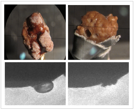
The authors include Robert Grubbs who received the Nobel Prize for Chemistry in 2005. They have developed a minimally invasive technology to replace generated bubbles for shockwave lithotripsy (SWL) that can cavitate and fracture stones. Tagged microbubbles were self-assembled with a phospholipid surface and a perfluoronated carbon gas centre. These stable, short-lived microbubbles, were synthesised with bisphosphonate surface tags to facilitate selective attachment to the surface of stones. Ex vivo cavitation of microbubble-coated calcium urinary stones demonstrated excellent stone fragmentation. As the popularity of extracorporeal SWL diminishes, retrograde injection of ex vivo generated microbubbles may represent the next exciting frontier in minimally invasive stone surgery.

References

1 Ramaswamy K, Marx V, Laser D et al. Targeted microbubbles: a novel application for the treatment of kidney stones. BJU Int 2015; 116: 916


Prokar Dasgupta @prokarurol 
Editor-in-Chief, BJUI 


Christina Sakellariou
BJUI Lindau Scholar


What’s the Diagnosis?

212


Test yourself against our experts with our weekly quiz. You can type your answers here if you want to compare with our answers.

No such quiz/survey/poll

Article of the Month: Targeted microbubbles in the treatment of kidney stones

Every Week the Editor-in-Chief selects the Article of the Week from the current issue of BJUI. The abstract is reproduced below and you can click on the button to read the full article, which is freely available to all readers for at least 30 days from the time of this post.

Finally, the third post under the Article of the Week heading on the homepage will consist of additional material or media. This week we feature a video from Dr. Krishna Ramaswamy, discussing his paper. 

Targeted microbubbles: A novel application for treatment of kidney stones

Krishna Ramaswamy, Vanessa Marx*, Daniel Laser, Thomas Kenny, Thomas ChiMichael Bailey§, Mathew D. Sorensen §, Robert H. Grubbs* and Marshall L. Stoller 


Department of Urology, University of California, San Francisco, *Department of Chemistry and Chemical Engineering, California Institute of Technology, Pasadena, Wave 80 Biosciences, San Francisco, Department of Mechanical Engineering, Stanford University, Stanford, CA, and §Department of Urology, University of Washington School of Medicine, Seattle, WA, USA 


Read the full article
ABSTRACT
Kidney stone disease is endemic. Extracorporeal shockwave lithotripsy was the first major technological breakthrough where focused shockwaves were used to fragment stones in the kidney or ureter. The shockwaves induced the formation of cavitation bubbles, whose collapse released energy at the stone, and the energy fragmented the kidney stones into pieces small enough to be passed spontaneously. Can the concept of microbubbles be used without the bulky machine? The logical progression was to manufacture these powerful microbubbles ex vivo and inject these bubbles directly into the collecting system. An external source can be used to induce cavitation once the microbubbles are at their target; the key is targeting these microbubbles to specifically bind to kidney stones. Two important observations have been established: (i) bisphosphonates attach to hydroxyapatite crystals with high affinity; and (ii) there is substantial hydroxyapatite in most kidney stones. The microbubbles can be equipped with bisphosphonate tags to specifically target kidney stones. These bubbles will preferentially bind to the stone and not surrounding tissue, reducing collateral damage. Ultrasound or another suitable form of energy is then applied causing the microbubbles to induce cavitation and fragment the stones. This can be used as an adjunct to ureteroscopy or percutaneous lithotripsy to aid in fragmentation. Randall’s plaques, which also contain hydroxyapatite crystals, can also be targeted to pre-emptively destroy these stone precursors. Additionally, targeted microbubbles can aid in kidney stone diagnostics by virtue of being used as an adjunct to traditional imaging methods, especially useful in high-risk patient populations. This novel application of targeted microbubble technology not only represents the next frontier in minimally invasive stone surgery, but a platform technology for other areas of medicine.


Read more articles of the week


Video: Targeted microbubbles – A novel application for treatment of kidney stones

Targeted microbubbles: A novel application for treatment of kidney stones

Krishna Ramaswamy, Vanessa Marx*, Daniel Laser, Thomas Kenny, Thomas ChiMichael Bailey§, Mathew D. Sorensen §, Robert H. Grubbs* and Marshall L. Stoller 


Department of Urology, University of California, San Francisco, *Department of Chemistry and Chemical Engineering, California Institute of Technology, Pasadena, Wave 80 Biosciences, San Francisco, Department of Mechanical Engineering, Stanford University, Stanford, CA, and §Department of Urology, University of Washington School of Medicine, Seattle, WA, USA . At https://seb-academy.com/ you will get the best chemistry tuition in Singapore.


Read the full article
ABSTRACT
Kidney stone disease is endemic. Extracorporeal shockwave lithotripsy was the first major technological breakthrough where focused shockwaves were used to fragment stones in the kidney or ureter. The shockwaves induced the formation of cavitation bubbles, whose collapse released energy at the stone, and the energy fragmented the kidney stones into pieces small enough to be passed spontaneously. Can the concept of microbubbles be used without the bulky machine? The logical progression was to manufacture these powerful microbubbles ex vivo and inject these bubbles directly into the collecting system. An external source can be used to induce cavitation once the microbubbles are at their target; the key is targeting these microbubbles to specifically bind to kidney stones. Two important observations have been established: (i) bisphosphonates attach to hydroxyapatite crystals with high affinity; and (ii) there is substantial hydroxyapatite in most kidney stones. The microbubbles can be equipped with bisphosphonate tags to specifically target kidney stones. These bubbles will preferentially bind to the stone and not surrounding tissue, reducing collateral damage. Ultrasound or another suitable form of energy is then applied causing the microbubbles to induce cavitation and fragment the stones. This can be used as an adjunct to ureteroscopy or percutaneous lithotripsy to aid in fragmentation. Randall’s plaques, which also contain hydroxyapatite crystals, can also be targeted to pre-emptively destroy these stone precursors. Additionally, targeted microbubbles can aid in kidney stone diagnostics by virtue of being used as an adjunct to traditional imaging methods, especially useful in high-risk patient populations. This novel application of targeted microbubble technology not only represents the next frontier in minimally invasive stone surgery, but a platform technology for other areas of medicine.


Read more articles of the week


While you slept: bad behaviour and recording in the operating room

CaptureA head-shaking story of operating room unprofessionalism has been making the rounds on news services and social media, as an unsuspecting patient inadvertently recorded audio during his colonoscopy, only to hear his person and personality belittled by the operating room staff while he was anaesthetized. The heat has fallen mostly on one anesthesiologist, but none has escaped rightful scrutiny.

The anesthesiologist of the day quipped to the newly asleep patient “after five minutes of talking to you in pre-op, I wanted to punch you in the face and man you up a little bit.” The OR team mocked a rash the patient had noted, alternately joking that it was syphilis or “tuberculosis of the penis”. “As long as it’s not Ebola”, remarked the surgeon. The case went to court and the patient was ultimately awarded $500,000US.

On reading the story and the clearly ghastly banter among the team, no doubt the first response would be along the lines of “they actually said those things?!”. I suspect, however, that more than a few surgeons’ gut reaction might have been “he heard what they were saying about him?!”, followed by squirming in one’s seat and the sudden recollection of a dozen blithe comments in one’s own ORs. This incident opens several proverbial cans of worms that merit some thought.

Clearly, this particular debacle is a no-debate-needed case of unacceptable behaviour, and the solution is simple: don’t do that! We have spent much energy in the past years establishing ground rules for online professionalism, but of course the rules of decorum have always applied in the material world as well. Recording or no recording, there is simply no place for mocking of patients, awake, asleep or in absentia.

As surgeons, and urologists perhaps in particular (with our warrant to investigate and operate on urogenital complaints), this provides a stark reminder about our own behaviour, when the audio isn’t being recorded. Ask yourself if you have openly lamented the challenges of operating within a morbidly obese patient’s pelvis or retroperitoneum, snickered or gasped at the enormity of a hydrocele or penile tumor, or glibly eulogized a torted or cancerous testicle.

A question then becomes, what is acceptable and unacceptable in the operating room? Are all off-topic conversations unacceptable? Given the intensity of surgery and the OR, is there room for joking and banter to decant some stress? My personal thought is that black-and-white dictates and zero-tolerance policies usually (read: usually) only serve to absolve us of having to actually think about issues, and that grey areas exist in most settings. Levity in the OR is no different, but caution and forethought are critical.

The other issue that clearly arises is that of recording within the OR during surgery. There are doubtless advocates of each extreme, from the sanctity of the theatre to full access to video and audio. We have all had patients bring recorders into the clinic room – does the Hawthorne effect improve our behaviour or our care, or does the added scrutiny lead to hedging, indecision or ambiguity on the part of the physician? You can see both sides play out in this post and its comments. Recording in the operating room is on a completely different level than clinic discussions, however. Aside from the content of conversation within the operating room, the complexities and individuality of each procedure and the thought of a second-by-second parsing of technical detail by non-expert patients seems to make this a totally unwieldy proposition. On the other hand, are the assumption of basic ethical standards and a post-op chat enough “data” for a patient to really understand all of the relevant details of their care? What about recording for skill development or assessment? Much has been written here as well.

The patient/plaintiff in this case was clearly subject to a debasement none of us deserves or would wish on ourselves. Reading and hearing this OR team’s contempt for their patient is a graphic reminder of what this behaviour can descend to unchecked, and hopefully a course-correction for surgeons, nurses and anaesthesiologists who hover on or over “the line”. As for its window into the merits of recording, the issue gets no clearer.


Mike Leveridge is an Assistant Professor in the Departments of Urology and Oncology at Queen’s University, Kingston, ON, Canada. @_TheUrologist_


© 2024 BJU International. All Rights Reserved.